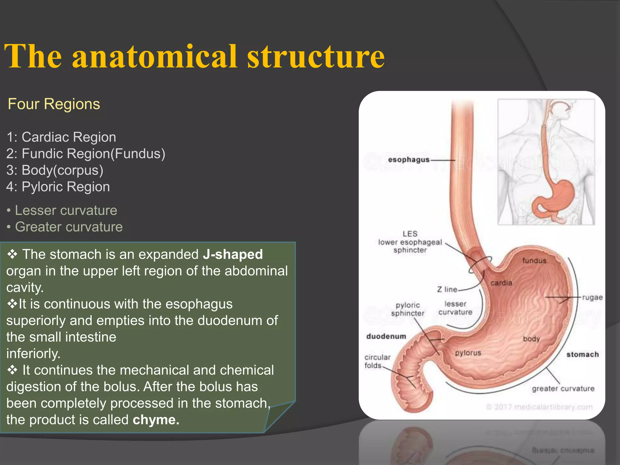
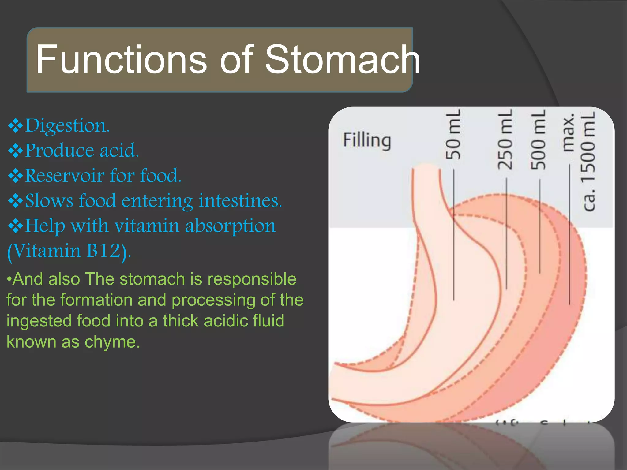
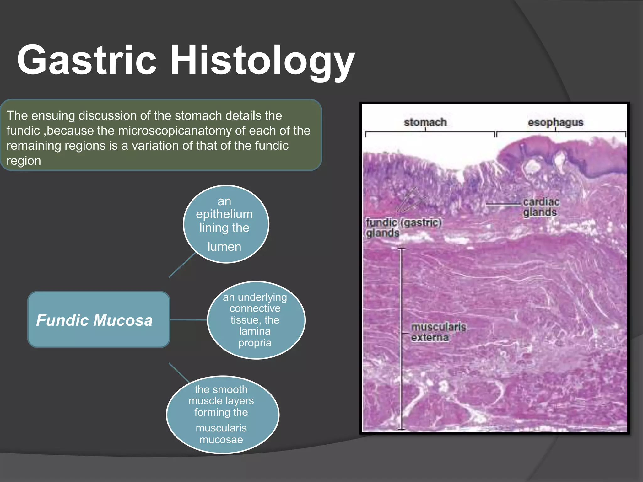
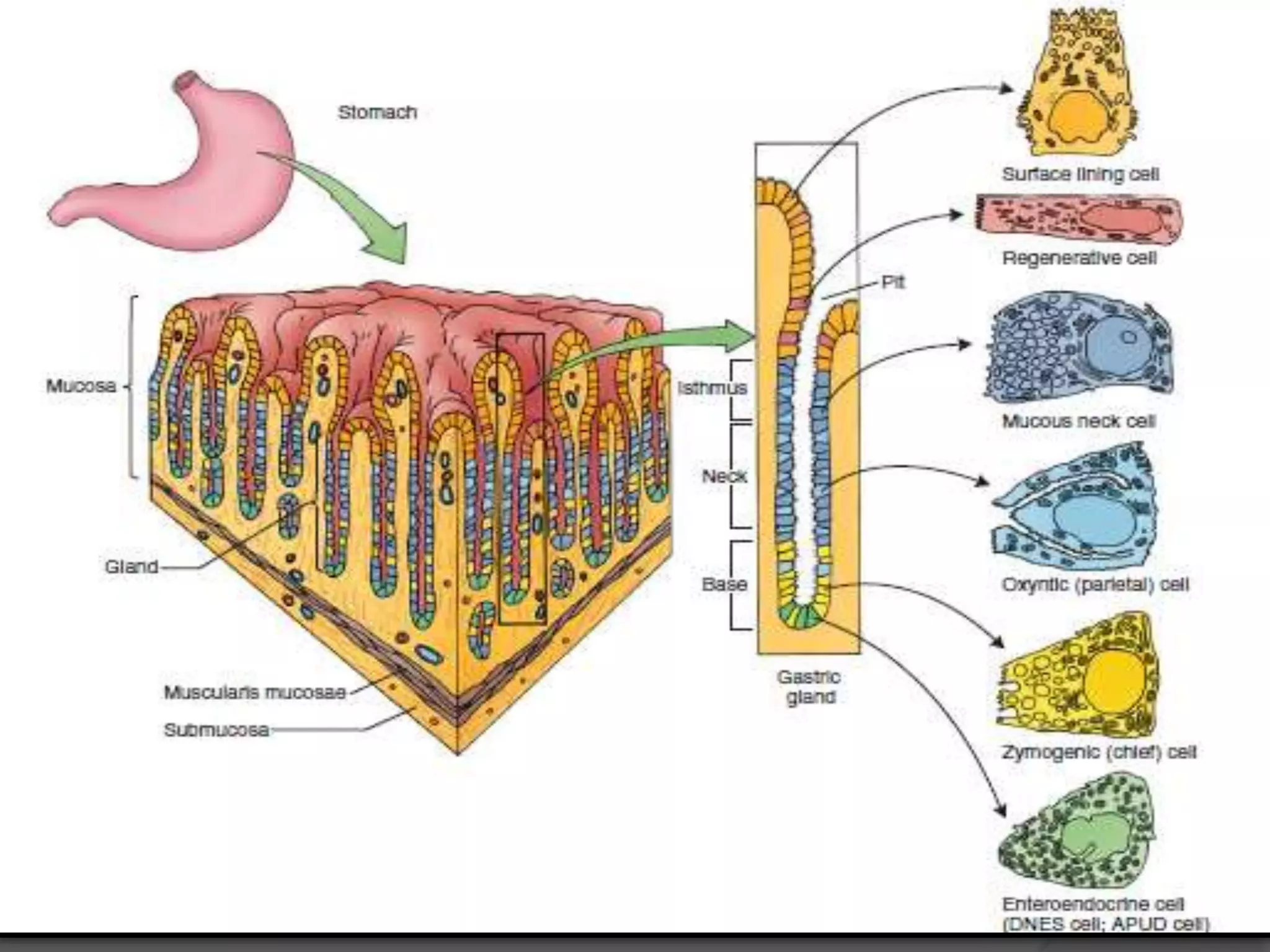
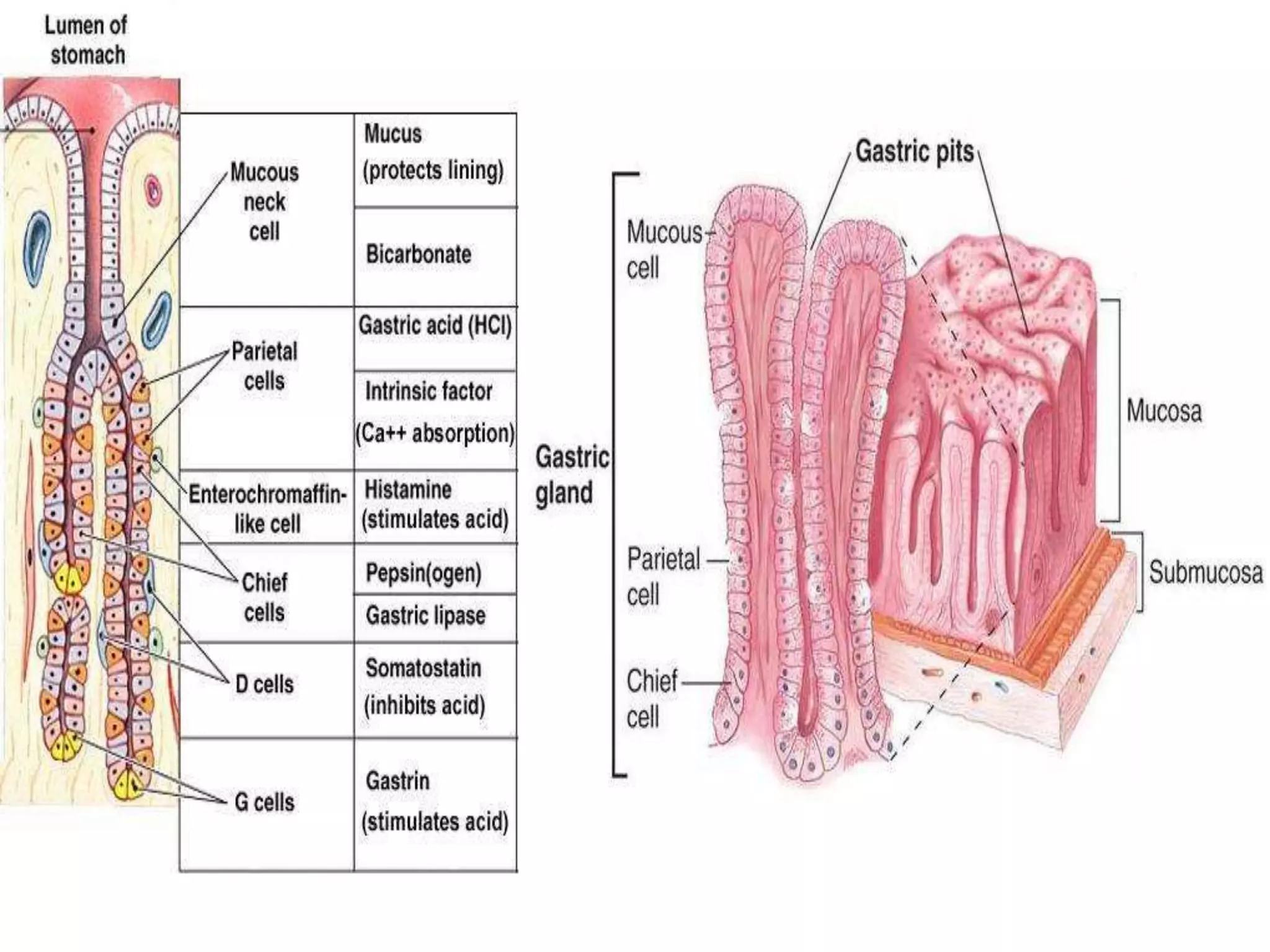
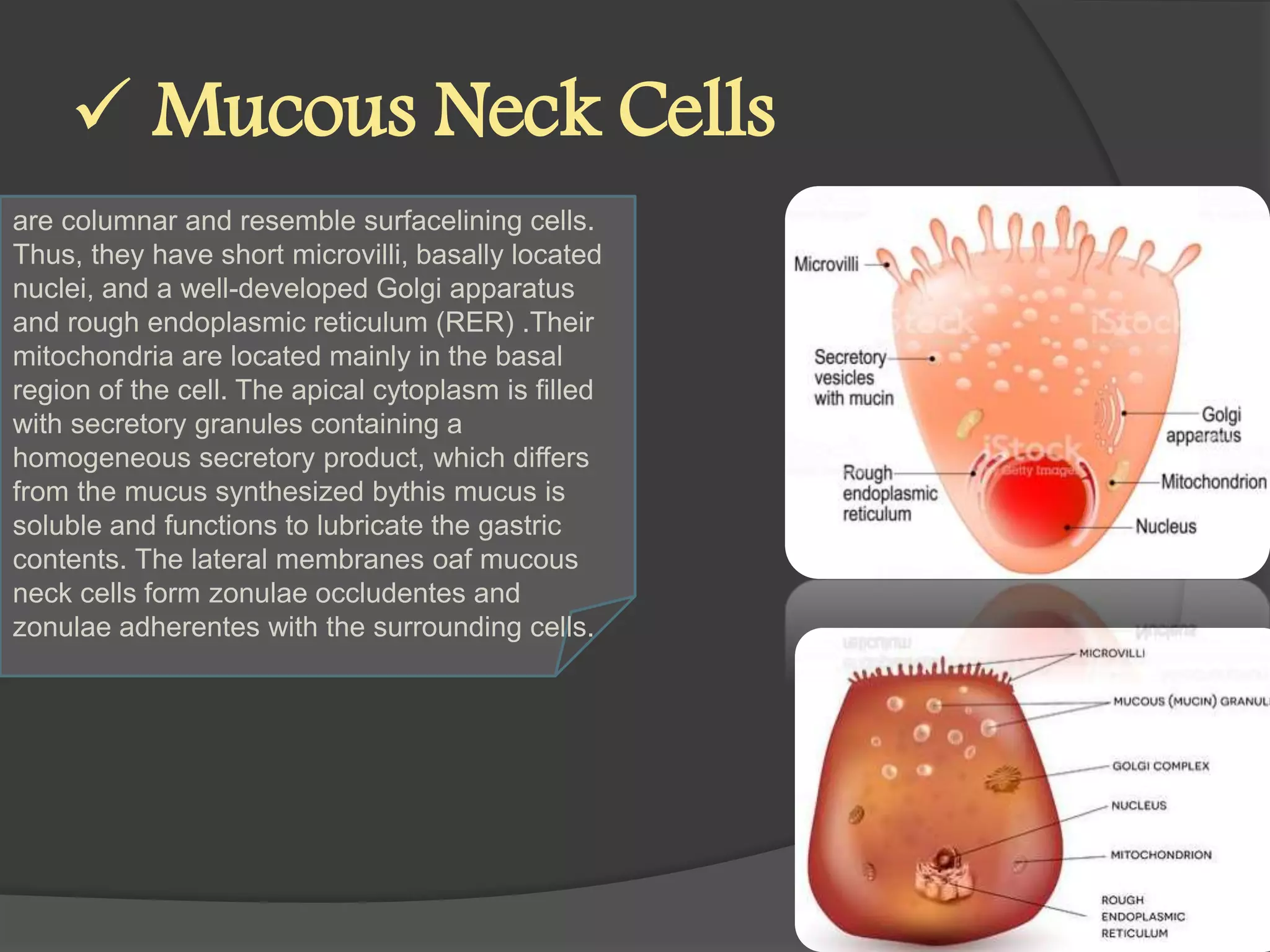
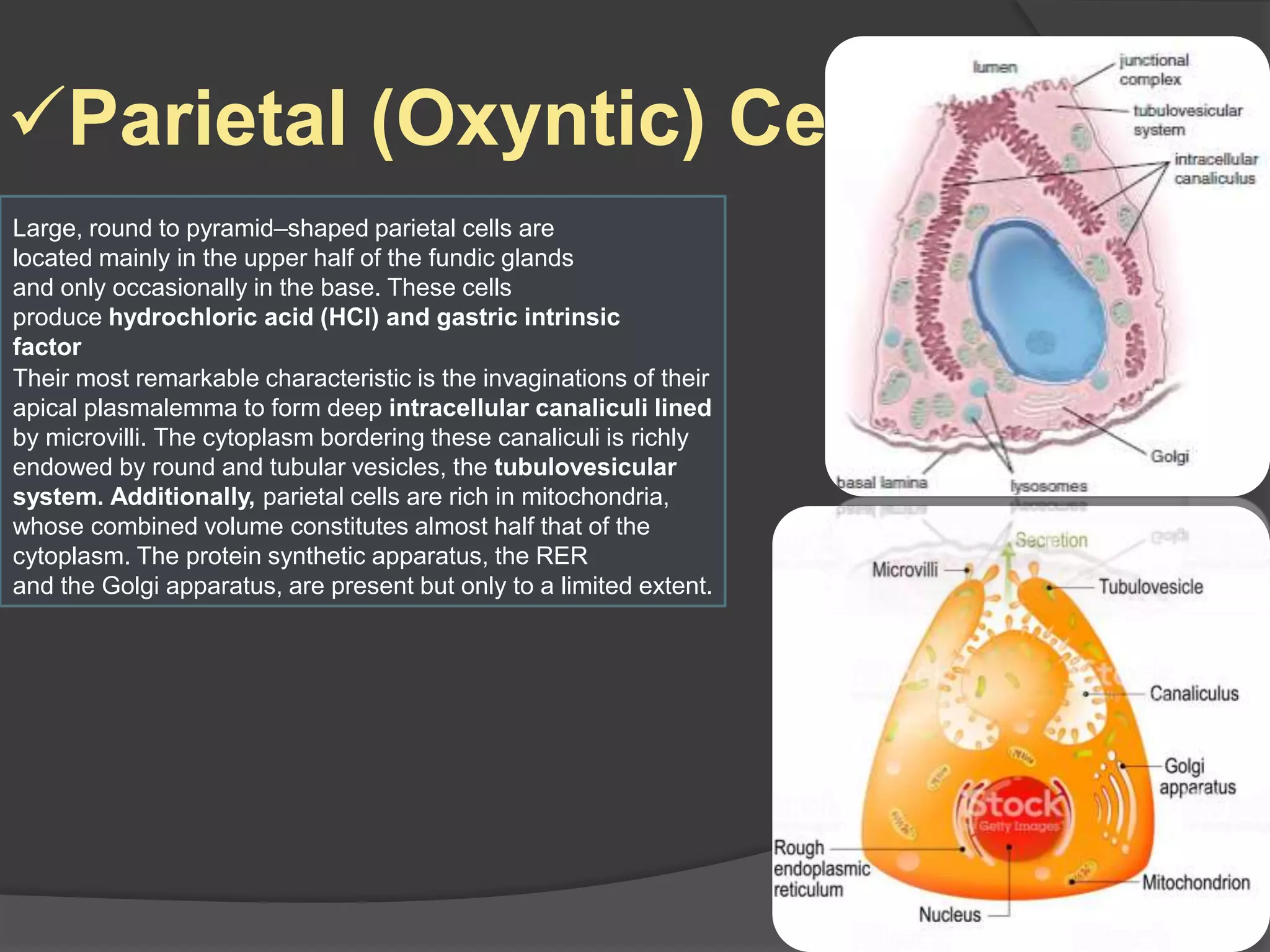
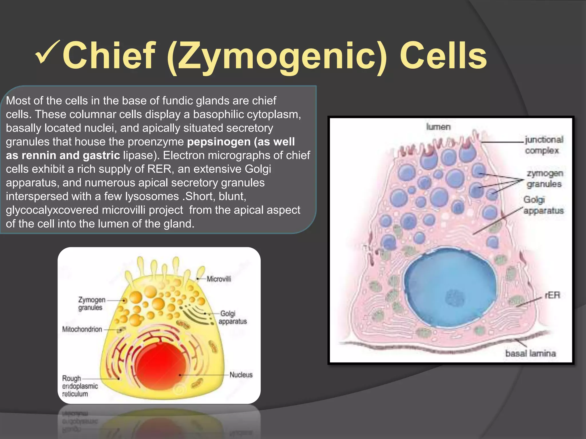
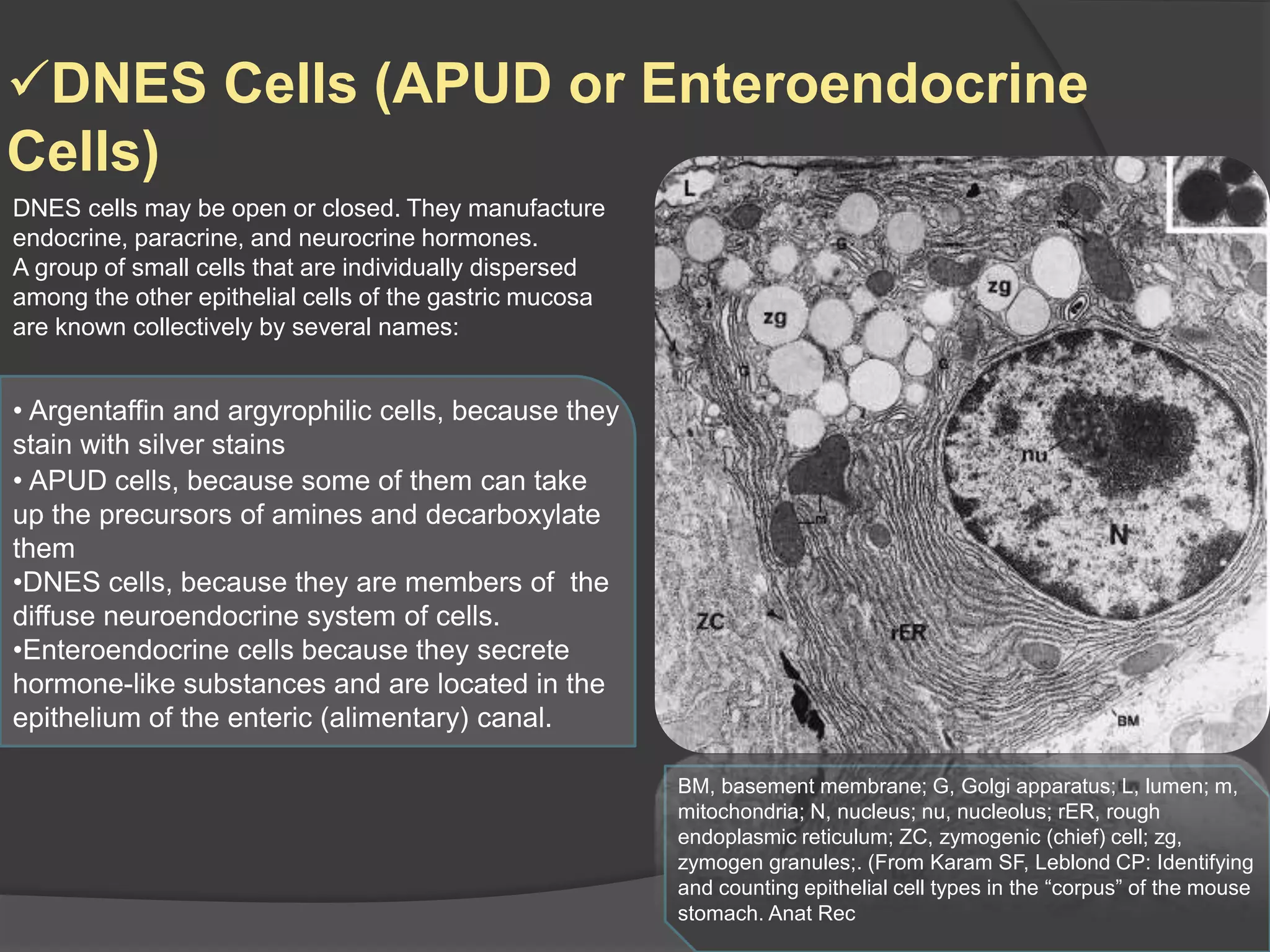
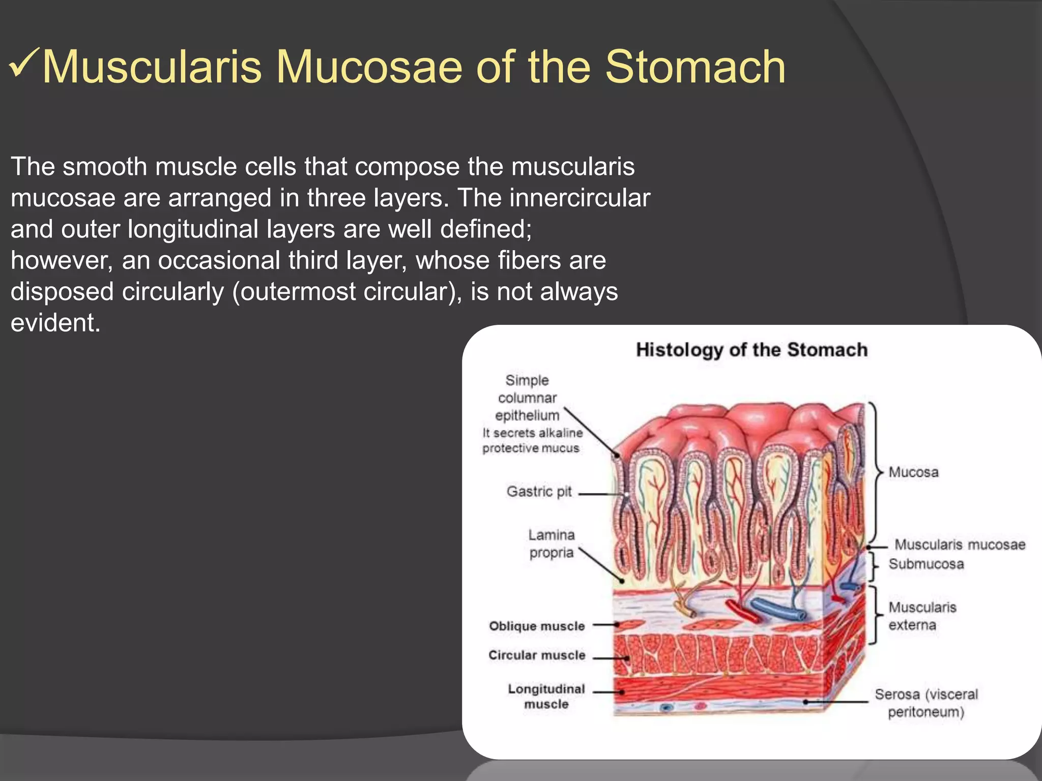
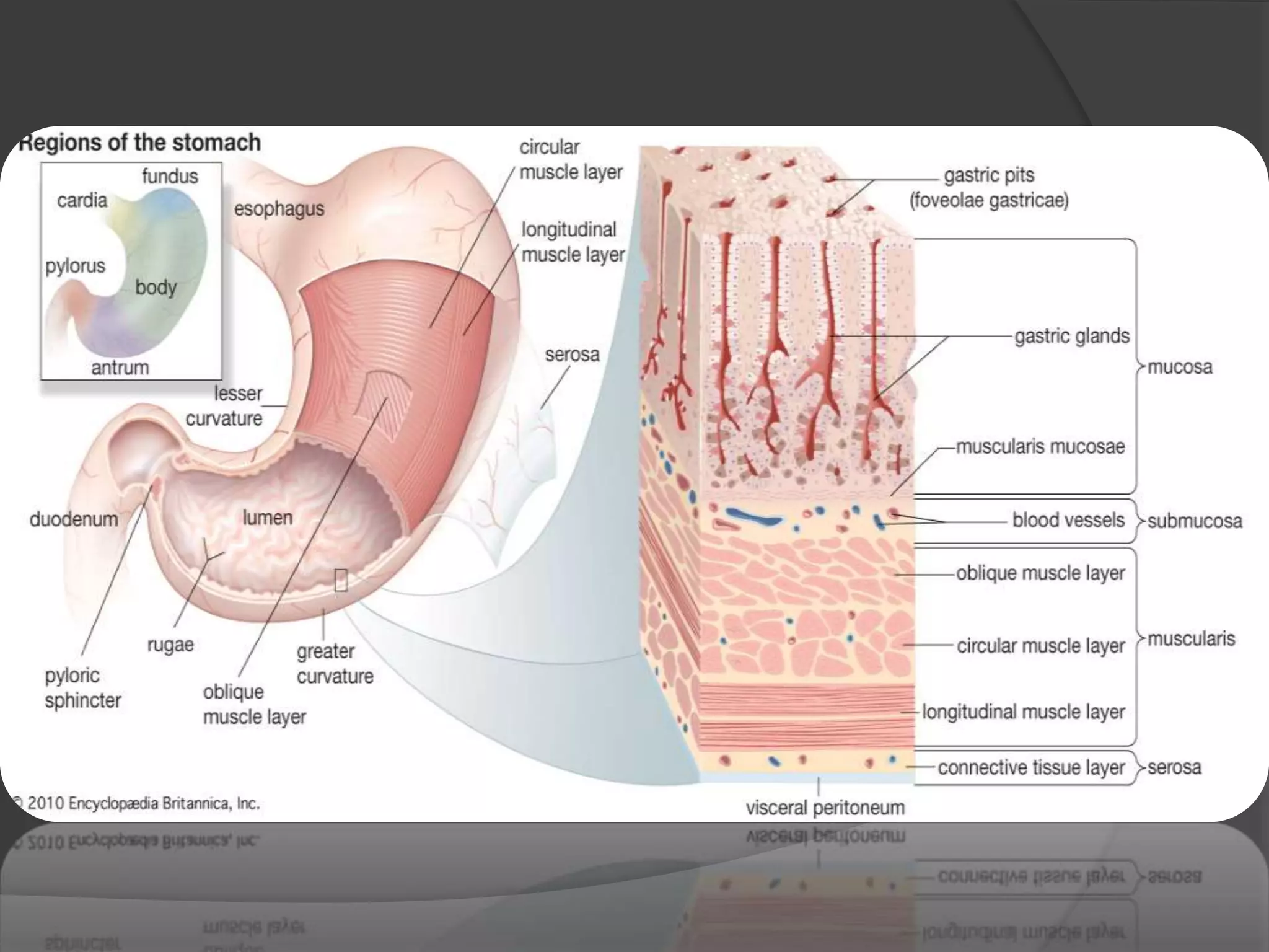
The Stomach
By :- Ali Laith Al-Alwani & Saja Ali
Group :-117 i-6
The anatomical structure
Four Regions
1: Cardiac Region
2: Fundic Region(Fundus)
3: Body(corpus)
4: Pyloric Region
• Lesser curvature
• Greater curvature
 The stomach is an expanded J-shaped
organ in the upper left region of the abdominal
cavity.
It is continuous with the esophagus
superiorly and empties into the duodenum of
the small intestine
inferiorly.
 It continues the mechanical and chemical
digestion of the bolus. After the bolus has
been completely processed in the stomach,
the product is called chyme.
Functions of Stomach
Digestion.
Produce acid.
Reservoir for food.
Slows food entering intestines.
Help with vitamin absorption
(Vitamin B12).
•And also The stomach is responsible
for the formation and processing of the
ingested food into a thick acidic fluid
known as chyme.
Gastric Histology
The ensuing discussion of the stomach details the
fundic ,because the microscopicanatomy of each of the
remaining regions is a variation of that of the fundic
region
an
epithelium
lining the
lumen
an underlying
connective
tissue, the
lamina
propria
the smooth
muscle layers
forming the
muscularis
mucosae
Fundic Mucosa
FUNDIC GLANDS
isthmus,
baseneck
The simple columnar epithelium
constituting the fundic gland is composed
of six cell types:-
(1) surface-lining cells,
(2) mucous neck cells,
(3) regenerative (stem) cells,
(4) parietal (oxyntic) cells,
(5) chief (zymogenic) cells,
(6) diffuse neuroendocrine system (DNES)
cells
 Mucous Neck Cells
are columnar and resemble surfacelining cells.
Thus, they have short microvilli, basally located
nuclei, and a well-developed Golgi apparatus
and rough endoplasmic reticulum (RER) .Their
mitochondria are located mainly in the basal
region of the cell. The apical cytoplasm is filled
with secretory granules containing a
homogeneous secretory product, which differs
from the mucus synthesized bythis mucus is
soluble and functions to lubricate the gastric
contents. The lateral membranes oaf mucous
neck cells form zonulae occludentes and
zonulae adherentes with the surrounding cells.
Regenerative (Stem)
Cells
These columnar stem cells do not have many
organelles but do have a rich supply of ribosomes.
Their nuclei are basally located, have little
heterochromatin, and display a large nucleolus. The
lateral cell membranes of these cells also form zonulae
occludentes and zonulae adherentes with those of
neighboring cells. Regenerative cells proliferate to
replace all of the specialized cells lining the fundic
glands, gastric pits, and luminal surface. Newly formed
cells migrate to their new locations either deep into the
gland or up into the gastric pit and gastric lining.
Surface-lining cells, DNES cells, and mucous neck cells
are replaced every 5 to 7 days; thus, regenerative cells
have a high proliferative rate.
Parietal (Oxyntic) Cells
Large, round to pyramid–shaped parietal cells are
located mainly in the upper half of the fundic glands
and only occasionally in the base. These cells
produce hydrochloric acid (HCl) and gastric intrinsic
factor
Their most remarkable characteristic is the invaginations of their
apical plasmalemma to form deep intracellular canaliculi lined
by microvilli. The cytoplasm bordering these canaliculi is richly
endowed by round and tubular vesicles, the tubulovesicular
system. Additionally, parietal cells are rich in mitochondria,
whose combined volume constitutes almost half that of the
cytoplasm. The protein synthetic apparatus, the RER
and the Golgi apparatus, are present but only to a limited extent.
Chief (Zymogenic) Cells
Most of the cells in the base of fundic glands are chief
cells. These columnar cells display a basophilic cytoplasm,
basally located nuclei, and apically situated secretory
granules that house the proenzyme pepsinogen (as well
as rennin and gastric lipase). Electron micrographs of chief
cells exhibit a rich supply of RER, an extensive Golgi
apparatus, and numerous apical secretory granules
interspersed with a few lysosomes .Short, blunt,
glycocalyxcovered microvilli project from the apical aspect
of the cell into the lumen of the gland.
DNES Cells (APUD or Enteroendocrine
Cells)
DNES cells may be open or closed. They manufacture
endocrine, paracrine, and neurocrine hormones.
A group of small cells that are individually dispersed
among the other epithelial cells of the gastric mucosa
are known collectively by several names:
• Argentaffin and argyrophilic cells, because they
stain with silver stains
• APUD cells, because some of them can take
up the precursors of amines and decarboxylate
them
•DNES cells, because they are members of the
diffuse neuroendocrine system of cells.
•Enteroendocrine cells because they secrete
hormone-like substances and are located in the
epithelium of the enteric (alimentary) canal.
BM, basement membrane; G, Golgi apparatus; L, lumen; m,
mitochondria; N, nucleus; nu, nucleolus; rER, rough
endoplasmic reticulum; ZC, zymogenic (chief) cell; zg,
zymogen granules;. (From Karam SF, Leblond CP: Identifying
and counting epithelial cell types in the “corpus” of the mouse
stomach. Anat Rec
Muscularis Mucosae of the Stomach
The smooth muscle cells that compose the muscularis
mucosae are arranged in three layers. The innercircular
and outer longitudinal layers are well defined;
however, an occasional third layer, whose fibers are
disposed circularly (outermost circular), is not always
evident.
Differences in the Mucosa of the Cardiac
and Pyloric Regions
The mucosa of the cardiac region of the stomach
differs from that of the fundic region, in that the gastric pits are
shallower and the base of its glands is highly
coiled. The cell population of these cardiac glands
composed mostly of surface-lining cells, some mucous
neck cells, a few DNES cells and parietal cells, and no
chief cells
The glands of the pyloric region contain the same
cell types as those in the cardiac region, but the predominant
cell type in the pylorus is the mucous neck
cell. In addition to producing mucus, these cells secrete
lysozyme, a bactericidal enzyme. Pyloric glands are
highly convoluted and tend to branch. Additionally, the
gastric pits of the pyloric region are deeper than in both
the cardiac and pyloric regions, extending approximately
halfway down into the lamina propria
Submucosa of the
StomachThe dense, irregular collagenous connective tissue
of the gastric submucosa has a rich vascular and lymphatic
network that supplies and drains the vessels of
the lamina propria. The cell population of the submucosa
resembles that of any connective tissue proper.
The submucosal plexus is in its accustomed location,
within the submucosa in the vicinity of the muscularis
externa.
Muscularis Externa
The smooth muscle cells of the gastric muscularis
externa are arranged in three layers. The innermost
oblique layer is not well defined except in the
cardiacregion. The middle circular layer is clearly evident
along the entire stomach and is especially pronounced
in the pyloric region, where it forms the pyloric
sphincter. The outer longitudinal muscle layer is
most evident in the cardiac region and the body of the
stomach but is poorly developed in the pylorus. The
myenteric plexus is located between the middle circular
and outer longitudinal layers of smooth muscle.
The serosa of the stomach is as
described above for the alimentary
canal in general. It is continuous with the
parietal peritoneum of the abdominal cavity
via the greater omentum and with visceral
peritoneum of the liver at the lesser
omentum. Otherwise, it exhibits no special
features.
Stomach anatomy and histology

Stomach anatomy and histology

  • 1.
    The Stomach By :-Ali Laith Al-Alwani & Saja Ali Group :-117 i-6
  • 2.
    The anatomical structure FourRegions 1: Cardiac Region 2: Fundic Region(Fundus) 3: Body(corpus) 4: Pyloric Region • Lesser curvature • Greater curvature  The stomach is an expanded J-shaped organ in the upper left region of the abdominal cavity. It is continuous with the esophagus superiorly and empties into the duodenum of the small intestine inferiorly.  It continues the mechanical and chemical digestion of the bolus. After the bolus has been completely processed in the stomach, the product is called chyme.
  • 4.
    Functions of Stomach Digestion. Produceacid. Reservoir for food. Slows food entering intestines. Help with vitamin absorption (Vitamin B12). •And also The stomach is responsible for the formation and processing of the ingested food into a thick acidic fluid known as chyme.
  • 5.
    Gastric Histology The ensuingdiscussion of the stomach details the fundic ,because the microscopicanatomy of each of the remaining regions is a variation of that of the fundic region an epithelium lining the lumen an underlying connective tissue, the lamina propria the smooth muscle layers forming the muscularis mucosae Fundic Mucosa
  • 7.
    FUNDIC GLANDS isthmus, baseneck The simplecolumnar epithelium constituting the fundic gland is composed of six cell types:- (1) surface-lining cells, (2) mucous neck cells, (3) regenerative (stem) cells, (4) parietal (oxyntic) cells, (5) chief (zymogenic) cells, (6) diffuse neuroendocrine system (DNES) cells
  • 11.
     Mucous NeckCells are columnar and resemble surfacelining cells. Thus, they have short microvilli, basally located nuclei, and a well-developed Golgi apparatus and rough endoplasmic reticulum (RER) .Their mitochondria are located mainly in the basal region of the cell. The apical cytoplasm is filled with secretory granules containing a homogeneous secretory product, which differs from the mucus synthesized bythis mucus is soluble and functions to lubricate the gastric contents. The lateral membranes oaf mucous neck cells form zonulae occludentes and zonulae adherentes with the surrounding cells.
  • 12.
    Regenerative (Stem) Cells These columnarstem cells do not have many organelles but do have a rich supply of ribosomes. Their nuclei are basally located, have little heterochromatin, and display a large nucleolus. The lateral cell membranes of these cells also form zonulae occludentes and zonulae adherentes with those of neighboring cells. Regenerative cells proliferate to replace all of the specialized cells lining the fundic glands, gastric pits, and luminal surface. Newly formed cells migrate to their new locations either deep into the gland or up into the gastric pit and gastric lining. Surface-lining cells, DNES cells, and mucous neck cells are replaced every 5 to 7 days; thus, regenerative cells have a high proliferative rate.
  • 13.
    Parietal (Oxyntic) Cells Large,round to pyramid–shaped parietal cells are located mainly in the upper half of the fundic glands and only occasionally in the base. These cells produce hydrochloric acid (HCl) and gastric intrinsic factor Their most remarkable characteristic is the invaginations of their apical plasmalemma to form deep intracellular canaliculi lined by microvilli. The cytoplasm bordering these canaliculi is richly endowed by round and tubular vesicles, the tubulovesicular system. Additionally, parietal cells are rich in mitochondria, whose combined volume constitutes almost half that of the cytoplasm. The protein synthetic apparatus, the RER and the Golgi apparatus, are present but only to a limited extent.
  • 14.
    Chief (Zymogenic) Cells Mostof the cells in the base of fundic glands are chief cells. These columnar cells display a basophilic cytoplasm, basally located nuclei, and apically situated secretory granules that house the proenzyme pepsinogen (as well as rennin and gastric lipase). Electron micrographs of chief cells exhibit a rich supply of RER, an extensive Golgi apparatus, and numerous apical secretory granules interspersed with a few lysosomes .Short, blunt, glycocalyxcovered microvilli project from the apical aspect of the cell into the lumen of the gland.
  • 15.
    DNES Cells (APUDor Enteroendocrine Cells) DNES cells may be open or closed. They manufacture endocrine, paracrine, and neurocrine hormones. A group of small cells that are individually dispersed among the other epithelial cells of the gastric mucosa are known collectively by several names: • Argentaffin and argyrophilic cells, because they stain with silver stains • APUD cells, because some of them can take up the precursors of amines and decarboxylate them •DNES cells, because they are members of the diffuse neuroendocrine system of cells. •Enteroendocrine cells because they secrete hormone-like substances and are located in the epithelium of the enteric (alimentary) canal. BM, basement membrane; G, Golgi apparatus; L, lumen; m, mitochondria; N, nucleus; nu, nucleolus; rER, rough endoplasmic reticulum; ZC, zymogenic (chief) cell; zg, zymogen granules;. (From Karam SF, Leblond CP: Identifying and counting epithelial cell types in the “corpus” of the mouse stomach. Anat Rec
  • 17.
    Muscularis Mucosae ofthe Stomach The smooth muscle cells that compose the muscularis mucosae are arranged in three layers. The innercircular and outer longitudinal layers are well defined; however, an occasional third layer, whose fibers are disposed circularly (outermost circular), is not always evident.
  • 19.
    Differences in theMucosa of the Cardiac and Pyloric Regions The mucosa of the cardiac region of the stomach differs from that of the fundic region, in that the gastric pits are shallower and the base of its glands is highly coiled. The cell population of these cardiac glands composed mostly of surface-lining cells, some mucous neck cells, a few DNES cells and parietal cells, and no chief cells The glands of the pyloric region contain the same cell types as those in the cardiac region, but the predominant cell type in the pylorus is the mucous neck cell. In addition to producing mucus, these cells secrete lysozyme, a bactericidal enzyme. Pyloric glands are highly convoluted and tend to branch. Additionally, the gastric pits of the pyloric region are deeper than in both the cardiac and pyloric regions, extending approximately halfway down into the lamina propria
  • 20.
    Submucosa of the StomachThedense, irregular collagenous connective tissue of the gastric submucosa has a rich vascular and lymphatic network that supplies and drains the vessels of the lamina propria. The cell population of the submucosa resembles that of any connective tissue proper. The submucosal plexus is in its accustomed location, within the submucosa in the vicinity of the muscularis externa. Muscularis Externa The smooth muscle cells of the gastric muscularis externa are arranged in three layers. The innermost oblique layer is not well defined except in the cardiacregion. The middle circular layer is clearly evident along the entire stomach and is especially pronounced in the pyloric region, where it forms the pyloric sphincter. The outer longitudinal muscle layer is most evident in the cardiac region and the body of the stomach but is poorly developed in the pylorus. The myenteric plexus is located between the middle circular and outer longitudinal layers of smooth muscle. The serosa of the stomach is as described above for the alimentary canal in general. It is continuous with the parietal peritoneum of the abdominal cavity via the greater omentum and with visceral peritoneum of the liver at the lesser omentum. Otherwise, it exhibits no special features.