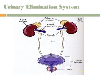
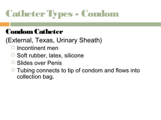
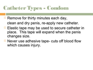
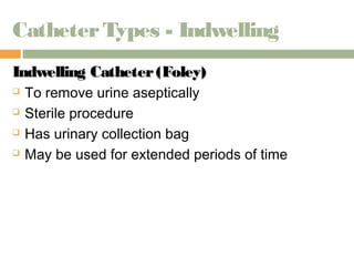
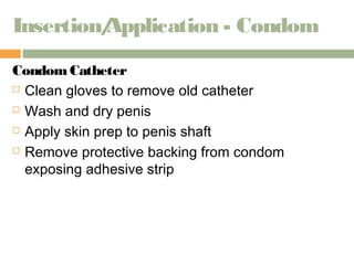
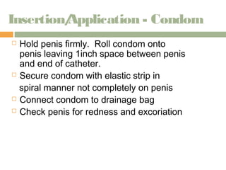
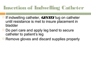
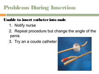
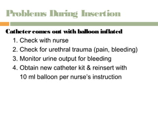
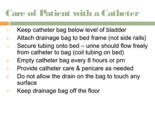
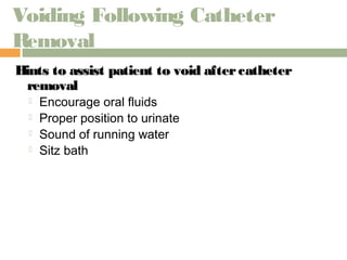
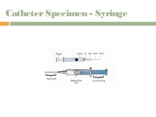
URINARY ELIMINATION 
& CATHETERS
Objectives 
 Demonstrate correct technique for inserting 
indwelling and straight catheters 
 Recognizes signs and symptoms of infection 
related to urinary catheters 
 Demonstrate aseptic routine care of a patient with 
an indwelling catheter 
 Demonstrate correct technique for removing an 
indwelling catheter. 
 Demonstrate correct procedure for application of 
condom catheter 
 Document and report to RN care related to urinary 
drainage
Urinary Elimination System
Anatomy 
Kidney - Urine p ro duc tio n 
Ureters - Tube s which c o nne c t the kidne y s to the 
bla d d e r. 
Bladder - Re s e rvo ir fo r urine until the urg e to vo id 
ta ke s p la c e . 
Urethra - Tube fro m the bla d d e r to the o uts id e o f the 
bo d y in no rm a l a na to m y . 
Male = 20 cm long 
Female = 4 cm long
Anatomy Facts 
 A continuous layer of mucous membrane lines 
the kidney pelvis, ureters, urethra & bladder 
 Because there is no break in this lining, bacteria 
introduced into this normally sterile environment 
can quickly spread throughout the entire system 
 When the bladder is empty it falls into folds which 
provide pockets where bacteria can multiply 
 Because this membrane layer is highly vascular, 
bacteria can easily enter the blood stream & 
cause septicemia
Catheterization - Reasons 
 Collect a sterile specimen 
 Check residual urine 
 Inability to void related to post-op complications 
 Before, during or after surgery to empty bladder 
 Temporary/Permanent Incontinence: 
a. Total- c o ntinuo us lo s s o f urine fro m bla d d e r 
b. Stress- le a ka g e o f sm a ll am o unts o f urine with 
phy s ic a l s tre s s i. e . c o ug hing o r s ne e z ing
Catheterization - Reasons 
 Urge - Invo lunta ry p a s s a g e o f urine a fte r fe e ling 
s tro ng ne e d to vo id . Una ble to s to p urina ting a nd 
c a n’t g e t to ba thro o m o n tim e . Se e n in the 
e ld e rly , with UTIs a nd tum o rs . 
 Functional - Invo lunta ry , unp re d ic ta ble p a s s a g e 
o f urine re la te d to no c a ll lig ht o r im m o bility , e tc .
Alternative to Catheterization 
 Bladder Scan 
 Estimates residual urine 
 Increased use over straight catheterization 
 90% accuracy 
 Decreases risk of infection and discomfort to patient
PRIOR to Catheterization 
KKnnooww aanndd CCoonnffiirrmm 
 Doctor’s order 
 Last void 
 Patient’s level of consciousness and understanding 
 Mobility, physical limitations, ability to cooperate 
 Sex, age, catheter size 
 Kids 6-10 
 Female 14-16 
 Male 16-18) 
 Allergies to antiseptics, tape, rubber, latex 
 Adverse conditions 
 Impaired passage such as enlarge prostate
Catheter Types - Condom 
CCoonnddoomm CCaatthheetteerr 
(External, Texas, Urinary Sheath) 
 Incontinent men 
 Soft rubber, latex, silicone 
 Slides over Penis 
 Tubing connects to tip of condom and flows into 
collection bag.
Catheter Types - Condom 
 Remove for thirty minutes each day, 
clean and dry penis, re-apply new catheter. 
 Elastic tape may be used to secure catheter in 
place. This tape will expand when the penis 
changes size. 
 Never use adhesive tape- cuts off blood flow 
which causes injury.
Catheter Types - Straight 
SSttrraaiigghhtt CCaatthheetteerr 
 Removes urine from bladder aseptically due to 
1. Incontinence 
2. Post void residuals 
3. Sterile specimen 
 Do e s no t a tta ch to a c o lle c tio n ba g 
 Co ns id e re d a s te rile p ro c e dure
Catheter Types - Indwelling 
Indwelling CCaatthheetteerr ((FFoolleeyy)) 
 To remove urine aseptically 
 Sterile procedure 
 Has urinary collection bag 
 May be used for extended periods of time
Catheter Types - Suprapubic 
SSuupprraappuubbiicc 
 Catheter inserted into the bladder through an 
opening in the lower abdomen (a bo ut 1 inch 
a bo ve the s ym phy s is p ubis ) 
 Placed under anesthesia 
 May be left in place for long periods of time 
 Sutured into place.
Rules for Catheterization 
1. Wash hands, use gloves 
2. Tubing without kinks/obstructions 
3. Drainage bag below bladder 
4. Attach to non-movable bed part 
5. Secure with leg strap 
6. Check system for leaks 
7. Perform perineal care BID, after all BMs &with cath care 
8. Watch I&O closely 
9. Report complaints of discomfort to nurse (pain, burning, 
irritation) 
10. Record amount, color, clarity, odor, particles
Insertion/Application - Condom 
Condom Catheter 
 Clean gloves to remove old catheter 
 Wash and dry penis 
 Apply skin prep to penis shaft 
 Remove protective backing from condom 
exposing adhesive strip
Insertion/Application - Condom 
 Hold penis firmly. Roll condom onto 
penis leaving 1inch space between penis 
and end of catheter. 
 Secure condom with elastic strip in 
spiral manner not completely on penis 
 Connect condom to drainage bag 
 Check penis for redness and excoriation
Insertion Tips – Straight/Foley 
 Have patient relax by taking slow deep 
breaths 
 Lubricate catheter well 
 To find female meatus 
 Look for the wink 
 Betadine pools in the meatus 
 Ask patient to bear down as if to void 
 If catheter enters the vagina, leave it
Insertion of Indwelling Catheter 
 If indwelling catheter, GENTLY tug on catheter 
until resistance is met to insure placement in 
bladder 
 Do peri care and apply leg band to secure 
catheter to patient’s leg 
 Remove gloves and discard supplies properly
Other Considerations 
When performing straight cath 
 Know policy & procedure for maximum amount 
of urine allowed to be withdrawn. 
To place catheter in an uncircumcised man 
 Carefully pull back foreskin before cleansing
Documentation 
DDooccuummeenntt 
 Type of procedure 
 Size of catheter and balloon 
 Amount of fluid actually instilled into balloon 
 Amount of urine returned 
 Characteristics of urine (color, odor, sediment) 
 Specimen sent if applicable 
 How patient tolerated procedure
Problems During Insertion 
Catheter inserted iinnttoo VVaaggiinnaa 
1. Leave in place 
2. Reposition fingers to re-visualize meatus 
3. Use new catheter 
4. Repeat procedure
Problems During Insertion 
Unable to iinnsseerrtt iinnttoo ffeemmaallee 
1. Ask patient or other team member to hold legs 
apart 
2. Observe urethral opening (before cleansing) for 
movement when pressure is applied 
3. Improve lighting 
4. Repeat insertion procedure
Problems During Insertion 
Unable to insert ccaatthheetteerr iinnttoo mmaallee 
1. Notify nurse 
2. Repeat procedure but change the angle of the 
penis 
3. Try an a coude catheter
Problems During Insertion 
Catheter comes out with bbaalllloooonn iinnffllaatteedd 
1. Check with nurse 
2. Check for urethral trauma (pain, bleeding) 
3. Monitor urine output for bleeding 
4. Obtain new catheter kit & reinsert with 
10 ml balloon per nurse’s instruction
Care of Patient with a Catheter 
1. Keep catheter bag below level of bladder 
2. Attach drainage bag to bed frame (not side rails) 
3. Secure tubing onto bed – urine should flow freely 
from catheter to bag (coil tubing on bed) 
4. Empty catheter bag every 8 hours or prn 
5. Provide catheter care & pericare as needed 
6. Do not allow the drain on the bag to touch any 
surface 
7. Keep drainage bag off the floor
Catheter Removal 
1. Physician’s order 
2. Gather supplies- 10cc syringe, gloves, chux 
3. Deflate balloon- note amount withdrawn 
4. Avoid pubic hair 
5. Pinch catheter; pull out on exhalation steadily & 
gently 
6. Measure output 
7. Discard catheter bag properly 
8. Provide peri care 
9. Monitor patient for output 
10. If not voided in 4-6 hours, notify nurse
Following Catheter Removal 
1. Patient to void within 4-6 hours 
2. Urine amount 120-240 ml (or per policy) 
3. Observe bladder for distention 
4. Monitor patient for any complaints of painful 
urination 
5. Monitor patient for hematuria
Catheter Removal 
Documentation 
Document : 
 Size of catheter removed 
 Size and amount of fluid in balloon 
 Patient tolerance 
 Output in catheter bag 
 Description of urine 
 Peri care 
 Time of removal
Voiding Following Catheter 
Removal 
Hints to assist patient to void aafftteerr ccaatthheetteerr 
rreemmoovvaall 
 Encourage oral fluids 
 Proper position to urinate 
 Sound of running water 
 Sitz bath
Catheter Specimen - Syringe
Urinary Diversions 
DDeeffiinniittiioonn 
Removal of bladder (cancer, bladder injuries) 
and making a new pathway for urine to exit the 
body. 
Two Ca te g o rie s 
1. Continent 
2. Noncontinent (incontinent)
Continent Urinary Diversions 
 Surgery creates an internal pouch where urine 
is stored (Kock or Indiana Pouch) 
 Do not need to wear an appliance 
 Taught to insert catheter into 
the stoma several times a day 
to drain the urine
Noncontinent Urinary Diversions 
 Patient cannot control urine 
 Must wear an external urinary ostomy 
pouch 
Types of non-continent uurriinnaarryy ddiivveerrssiioonnss 
1. Ureterostomy
Ureterostomy 
Ureterostomy 
The s urg ic a l c re a tio n o f a n a rtific ia l o p e ning 
be twe e n the ure te r a nd the a bd o m e n. 
 Both ureters may be brought through the skin 
onto the abdomen 
 Drains urine
Ileal Conduit 
 A small section of the ileum is resected from the 
intestine 
 One end is sutured closed 
 Ureters are implanted in the other end 
Open end of the ileum becomes the stoma 
Drains urine
Ureterostomies

Urinary elimination & Catheters lecture

  • 1.
  • 2.
    Objectives  Demonstratecorrect technique for inserting indwelling and straight catheters  Recognizes signs and symptoms of infection related to urinary catheters  Demonstrate aseptic routine care of a patient with an indwelling catheter  Demonstrate correct technique for removing an indwelling catheter.  Demonstrate correct procedure for application of condom catheter  Document and report to RN care related to urinary drainage
  • 3.
  • 4.
    Anatomy Kidney -Urine p ro duc tio n Ureters - Tube s which c o nne c t the kidne y s to the bla d d e r. Bladder - Re s e rvo ir fo r urine until the urg e to vo id ta ke s p la c e . Urethra - Tube fro m the bla d d e r to the o uts id e o f the bo d y in no rm a l a na to m y . Male = 20 cm long Female = 4 cm long
  • 5.
    Anatomy Facts A continuous layer of mucous membrane lines the kidney pelvis, ureters, urethra & bladder  Because there is no break in this lining, bacteria introduced into this normally sterile environment can quickly spread throughout the entire system  When the bladder is empty it falls into folds which provide pockets where bacteria can multiply  Because this membrane layer is highly vascular, bacteria can easily enter the blood stream & cause septicemia
  • 6.
    Catheterization - Reasons  Collect a sterile specimen  Check residual urine  Inability to void related to post-op complications  Before, during or after surgery to empty bladder  Temporary/Permanent Incontinence: a. Total- c o ntinuo us lo s s o f urine fro m bla d d e r b. Stress- le a ka g e o f sm a ll am o unts o f urine with phy s ic a l s tre s s i. e . c o ug hing o r s ne e z ing
  • 7.
    Catheterization - Reasons  Urge - Invo lunta ry p a s s a g e o f urine a fte r fe e ling s tro ng ne e d to vo id . Una ble to s to p urina ting a nd c a n’t g e t to ba thro o m o n tim e . Se e n in the e ld e rly , with UTIs a nd tum o rs .  Functional - Invo lunta ry , unp re d ic ta ble p a s s a g e o f urine re la te d to no c a ll lig ht o r im m o bility , e tc .
  • 8.
    Alternative to Catheterization  Bladder Scan  Estimates residual urine  Increased use over straight catheterization  90% accuracy  Decreases risk of infection and discomfort to patient
  • 9.
    PRIOR to Catheterization KKnnooww aanndd CCoonnffiirrmm  Doctor’s order  Last void  Patient’s level of consciousness and understanding  Mobility, physical limitations, ability to cooperate  Sex, age, catheter size  Kids 6-10  Female 14-16  Male 16-18)  Allergies to antiseptics, tape, rubber, latex  Adverse conditions  Impaired passage such as enlarge prostate
  • 10.
    Catheter Types -Condom CCoonnddoomm CCaatthheetteerr (External, Texas, Urinary Sheath)  Incontinent men  Soft rubber, latex, silicone  Slides over Penis  Tubing connects to tip of condom and flows into collection bag.
  • 11.
    Catheter Types -Condom  Remove for thirty minutes each day, clean and dry penis, re-apply new catheter.  Elastic tape may be used to secure catheter in place. This tape will expand when the penis changes size.  Never use adhesive tape- cuts off blood flow which causes injury.
  • 12.
    Catheter Types -Straight SSttrraaiigghhtt CCaatthheetteerr  Removes urine from bladder aseptically due to 1. Incontinence 2. Post void residuals 3. Sterile specimen  Do e s no t a tta ch to a c o lle c tio n ba g  Co ns id e re d a s te rile p ro c e dure
  • 13.
    Catheter Types -Indwelling Indwelling CCaatthheetteerr ((FFoolleeyy))  To remove urine aseptically  Sterile procedure  Has urinary collection bag  May be used for extended periods of time
  • 14.
    Catheter Types -Suprapubic SSuupprraappuubbiicc  Catheter inserted into the bladder through an opening in the lower abdomen (a bo ut 1 inch a bo ve the s ym phy s is p ubis )  Placed under anesthesia  May be left in place for long periods of time  Sutured into place.
  • 15.
    Rules for Catheterization 1. Wash hands, use gloves 2. Tubing without kinks/obstructions 3. Drainage bag below bladder 4. Attach to non-movable bed part 5. Secure with leg strap 6. Check system for leaks 7. Perform perineal care BID, after all BMs &with cath care 8. Watch I&O closely 9. Report complaints of discomfort to nurse (pain, burning, irritation) 10. Record amount, color, clarity, odor, particles
  • 16.
    Insertion/Application - Condom Condom Catheter  Clean gloves to remove old catheter  Wash and dry penis  Apply skin prep to penis shaft  Remove protective backing from condom exposing adhesive strip
  • 17.
    Insertion/Application - Condom  Hold penis firmly. Roll condom onto penis leaving 1inch space between penis and end of catheter.  Secure condom with elastic strip in spiral manner not completely on penis  Connect condom to drainage bag  Check penis for redness and excoriation
  • 18.
    Insertion Tips –Straight/Foley  Have patient relax by taking slow deep breaths  Lubricate catheter well  To find female meatus  Look for the wink  Betadine pools in the meatus  Ask patient to bear down as if to void  If catheter enters the vagina, leave it
  • 19.
    Insertion of IndwellingCatheter  If indwelling catheter, GENTLY tug on catheter until resistance is met to insure placement in bladder  Do peri care and apply leg band to secure catheter to patient’s leg  Remove gloves and discard supplies properly
  • 20.
    Other Considerations Whenperforming straight cath  Know policy & procedure for maximum amount of urine allowed to be withdrawn. To place catheter in an uncircumcised man  Carefully pull back foreskin before cleansing
  • 21.
    Documentation DDooccuummeenntt Type of procedure  Size of catheter and balloon  Amount of fluid actually instilled into balloon  Amount of urine returned  Characteristics of urine (color, odor, sediment)  Specimen sent if applicable  How patient tolerated procedure
  • 22.
    Problems During Insertion Catheter inserted iinnttoo VVaaggiinnaa 1. Leave in place 2. Reposition fingers to re-visualize meatus 3. Use new catheter 4. Repeat procedure
  • 23.
    Problems During Insertion Unable to iinnsseerrtt iinnttoo ffeemmaallee 1. Ask patient or other team member to hold legs apart 2. Observe urethral opening (before cleansing) for movement when pressure is applied 3. Improve lighting 4. Repeat insertion procedure
  • 24.
    Problems During Insertion Unable to insert ccaatthheetteerr iinnttoo mmaallee 1. Notify nurse 2. Repeat procedure but change the angle of the penis 3. Try an a coude catheter
  • 25.
    Problems During Insertion Catheter comes out with bbaalllloooonn iinnffllaatteedd 1. Check with nurse 2. Check for urethral trauma (pain, bleeding) 3. Monitor urine output for bleeding 4. Obtain new catheter kit & reinsert with 10 ml balloon per nurse’s instruction
  • 26.
    Care of Patientwith a Catheter 1. Keep catheter bag below level of bladder 2. Attach drainage bag to bed frame (not side rails) 3. Secure tubing onto bed – urine should flow freely from catheter to bag (coil tubing on bed) 4. Empty catheter bag every 8 hours or prn 5. Provide catheter care & pericare as needed 6. Do not allow the drain on the bag to touch any surface 7. Keep drainage bag off the floor
  • 27.
    Catheter Removal 1.Physician’s order 2. Gather supplies- 10cc syringe, gloves, chux 3. Deflate balloon- note amount withdrawn 4. Avoid pubic hair 5. Pinch catheter; pull out on exhalation steadily & gently 6. Measure output 7. Discard catheter bag properly 8. Provide peri care 9. Monitor patient for output 10. If not voided in 4-6 hours, notify nurse
  • 28.
    Following Catheter Removal 1. Patient to void within 4-6 hours 2. Urine amount 120-240 ml (or per policy) 3. Observe bladder for distention 4. Monitor patient for any complaints of painful urination 5. Monitor patient for hematuria
  • 29.
    Catheter Removal Documentation Document :  Size of catheter removed  Size and amount of fluid in balloon  Patient tolerance  Output in catheter bag  Description of urine  Peri care  Time of removal
  • 30.
    Voiding Following Catheter Removal Hints to assist patient to void aafftteerr ccaatthheetteerr rreemmoovvaall  Encourage oral fluids  Proper position to urinate  Sound of running water  Sitz bath
  • 31.
  • 32.
    Urinary Diversions DDeeffiinniittiioonn Removal of bladder (cancer, bladder injuries) and making a new pathway for urine to exit the body. Two Ca te g o rie s 1. Continent 2. Noncontinent (incontinent)
  • 33.
    Continent Urinary Diversions  Surgery creates an internal pouch where urine is stored (Kock or Indiana Pouch)  Do not need to wear an appliance  Taught to insert catheter into the stoma several times a day to drain the urine
  • 34.
    Noncontinent Urinary Diversions  Patient cannot control urine  Must wear an external urinary ostomy pouch Types of non-continent uurriinnaarryy ddiivveerrssiioonnss 1. Ureterostomy
  • 35.
    Ureterostomy Ureterostomy Thes urg ic a l c re a tio n o f a n a rtific ia l o p e ning be twe e n the ure te r a nd the a bd o m e n.  Both ureters may be brought through the skin onto the abdomen  Drains urine
  • 36.
    Ileal Conduit A small section of the ileum is resected from the intestine  One end is sutured closed  Ureters are implanted in the other end Open end of the ileum becomes the stoma Drains urine
  • 37.