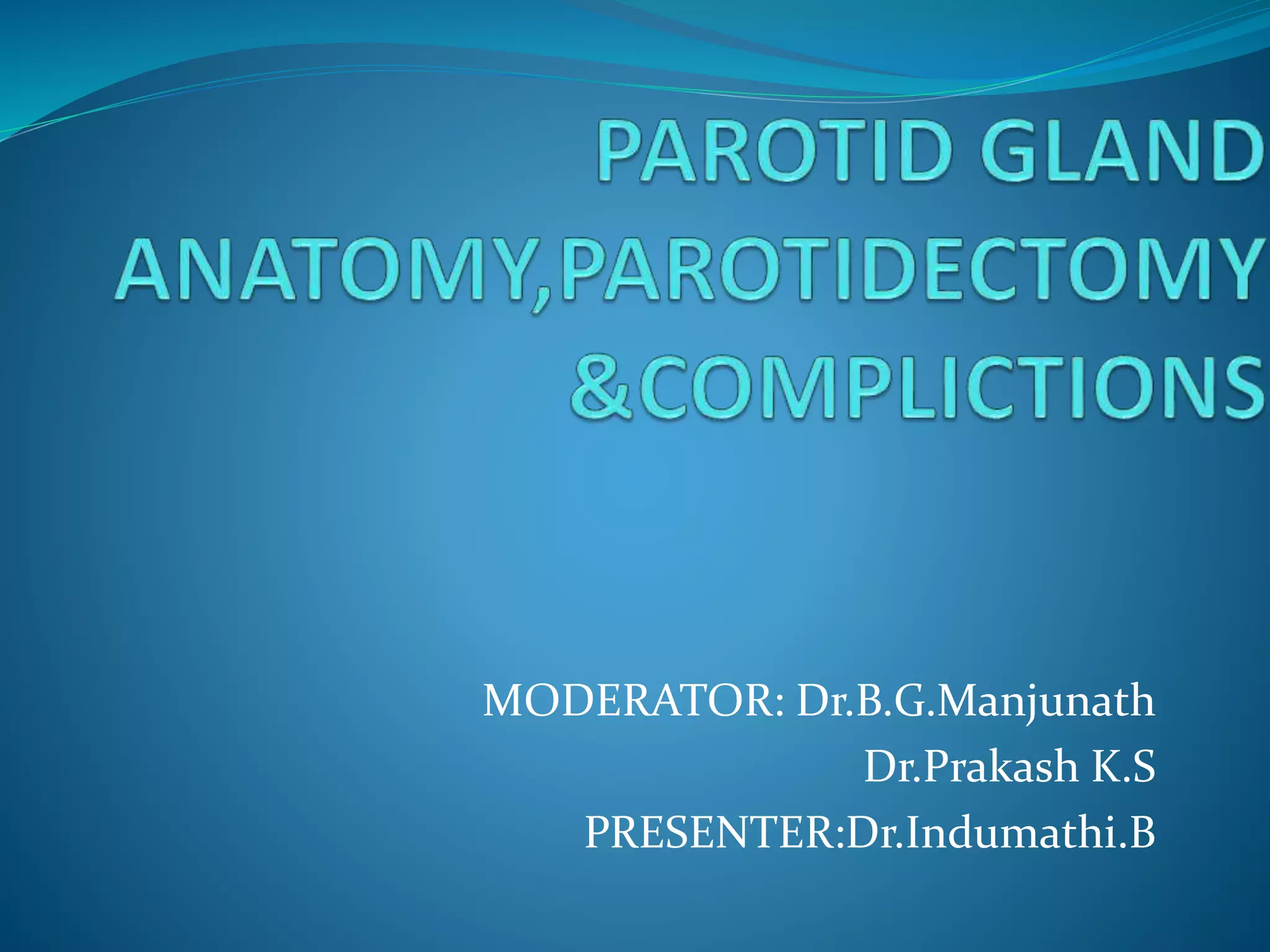
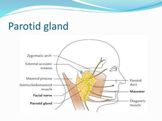
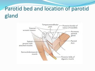
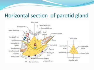
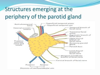
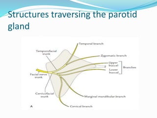
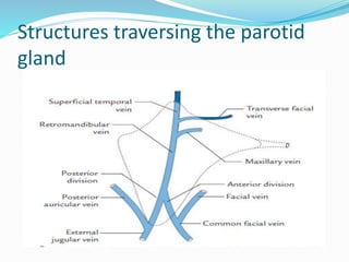
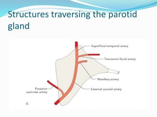
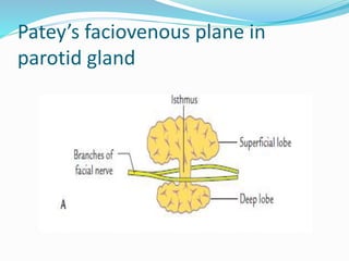
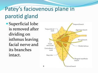
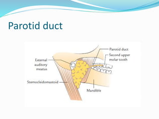
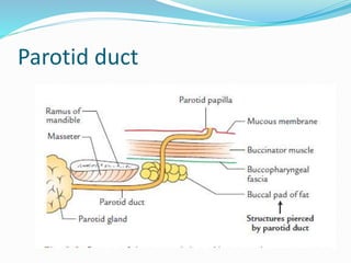
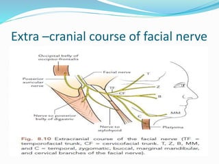
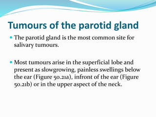
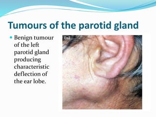
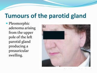
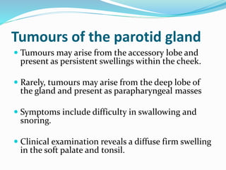
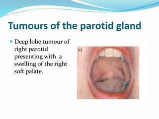
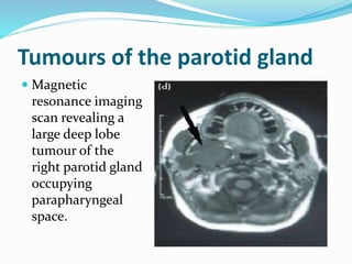
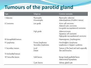
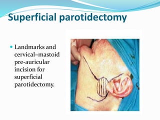
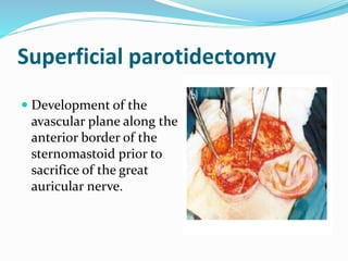
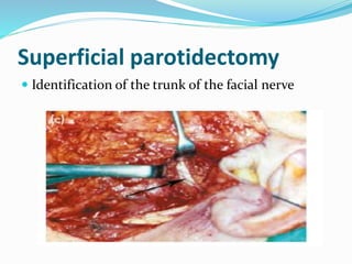
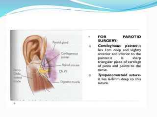
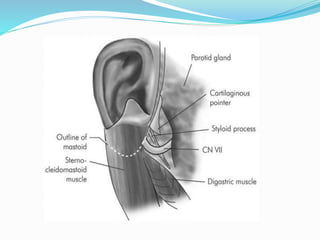
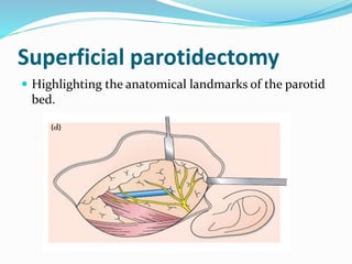
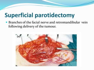
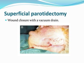
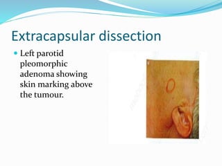
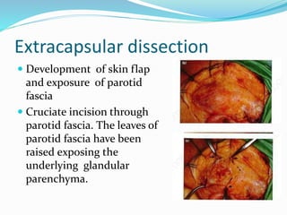
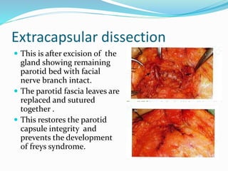
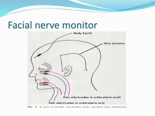
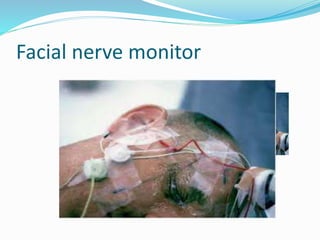
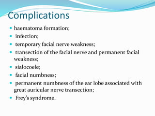
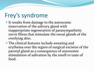
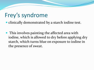
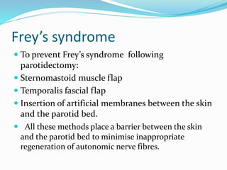
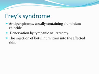
MODERATOR: Dr.B.G.Manjunath
Dr.Prakash K.S
PRESENTER:Dr.Indumathi.B
Parotid gland
Parotid bed and location of parotid
gland
Parotid capsule
Horizontal section of parotid gland
Structures emerging at the
periphery of the parotid gland
Structures traversing the parotid
gland
Structures traversing the parotid
gland
Structures traversing the parotid
gland
Patey’s faciovenous plane in
parotid gland
Patey’s faciovenous plane in
parotid gland
 Superficial lobe
is removed after
dividing on
isthmus leaving
facial nerve and
its branches
intact.
Parotid duct
Parotid duct
Extra –cranial course of facial nerve
EMBRYOLOGY
HISTOLOGY
Tumours of the parotid gland
 The parotid gland is the most common site for
salivary tumours.
 Most tumours arise in the superficial lobe and
present as slowgrowing, painless swellings below
the ear (Figure 50.21a), infront of the ear (Figure
50.21b) or in the upper aspect of the neck.
Tumours of the parotid gland
 Benign tumour
of the left
parotid gland
producing
characteristic
deflection of
the ear lobe.
Tumours of the parotid gland
 Pleomorphic
adenoma arising
from the upper
pole of the left
parotid gland
producing a
preauricular
swelling.
Tumours of the parotid gland
 Tumours may arise from the accessory lobe and
present as persistent swellings within the cheek.
 Rarely, tumours may arise from the deep lobe of
the gland and present as parapharyngeal masses
 Symptoms include difficulty in swallowing and
snoring.
 Clinical examination reveals a diffuse firm swelling
in the soft palate and tonsil.
Tumours of the parotid gland
 Deep lobe tumour of
right parotid
presenting with a
swelling of the right
soft palate.
Tumours of the parotid gland
 Magnetic
resonance imaging
scan revealing a
large deep lobe
tumour of the
right parotid gland
occupying
parapharyngeal
space.
Tumours of the parotid gland
Superficial parotidectomy
 Landmarks and
cervical–mastoid
pre-auricular
incision for
superficial
parotidectomy.
Superficial parotidectomy
 Development of the
avascular plane along the
anterior border of the
sternomastoid prior to
sacrifice of the great
auricular nerve.
Superficial parotidectomy
 Identification of the trunk of the facial nerve
Superficial parotidectomy
 Highlighting the anatomical landmarks of the parotid
bed.
Superficial parotidectomy
 Branches of the facial nerve and retromandibular vein
following delivery of the tumour.
Superficial parotidectomy
 Wound closure with a vacuum drain.
Extracapsular dissection
 Left parotid
pleomorphic
adenoma showing
skin marking above
the tumour.
Extracapsular dissection
 Development of skin flap
and exposure of parotid
fascia
 Cruciate incision through
parotid fascia. The leaves of
parotid fascia have been
raised exposing the
underlying glandular
parenchyma.
Extracapsular dissection
 Exposure of the
tumour showing
branches of facial
nerve.
 Gradual
mobilisation of the
tumour with
preservation of
facial nerve.
Extracapsular dissection
 This is after excision of the
gland showing remaining
parotid bed with facial
nerve branch intact.
 The parotid fascia leaves are
replaced and sutured
together .
 This restores the parotid
capsule integrity and
prevents the development
of freys syndrome.
Facial nerve monitor
Complications
 haematoma formation;
 infection;
 temporary facial nerve weakness;
 transection of the facial nerve and permanent facial
weakness;
 sialocoele;
 facial numbness;
 permanent numbness of the ear lobe associated with
great auricular nerve transection;
 Frey’s syndrome.
Frey’s syndrome
 It results from damage to the autonomic
innervation of the salivary gland with
inappropriate regeneration of parasympathetic
nerve fibres that stimulate the sweat glands of the
overlying skin.
 The clinical features include sweating and
erythema over the region of surgical excision of the
parotid gland as a consequence of autonomic
stimulation of salivation by the smell or taste of
food.
Frey’s syndrome
 clinically demonstrated by a starch iodine test.
 This involves painting the affected area with
iodine, which is allowed to dry before applying dry
starch, which turns blue on exposure to iodine in
the presence of sweat.
Frey’s syndrome
 To prevent Frey’s syndrome following
parotidectomy:
 Sternomastoid muscle flap
 Temporalis fascial flap
 Insertion of artificial membranes between the skin
and the parotid bed.
 All these methods place a barrier between the skin
and the parotid bed to minimise inappropriate
regeneration of autonomic nerve fibres.
Frey’s syndrome
 Antiperspirants, usually containing aluminium
chloride
 Denervation by tympanic neurectomy.
 The injection of botulinum toxin into the affected
skin.
 THANK YOU.

Parotid gland