Downloaded 449 times

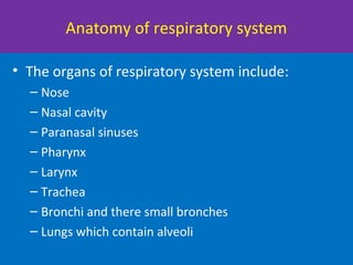

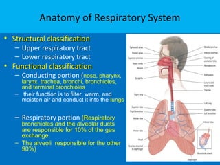

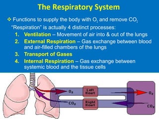
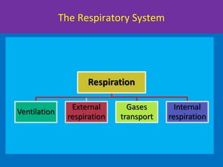
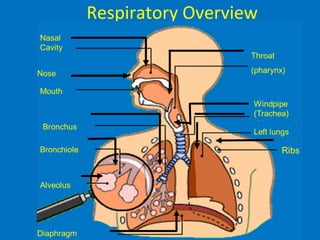


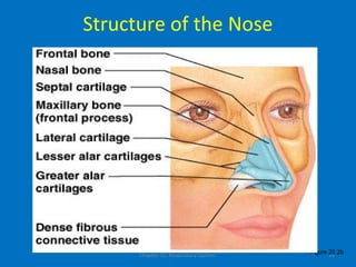
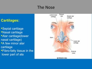


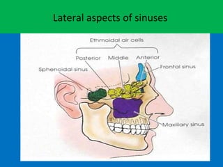
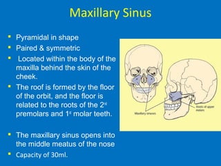

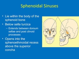

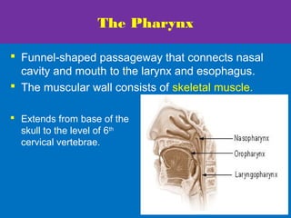


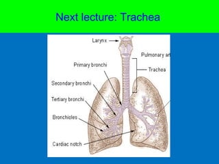


The document discusses the anatomy and functions of the respiratory system. It describes the major organs that make up the respiratory system including the nose, pharynx, larynx, trachea, lungs and diaphragm. It examines the role of these organs in oxygen intake, carbon dioxide removal, and air conduction throughout the respiratory tract. Key functions like ventilation, gas exchange, and transport are summarized.


![L12__Respiratory_system_anatomy[1].pptx](https://cdn.slidesharecdn.com/ss_thumbnails/l12respiratorysystemanatomy1-230531143920-02738076-thumbnail.jpg?width=640&height=640&fit=bounds)


