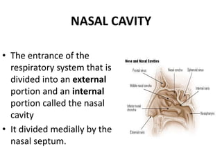
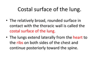
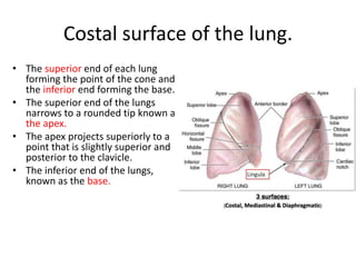
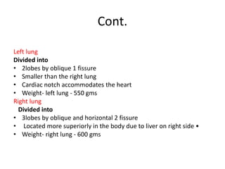
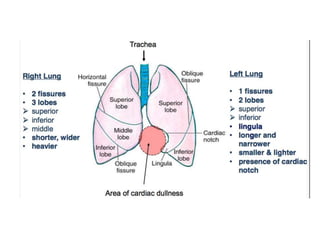
Here are the answers to your questions:
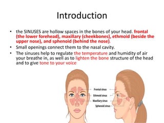
1. The sinuses are the frontal (the lower forehead), maxillary (cheekbones), ethmoid (beside the upper nose), and sphenoid (behind the nose).
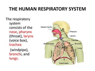
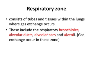
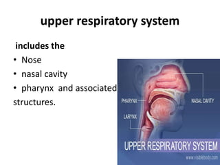
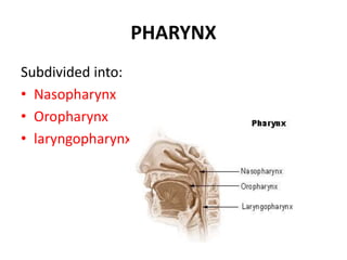
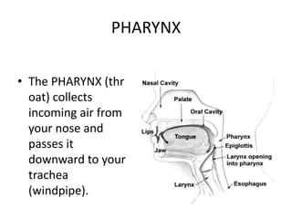
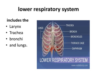
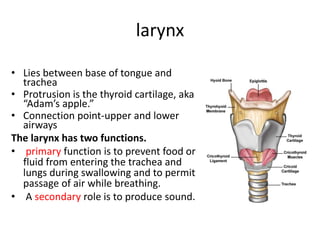
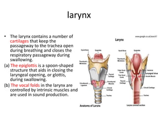
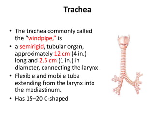
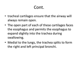
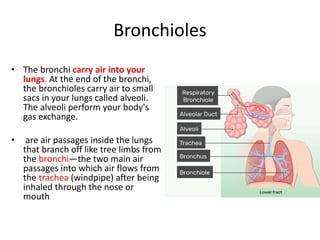
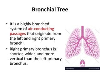
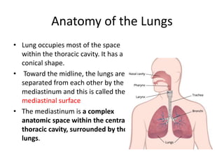
2. The human respiratory system consists of the nose, pharynx (throat), larynx (voice box), trachea (windpipe), bronchi, and lungs.
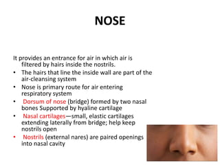
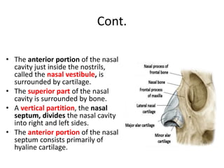
3. The nasal cartilages are small, elastic cartilages extending laterally from the nasal bridge that help keep the nostrils open.
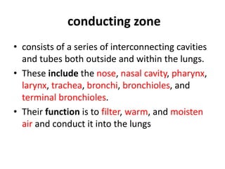
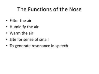
4. The three main functions of the nose are to filter the air, humidify the air, and warm the air.
5. The left lung