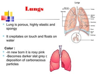
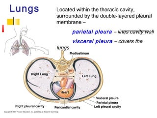
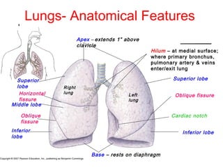
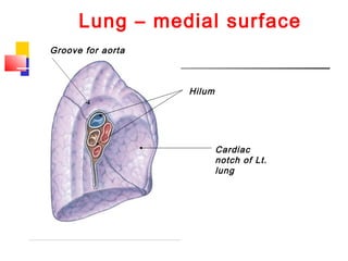
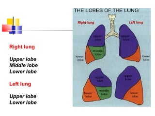
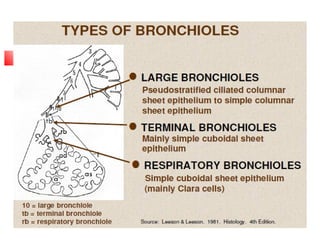
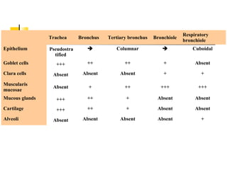
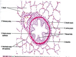
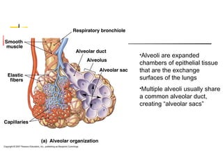
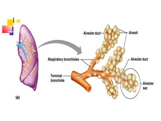
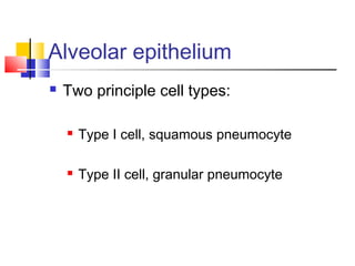
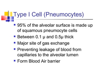
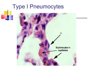
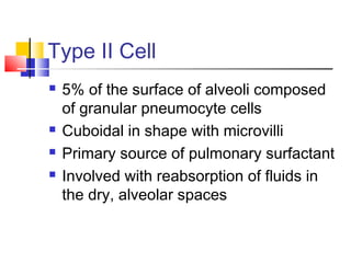
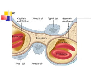
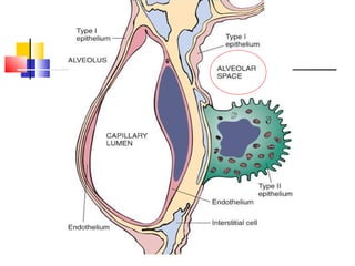
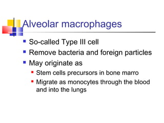
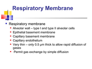
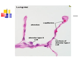
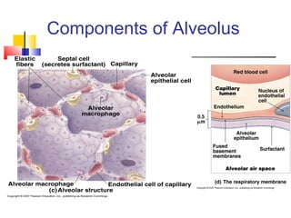
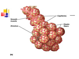
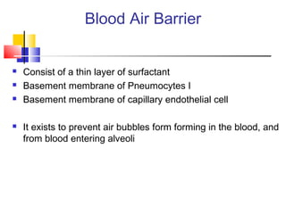
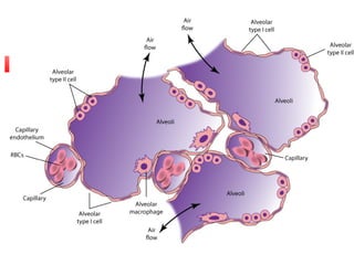
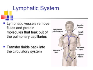
The lungs are located in the thoracic cavity and surrounded by the pleural membranes. They have an apex, base, lobes, and hilum where structures enter and exit. The lungs contain branching airways down to terminal bronchioles and alveoli, which are the gas exchange surfaces. Alveoli are made of type I and II cells and contain surfactant. The blood-air barrier is very thin to allow for gas exchange by diffusion. The lungs receive blood flow from the pulmonary and bronchial circulations and have lymphatic drainage.
Anatomy of Tracheobronchial Tree and Bronchopulmonary Segments with summary o...Jega Subramaniam
Edited version of my Presentation in College.
Hope its useful for you rather than sleeping in my desktop.
Sorry if there is any mistakes.
Thanks and god bless.
Anatomy of Tracheobronchial Tree and Bronchopulmonary Segments with summary o...Jega Subramaniam
Edited version of my Presentation in College.
Hope its useful for you rather than sleeping in my desktop.
Sorry if there is any mistakes.
Thanks and god bless.
Gas exchange between the alveoli and the pulmonary capillary blood occurs by diffusion, as will be discussed in the next chapter. Diffusion of oxygen and carbon dioxide occurs passively, according to their concentration differences across the alveolar-capillary barrier. These concentration differences must be maintained by ventilation of the alveoli and perfusion of the pulmonary capillaries.
Alveolar ventilation brings oxygen into the lung and removes carbon dioxide from it. Similarly, the mixed venous blood brings carbon dioxide into the lung and takes up alveolar oxygen. The alveolar Image not available. and Image not available. are thus determined by the relationship between alveolar ventilation and pulmonary capillary perfusion. Alterations in the ratio of ventilation to perfusion, called the Image not available., will result in changes in the alveolar Image not available. and Image not available., as well as in gas delivery to or removal from the lung.
Alveolar ventilation is normally about 4 to 6 L/min and pulmonary blood flow (which is equal to cardiac output) has a similar range, and so the Image not available. for the whole lung is in the range of 0.8 to 1.2. Image not available. However, ventilation and perfusion must be matched on the alveolar-capillary level, and the Image not available. for the whole lung is really of interest only as an approximation of the situation in all the alveolar-capillary units of the lung. For instance, suppose that all 5 L/min of the cardiac output went to the left lung and all 5 L/min of alveolar ventilation went to the right lung. The whole lung Image not available. would be 1.0, but there would be no gas exchange because there could be no gas diffusion between the ventilated alveoli and the perfused pulmonary capillaries.
Oxygen is delivered to the alveolus by alveolar ventilation, is removed from the alveolus as it diffuses into the pulmonary capillary blood, and is carried away by blood flow. Similarly, carbon dioxide is delivered to the alveolus in the mixed venous blood and diffuses into the alveolus in the pulmonary capillary. The carbon dioxide is removed from the alveolus by alveolar ventilation. As will be discussed in Chapter 6, at resting cardiac outputs the diffusion of both oxygen and carbon dioxide is normally limited by pulmonary perfusion. Thus, the alveolar partial pressures of both oxygen and carbon dioxide are determined by the Image not available. If the Image not available. in an alveolar-capillary unit increases, the delivery of oxygen relative to its removal will increase, as will the removal ...
Slideshow is from the University of Michigan Medical
School's M1 Infectious Disease / Microbiology sequence
View additional course materials on Open.Michigan:
openmi.ch/med-M1IDM
An approach to Interstitial Lung Disease / Diffuse Parenchymal Lung DiseaseThomas Kurian
YouTube link: https://youtu.be/gPr31qrivUc
An approach to Diffuse Parenchymal Lung disease / Interstitial Lung disease with emphasis on the idiopathic causes.
This PPT covers Anatomy and Physiology of respiratory system. Anatomy of respiratory organs, Mechanism of respiration, Internal Respiration, external respiration, Transport of oxygen in blood, Transport of carbon dioxide in blood, Regulation of respiration, lung volume and lung capacities are explained.
Introduction to AI for Nonprofits with Tapp NetworkTechSoup
Dive into the world of AI! Experts Jon Hill and Tareq Monaur will guide you through AI's role in enhancing nonprofit websites and basic marketing strategies, making it easy to understand and apply.
Embracing GenAI - A Strategic ImperativePeter Windle
Artificial Intelligence (AI) technologies such as Generative AI, Image Generators and Large Language Models have had a dramatic impact on teaching, learning and assessment over the past 18 months. The most immediate threat AI posed was to Academic Integrity with Higher Education Institutes (HEIs) focusing their efforts on combating the use of GenAI in assessment. Guidelines were developed for staff and students, policies put in place too. Innovative educators have forged paths in the use of Generative AI for teaching, learning and assessments leading to pockets of transformation springing up across HEIs, often with little or no top-down guidance, support or direction.
This Gasta posits a strategic approach to integrating AI into HEIs to prepare staff, students and the curriculum for an evolving world and workplace. We will highlight the advantages of working with these technologies beyond the realm of teaching, learning and assessment by considering prompt engineering skills, industry impact, curriculum changes, and the need for staff upskilling. In contrast, not engaging strategically with Generative AI poses risks, including falling behind peers, missed opportunities and failing to ensure our graduates remain employable. The rapid evolution of AI technologies necessitates a proactive and strategic approach if we are to remain relevant.
How to Make a Field invisible in Odoo 17Celine George
It is possible to hide or invisible some fields in odoo. Commonly using “invisible” attribute in the field definition to invisible the fields. This slide will show how to make a field invisible in odoo 17.
Operation “Blue Star” is the only event in the history of Independent India where the state went into war with its own people. Even after about 40 years it is not clear if it was culmination of states anger over people of the region, a political game of power or start of dictatorial chapter in the democratic setup.
The people of Punjab felt alienated from main stream due to denial of their just demands during a long democratic struggle since independence. As it happen all over the word, it led to militant struggle with great loss of lives of military, police and civilian personnel. Killing of Indira Gandhi and massacre of innocent Sikhs in Delhi and other India cities was also associated with this movement.
Acetabularia Information For Class 9 .docxvaibhavrinwa19
Acetabularia acetabulum is a single-celled green alga that in its vegetative state is morphologically differentiated into a basal rhizoid and an axially elongated stalk, which bears whorls of branching hairs. The single diploid nucleus resides in the rhizoid.
Francesca Gottschalk - How can education support child empowerment.pptxEduSkills OECD
Francesca Gottschalk from the OECD’s Centre for Educational Research and Innovation presents at the Ask an Expert Webinar: How can education support child empowerment?
2. Lung is porous, highly elastic and
spongy
It crepitates on touch and floats on
water
Color :
-In new born it is rosy pink
-Becomes darker slat grey due to
deposition of carbonacious
particles
Lungs
3. Lungs Located within the thoracic cavity,
surrounded by the double-layered pleural
membrane –
parietal pleura – lines cavity wall
visceral pleura – covers the
lungs
4. Lungs- Anatomical Features
Apex – extends 1” above
clavicle
Base – rests on diaphragm
Right
lung
Left
lung
Superior
lobe
Middle lobe
Inferior
lobe
Horizontal
fissure
Oblique
fissure
Superior lobe
Inferior lobe
Oblique fissure
Cardiac notch
Hilum – at medial surface;
where primary bronchus,
pulmonary artery & veins
enter/exit lung
7. Each lung has a primary
bronchus entering at the
Hilus.
Each lobe of a lung has a
secondary (a.k.a. lobar)
bronchus
Lobes are functionally divided
into bronchopulmonary
segments & each segment has
a tertiary (segmental)
bronchus
Segments are functionally
divided into many lobules &
each lobule receives a terminal
bronchiole
Airways within Lungs
10. Terminal Bronchioles
16th
to 19th
generation
Average diameter is 0.5 mm
Cilia and mucous glands begin to
disappear totally
End of the conducting airway
Canals of Lambert-interconnect this
generation,provide collateral ventilation
11.
12. Respiratory Zone
Defined by the presence of alveoli; begins as terminal
bronchioles feed into Respiratory bronchioles
Respiratory bronchioles lead to alveolar ducts, then to
terminal clusters of alveolar sacs composed of alveoli
Approximately 300 million alveoli:
Account for most of the lungs’ volume
Provide tremendous surface area for gas exchange
13. Alveoli
200-300 million in a normal lung
Between 75 µ to 300 µ in diameter- Total
area-75 square meters
Most gas exchange takes place at alveolar-
capillary membrane
85-95% of alveoli covered by small
pulmonary capillaries
The cross-sectional area or surface area is
approximately 70m2
14. Alveoli are expanded
chambers of epithelial tissue
that are the exchange
surfaces of the lungs
Multiple alveoli usually share
a common alveolar duct,
creating “alveolar sacs”
15. Acinus or Lobule
Each acinus (unit) is approximately 3.5 mm
in diameter
Each contains about 2000 aveloli
Approximately 130,000 primary lobules in
the lung
18. Alveolar epithelium
Two principle cell types:
Type I cell, squamous pneumocyte
Type II cell, granular pneumocyte
19. Type I Cell (Pneumocytes)
95% of the alveolar surface is made up
of squamous pneumocyte cells
Between 0.1 µ and 0.5µ thick
Major site of gas exchange
Preventing leakage of blood from
capillaries to the alveolar lumen
Form Blood Air barrier
21. Type II Cell
5% of the surface of alveoli composed
of granular pneumocyte cells
Cuboidal in shape with microvilli
Primary source of pulmonary surfactant
Involved with reabsorption of fluids in
the dry, alveolar spaces
22.
23. Type II pneumocytes
Also known as Septal cells
Rounded or cuboidal secretory cells with microvilli
Secretory granules are made of several layers- Multilamellar
bodies.
Is constantly renewed.
Pulmonary Surfactant – is the fluid secreted that spreads
over the alveolar surface.
24. Pulmonary Surfactant
Surfactant contains
phospholipids, proteins and
glycosaminoglycans, reduces
the surface tension and prevents
collapse of the alveolus during
expiration.
The reduced surface tension in
the alveoli decreases the force
that is needed to inflate alveoli
during inspiration.
Therefore surfactant
stabilizes the alveolar
diameters, facilitates their
expansion and prevents
their collapse by
minimizing the collapsing
forces.
Surfactant also has
bactericidal properties
25.
26. Canals of Lambert/Pores of Kohn
Provide for collateral ventilation of
difference acinii or primary lobules
Additional ventilation of blocked units
May explain why diseases spread so
quickly at the lung tissue (paremchymal)
level
27. Alveolar macrophages
So-called Type III cell
Remove bacteria and foreign particles
May originate as
Stem cells precursors in bone marro
Migrate as monocytes through the blood
and into the lungs
28. Intersitium/interstial space
Surround, supports, and shapes the
alveoli and capillaries
Composed of a gel like substance and
collagen fibers
Contains tight space and loose space
areas
29. Interstitium
Water content in loose space can increase
by 30% before there is a significant change
in pulmonary capillary pressure
Lymphatic drainage easily exceeded
Collagen limits alveolar distensibility
30. Respiratory Membrane
Respiratory membrane
Alveolar wall – type I and type II alveolar cells
Epithelial basement membrane
Capillary basement membrane
Capillary endothelium
Very thin – only 0.5 µm thick to allow rapid diffusion of
gases
Permit gas exchange by simple diffusion
34. Blood Air Barrier
Consist of a thin layer of surfactant
Basement membrane of Pneumocytes I
Basement membrane of capillary endothelial cell
It exists to prevent air bubbles form forming in the blood, and
from blood entering alveoli
35.
36. Nutrition of the lung
The lung gets nutrition from two sources:
1.Conducting part up to the beginning of respiratory
bronchiole is supplied by Bronchial artery
2. Respiratory part is supplied by pulmonary artery via
Pulmonary capillary plexus
•Primary purpose is to deliver blood to lungs for gas exchange
•Right lung has one bronchial artery and left lung has two
Bronchial artery
37. Bronchial arteries
Also nourish
Mediastinal lymph nodes
Pulmonary nerves
Some muscular pulmonary arteries and
veins
Portions of the esophagus
Visceral pleura
38. Bronchial venous system
1/3 blood returns to right heart
Azygous
Hemiazygous
Intercostal veins
This blood comes form the first two or
three generations of bronchi
39. Bronchial venous return
2/3 of blood flowing to terminal bronchioles drains
into pulmonary circulation via “bronchopulmonary
anastomoses”
Then flows to left atrium via pulmonary veins
Contributes to “venous admixture” or “anatomic
shunt” (ca. 5% of C.O.)
40. Pulmonary Capillaries
Walls are les than 0.1µ thick
Total external thickness is about 10µ
Selective permeability to water,
electrolytes, sugars
Produce and destroy biologically active
substances
41. Lymphatic System
Lymphatic vessels remove
fluids and protein
molecules that leak out of
the pulmonary capillaries
Transfer fluids back into
the circulatory system
42. Lymphatics
Lymphatic vessels arise within loose spaces of
connective tissue, not in the walls of the alveoli.
Vessels then follow bronchial airways,
pulmonary airways, pulmonary arteries and
veins to the hilum
Vessels end in pulmonary and
bronchopulmonary lymph nodes within and
outside of lung parenchyma