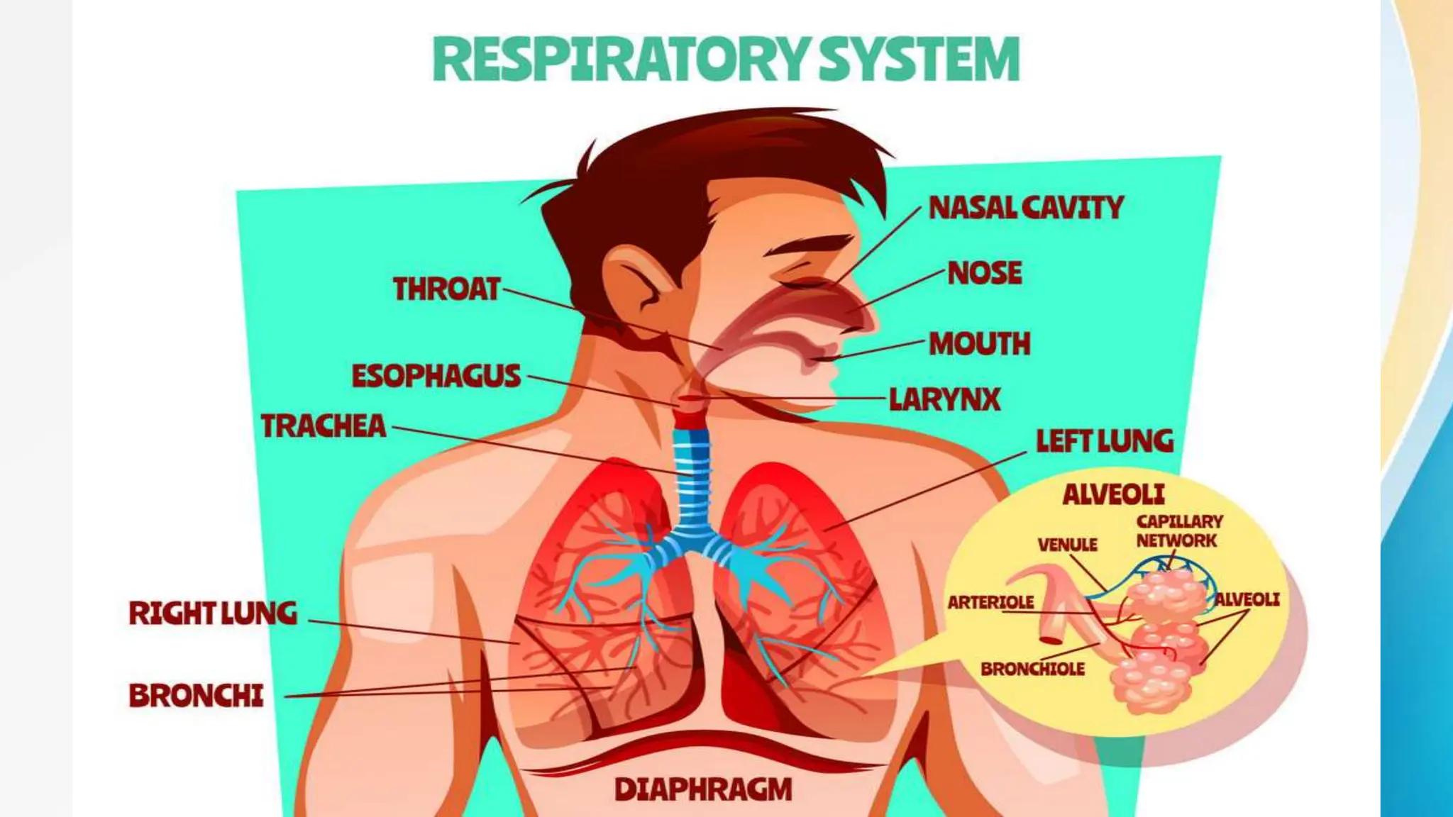
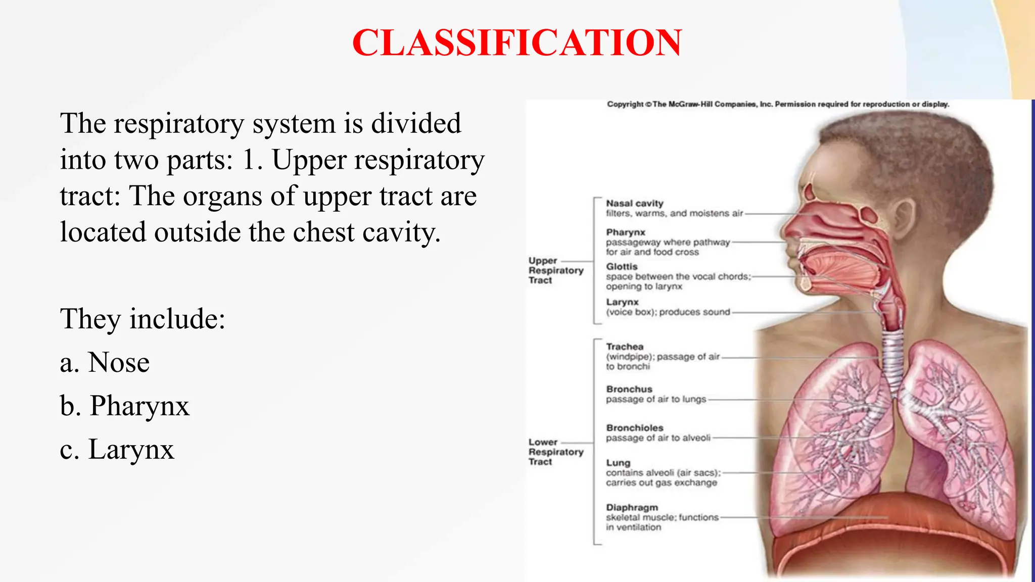
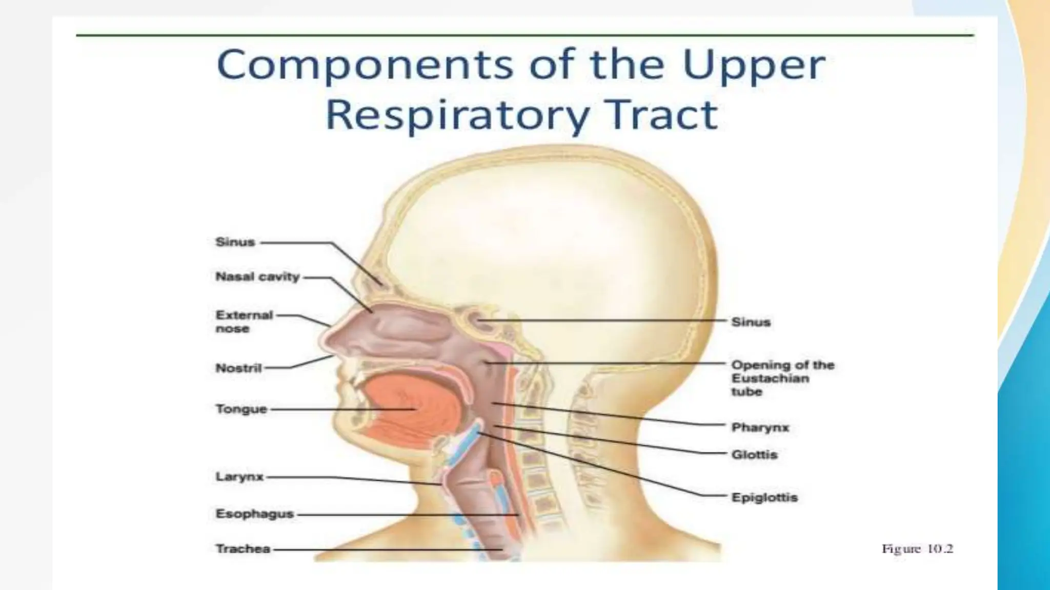
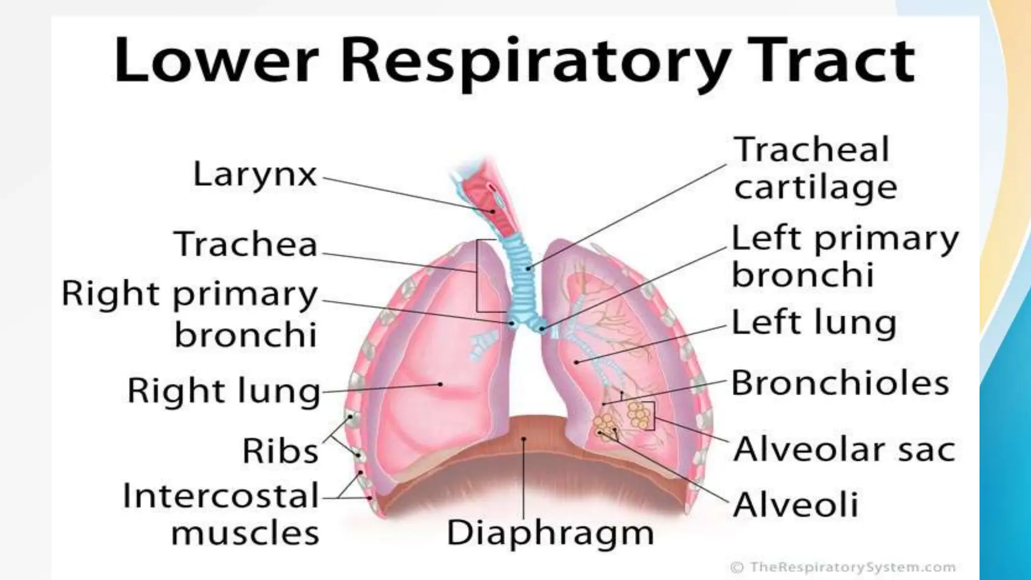
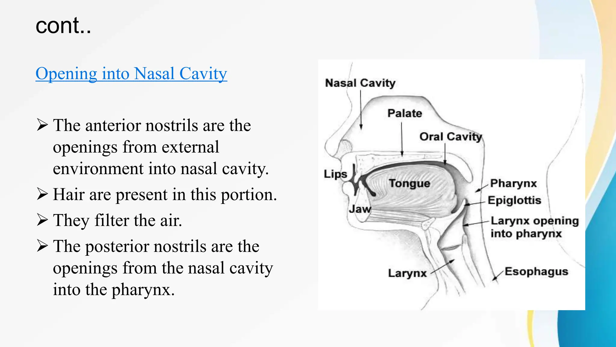
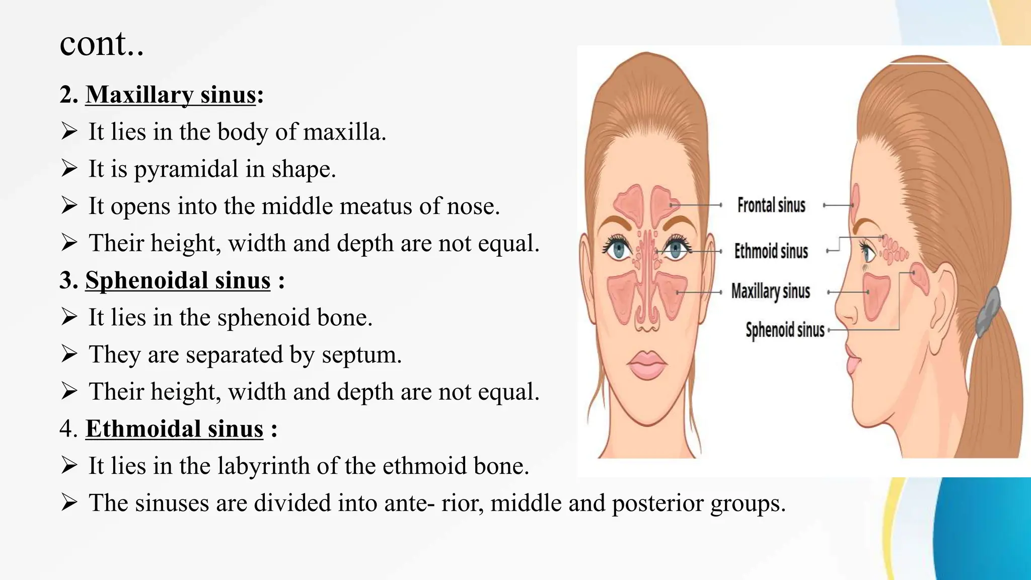
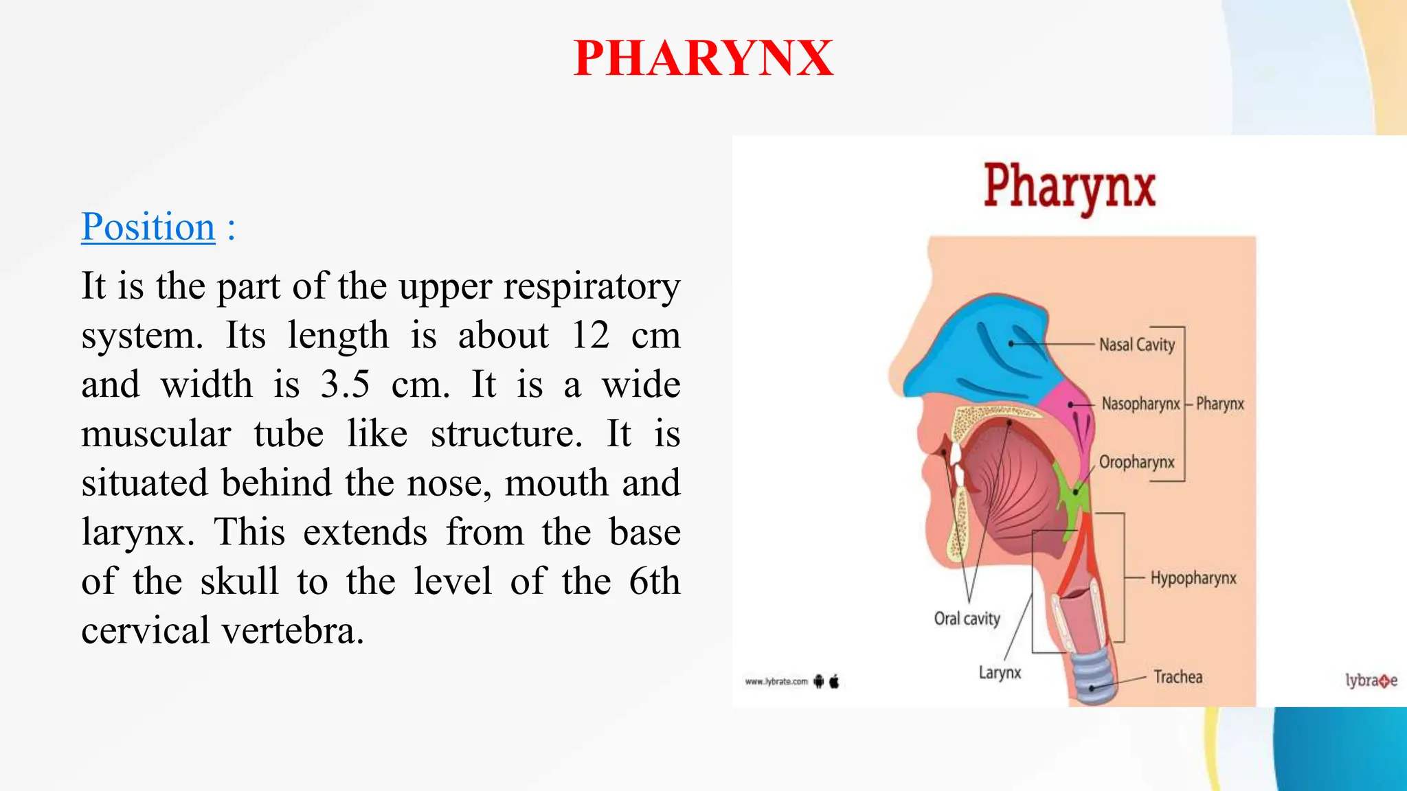
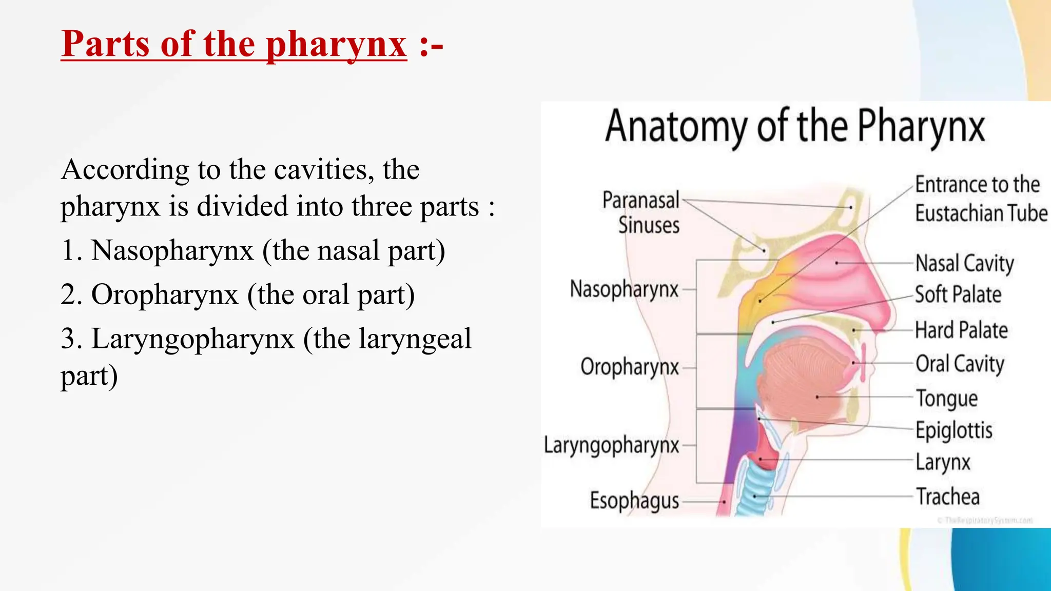
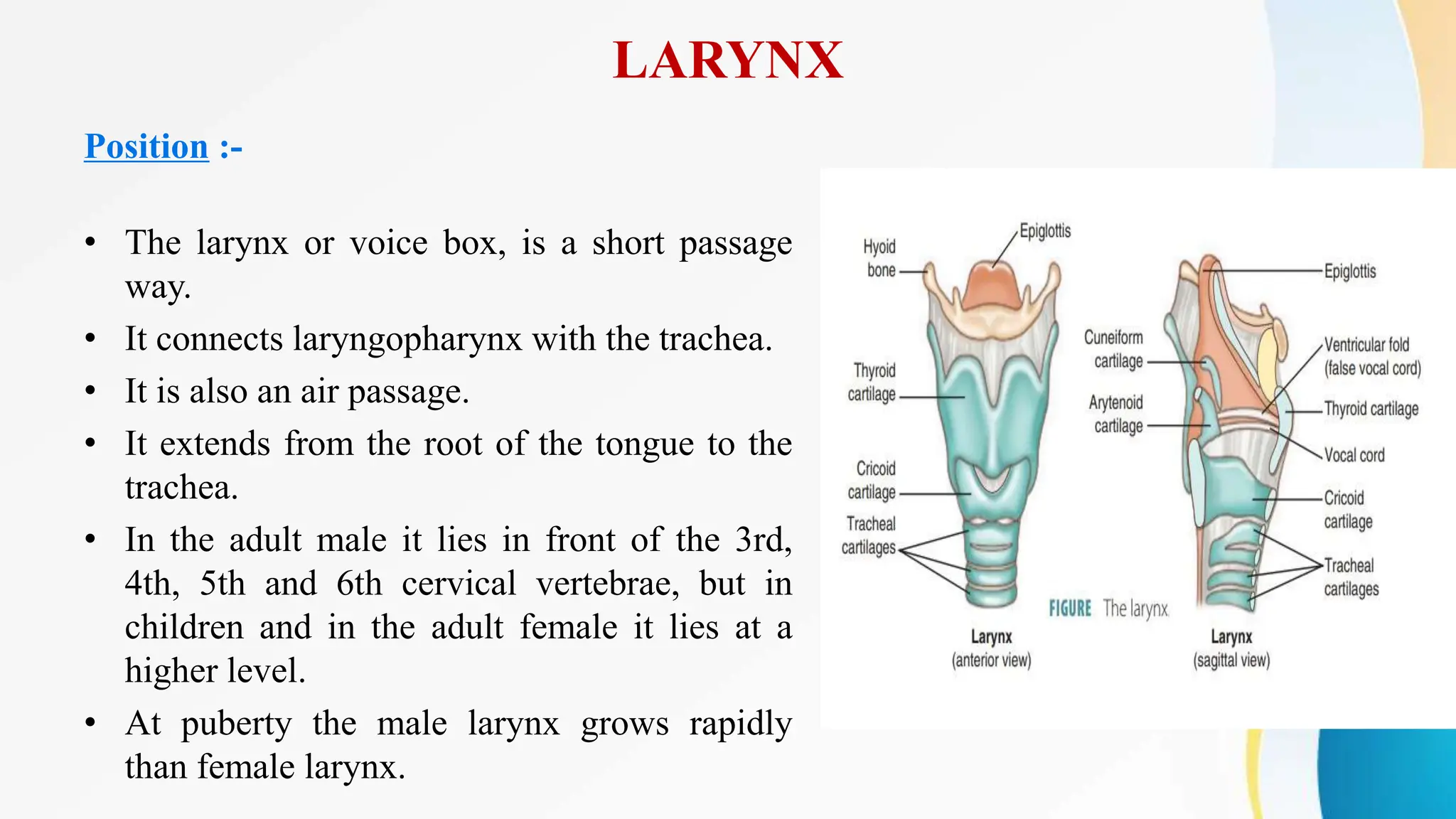
2. INTRODUCTION
Respiration is the exchange of gases which occurs between the atmosphere,
blood and cells. In this process oxygen (O2) is inhaled (taken inside) and
carbondioxide is exhaled (expelled out).
Respiration is of two types :
1. External respiration: It is the exchange of gases between the
blood and lungs.
2. Internal respiration: It is the exchange of gases between the
blood and cells.
3.
4. CLASSIFICATION
The respiratory system is divided
into two parts: 1. Upper respiratory
tract: The organs of upper tract are
located outside the chest cavity.
They include:
a. Nose
b. Pharynx
c. Larynx
5. Cont..
2. Lower respiratory tract: The organs of lower tract are located inside the
chest cavity. They include:
a. Trachea
b. Two bronchi (one bronchus to each lung)
c. Bronchioles and smaller air passages
d. Two lungs and their covering, the pleura
e. Muscle of respiration: the intercostal muscles and the diaphragm
6. UPPER RESPIRATORY TRACT
Nose and nasal cavity
Nose Position and Structure :- Nose is the first respiratory organ. Nose has
two portions:
External Portion :
• It is made up of bone and hyaline cartilage.
• It is covered with skin.
• It is lined by mucous membrane.
• It is flexible.
• External nose has two openings called the nostrils or external nares.
• The nares are separated by the septum bone.
7.
8. cont..
Internal Portion :
The internal portion of the nose is a large cavity. It lies over the roof of
mouth.
• The roof of the nose is separated from the cranial cavity by ethmoid
bones called cribriform plate.
• The roof of nose is formed by cribriform plate, sphenoid bone, frontal
bone and nasal bones.
• The floor of nose is formed by hard and soft palate. The medial wall is
formed by the septum.
9. cont..
• The lateral walls are formed by the maxilla, ethmoid bone and the
inferior conchae. The posterior wall is formed by the posterior wall of
the pharynx.
Lining of the Nose
The inside of nose is lined with mucous membrane.
The mucous membrane is made up of pseudo stratified ciliated columnar
epithelium.
This is rich in goblet cells.
The goblet cells secrete the mucus.
10.
11. cont..
Opening into Nasal Cavity
The anterior nostrils are the
openings from external
environment into nasal cavity.
Hair are present in this portion.
They filter the air.
The posterior nostrils are the
openings from the nasal cavity
into the pharynx.
12. cont..
Paranasal sinuses : These are air filled
spaces. These are present within some
bones around the nasal cavities. There
are four pairs of paranasal sinuses.
1. Frontal sinus:
It lies in frontal bone.
It opens into the middle meatus of
nose.
Their average height, width and depth
are equal.
13. cont..
2. Maxillary sinus:
It lies in the body of maxilla.
It is pyramidal in shape.
It opens into the middle meatus of nose.
Their height, width and depth are not equal.
3. Sphenoidal sinus :
It lies in the sphenoid bone.
They are separated by septum.
Their height, width and depth are not equal.
4. Ethmoidal sinus :
It lies in the labyrinth of the ethmoid bone.
The sinuses are divided into ante- rior, middle and posterior groups.
14. RESPIRATORY FUNCTION OF NOSE
The nose is a part of respiratory system. The exchange of gases i.e. CO, and
O, takes place through the nose. The main functions of the nose are given
below:
Filtering :
The air enters into the nostrils and passes through the vestibule.
The vestibule is lined by skin and hair are present.
These hair filter out large dust particles.
These dust particles adhere to the mucus.
The mucus also prevents the drying of the epithelium of nose.
15. cont..
Cleaning of Air :
The hair filter the air, so the air reaching to the respiratory system would be
clean.
Exchange of Gases :
The nose helps in exchange of gases (that is intake of oxygen for
delivery to body cells and elimination of carbon dioxide produced by
body cells).
Respiration takes place in three basic steps- pulmonary ventilation,
external respiration and inter- nal respiration.
16. cont..
Warming :
The mucous membrane contains capillaries.
As the air strikes the concha and mucous membrane, it is warmed by
blood in the capillaries.
Humidification :
When the air passes over the mucosa, it permits the moistening of dry air.
If dry air enters into the respiratory passage, it cracks the mucus
membrane.
When dust gets collected in nose, the person sneezes or coughs to throw
the dust particles out, because they irritate the nose.
17. OLFACTORY FUNCTION OF NOSE
• The word olfactory means sense of smell.
• The nose is the organ of the of smell.
• There are nerve end- sense ings which detect the smell.
• The olfactory receptors lie in the membrane lining the superior nasal
conchae and adjacent septum.
• This region is called olfactory epithelium.
• This specialized membrane contains many olfactory nerve cells.
• These nerve cells are stimulated by chemical substances given off by
odourous material.
• These nerve impulses are conveyed by the olfac- tory nerves to the brain
where the sense of smell is perceived.
18. PHARYNX
Position :
It is the part of the upper respiratory
system. Its length is about 12 cm
and width is 3.5 cm. It is a wide
muscular tube like structure. It is
situated behind the nose, mouth and
larynx. This extends from the base
of the skull to the level of the 6th
cervical vertebra.
19. Structures Associated with the Pharynx
• Superiorly : Base of the skull, including the part of the body of the
sphenoid, occipital bone and front of the pharyngeal
tubercle.
• Inferiorly : The pharynx is continuous with the oesophagus at the level
of the 6th cerv- ical vertebra.
• Posteriorly : Areolar tissue, involuntary muscle, the pharynx glides
freely on the prevertebral fascia which separates it from the
cervical spine.
• Anteriorly : It is connected with the nasal cavity, oral cavity and the
larynx. So, the anterior wall is incomplete.
20. Parts of the pharynx :-
According to the cavities, the
pharynx is divided into three parts :
1. Nasopharynx (the nasal part)
2. Oropharynx (the oral part)
3. Laryngopharynx (the laryngeal
part)
21. Nasopharynx :
• As the name indicates, it is the nasal part of the pharynx.
• It lies behind the nose.
• This extends from the posterior nares to the level of the soft palate.
• There are five openings in its wall.
• It is lined by ciliated columnar epithelium.
• The nasopharynx also exchanges the small amount of air with the
auditory tube and so as to equalize air pressure between the pharynx and
middle ear.
22. Oropharynx :
• It is the oral part of the pharynx.
• It is situated behind the oral cavity.
• It communicates with the nasopharynx.
• It is located behind the mouth, from the soft palate above, to the level of
the hyoid bone below.
• It lies at the level of 3rd cervical vertebra.
• Oropharynx is lined by non- keratinized stratified squamous epithelium.
23. Laryngopharynx :
• It is the laryngeal part of the pharynx.
• It is situated behind the pharynx.
• It begins at the level of hyoid bone.
• It starts from oropharynx and ends at the beginning of oesophagus.
• It is lined by non-keratinized stratified squamous epithelium.
• It acts as both respiratory and a digestive pathway.
24. Structure of pharynx :-
The wall of the pharynx is composed of the following three layers from
inward to outward :
1. Mucous membrane
2. Fibrous tissue
3. Muscle tissue
25. Mucous Membrane :
• It is the innermost lining.
• It consists of ciliated columnar epithelial tissue in the nasopharynx.
• In the oropharynx and laryngopharynx it is formed by stratified
squamous epithelium.
• So, it is thick at the level of oropharynx and laryngopharynx, which is
continuous with the lining of the mouth and oesophagus.
26. Fibrous Tissue :
It is the middle layer.
Fibrous layer is made up of collagen fibres.
It is thicker in the nasopharynx.
It becomes thinner towards the lower end.
Muscle Tissue :
It is the outermost layer.
This part consists of several involuntary or non-striated muscles.
It means these muscles are not under conscious con- trol.
These muscles help in swallowing.
27. Blood and Nerve supply :-
• The pharynx is supplied by the pharyngeal plexus of nerves.
• These are formed by parasympathetic and sympathetic nerves.
• Parasympathetic supply of the blood is by the vagus nerve and
sympathetic supply of blood is by pharyngeal branches of the superior
cervical sympathetic ganglion.
• Arteries supply blood to the pharynx.
• These are ascending carotid artery, lingual artery, palatine and maxillary
artery.
28. Function of Pharynx :-
Hearing :
The nasopharynx helps in equalising the pressure between the pharynx and
middle ear, which helps in hearing.
Protection :
The pharynx and larynx are covered with lymphatic tissue, which contain
white blood cells. They synthesise and secrete the specific antibodies
against microbes or micro-organisms.
Warming and Humidifying :
The mucous membrane warmed by blood in the capillaries. When the air
passes over the mucosa, it permits the moistening of dry inspired air.
29. cont..
Passageway for Air and Food :
The pharynx serves as a common pathway for the respiratory and digestive
tracts. Both food and air pass from the almost same structure before
reaching to its destination.
Taste :
In the oral and pharyngeal parts there are olfac tory nerve endings of the
sense of taste.
Speech :
The pharynx helps in speech. The pharynx can change its shape which
produces different vocal sounds and helps in speech.
30. LARYNX
Position :-
• The larynx or voice box, is a short passage
way.
• It connects laryngopharynx with the trachea.
• It is also an air passage.
• It extends from the root of the tongue to the
trachea.
• In the adult male it lies in front of the 3rd,
4th, 5th and 6th cervical vertebrae, but in
children and in the adult female it lies at a
higher level.
• At puberty the male larynx grows rapidly
than female larynx.
31. Structures Associated with the Larynx :-
• Superiorly : The hyoid bone and the root of the tongue.
• Inferiorly : It is continuous with the trachea.
• Anteriorly : The muscles attached to the hyoid bone and the muscle of
the neck.
• Posteriorly : The laryngopharynx and 3rd to 6th cervical vertebrae.
• Laterally : The lobes of the thyroid gland.
32. Structure of larynx :-
Cartilage :
The cartilages are formed by chondrocytes
cell.
These cells are of different shape.
These help in attachment with the help of
ligaments and membranes.
The contains 9 cartilages, of which 3 are
unpaired and 3 paired.
Unpaired Cartilages :
1. Thyroid
2. Cricoid
3. Epiglottis
33. cont..
Thyroid Cartilage :
It is an unpaired cartilage.
It is V- shaped.
It is larger in males than females.
It is made up of hyaline It is also called Adam's apple thyrohyoid
membrane.
It connects the thyroid cartilage with hyoid bone.
34. cont..
Cricoid Cartilage :
It is an unpaired cartilage.
It is also called signet ring, because its shape is like a ring.
It is also made up of hyaline cartilage.
It is a land mark for tracheostomy.
It is lined with ciliated columnar epithelium.
Cricothyroid ligament attaches the cricoid cartilage with thyroid
cartilage.
35. cont..
Epiglottis :
It is also a paired cartilage.
It is present above the tongue.
This is a leaf shaped cartilage.
This moves up during swallowing.
This is made up of elastic cartilage.
These are lined with stratified squamous epithelium.
It prevents food or liquids from entering the trachea.
37. cont..
Arytenoid Cartilages :
It is a paired cartilage.
It is triangular in shape.
It is made up of hyaline cartilage.
They attach to the vocal cord.
They provide attachment with muscles.
The base of each cartilage articulates with the superior border of the
cricoid cartilage.
The anterior angle of these cartilages provide a point for the attachment
of the vocal cord.
38. cont..
Corniculate Cartilage :
It is a horn-shaped cartilage.
It is made up of elastic cartilage.
It articulates at the apex of the arytenoid cartilages.
This cartilage is small as compared with other cartilages.
39. cont..
Cuneiform Cartilage :
It is a rod-shaped cartilage.
It is situated near the base of the epiglottis.
It resembles with the arytenoid cartilage.
It is made up of elastic cartilage.
It is present anterior to the corniculate cartilage.
It supports the vocal cords (vocal folds) and lateral aspect of the
epiglottis.
40. Blood and Nerve Supply :-
• Superior laryngeal artery and inferior laryngeal artery supply blood to
the larynx.
• This blood is drained into the superior thyroid vein and inferior thyroid
vein.
• All the muscles of the larynx are supplied by the laryngeal nerve except
cricothyroid, which is supplied by the external laryngeal nerve.
• The internal laryngeal nerve supplies the mucous membrane above the
level of the vocal folds.
• The recurrent laryngeal nerve supplies below the level of the vocal folds.