
Recommended
More Related Content
What's hot
What's hot (20)
Similar to Respiratorysystem 120105050542-phpapp01
Similar to Respiratorysystem 120105050542-phpapp01 (20)
Recently uploaded
Recently uploaded (20)
Respiratorysystem 120105050542-phpapp01
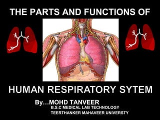
- 1. THE PARTS AND FUNCTIONS OF By…MOHD TANVEER B.S.C MEDICAL LAB TECHNOLOGY TEERTHANKER MAHAVEER UNIVERSTY
- 2. Primary Function of Respiratory System • The respiratory system supplies the blood with oxygen so that the blood can deliver oxygen to all parts of the body. • And also removes carbon dioxide waste that cells produce.
- 3. * It is the system, consisting of tubes and is responsible for the exchange of gases in Humans by filtering incoming air and transporting it into the microscopic alveoli where gases are exchanged * Your respiratory system provides the energy needed by cells of the body to funtion accroding to their designated tasks. THE HUMAN RESPIRATORY SYSTEM
- 4. THE HUMAN RESPIRATORY SYSTEM
- 5. The organs of the “Respiratory Tract” can be divided into two groups “STRUCTURALLY” ** The Upper Respiratory Tract ** The Lower Respiratory Tract * Nose * Nasal cavity * Sinuses * Pharynx * Larynx * Trachea * Bronchial Tree * Lungs
- 6. THE HUMAN RESPIRATORY SYSTEM
- 7. The organs of the “Respiratory Tract” can be divided into two groups “FUNCTIONALLY” ** The Conducting Portion - system of interconnecting cavities and tubes that conduct air into the lungs ** The Respiratory Portion - system where the exchange of respiratory gases occurs * Nose * Pharynx * Larynx * Trachea * Bronchi * Respiratory bronchioles * Alveolar Ducts * Alveoli
- 9. THE HUMAN RESPIRATORY SYSTEM I. N O S E A. N a s a l C a v i t y B. P a r a n a s a l S i n u s e s II. P H A R Y N X III. L A R Y N X A. E p I g i o t t i s B. V o c a l C o r d s IV. T R A C H E A v. B R O N C H I A. B r o n c h i a l T r e e VI. L U N G S A. L o b e s o f t h e L u n g s B. P l e u r a l C a v i t i e s C. A l v e o l i
- 10. THE NOSE
- 11. The Nose • Only externally visible part of the respiratory system. • Functions include: • Providing an airway for respiration • Moistening and warming air • Filtering inspired air • Serving as a resonating center for speech • Housing the olfactory receptors.
- 12. * It provides an entrance for air in which air is filtered by coarse hairs inside the nostrils. * It has 2 portions : the external and internal * The external portion is supported by a framework of bone and cartilage covered with skin and lined with mucous membrane. * The internal portion is a large cavity in the skull, THE NOSE
- 13. The Nasal Cavity
- 14. The Nose • Only externally visible part of the respiratory system. • Functions include: • Providing an airway for respiration • Moistening and warming air • Filtering inspired air • Serving as a resonating center for speech • Housing the olfactory receptors.
- 15. * Interior area of the nose; lined with a sticky mucous membrane and contains tiny, surface hairs, cilia. divided medially by the nasal septum. * Nasal conchae divide the cavity into passageways that are lined with mucous membrane, and help increase the surface area available to warm and filter incoming air. •Particles trapped in the mucus are carried to the pharynx by ciliary action, swallowed, The Nasal Cavity
- 16. The Lateral Walls of Nasal Cavity 1. Inferior meatus: nasolacrimal duct 2. Middle meatus: • Maxillary sinus • Frontal sinus • Anterior ethmoid sinuses 1. Superior meatus: posterior ethmoid sinuses 2. Sphenoethmoidal recess: sphenoid sinus
- 17. The Medial Wall of Nasal Cavity The Nasal Septum Divides the nasal cavity into right and left halves It has osseous and Cartilaginous parts Nasal septum consists of the Perpendicular plate of the ethmoid bone (superior) The vomer (inferior) and Septial cartilage (anterior) Perpendicular Plate (ethmoid) Septal Cartilage Vomer
- 18. Blood Supply: External Nose: Dorsal nasal artery Angular artery Superior labial artery Internal Nose: Sphenopalatine artery from maxillary A. B. Ant. & Post ethmoidal A. from ophthalmic a. C. Branches of facial A. Sphenopalatine a. Maxillary a. Veins Ethmoidal vein------Ophthalmic v. Other branches---- Pterygoid venous plexus Facial vein
- 19. NERVE SUPPLY Olfactory nerve Anterior ethmoidal nerve Nasal branches of pterigo palatine ganglion Nasopalatine nerve External nose –Infra orbital nerve, Infra trochlear, External nasal nerve. Nerve Supply of the Nasal Cavity
- 21. * Sinuses are air-filled spaces within the maxillary, frontal, ethmoid, and sphenoid bones of the skull. * These spaces open to the nasal cavity and are lined with mucus membrane that is continuous with that lining the nasal cavity. * The sinuses reduce the weight of the skull and serve as a resonant chamber to affect the quality of the voice. Paranasal Sinuses
- 22. The Paranasal Sinuses • Paranasal Sinuses – Cavities in cranial bones • Functions – Lighten skull bones – Produce mucus – Resonate during sound production – help warm and moisten air Each sinus is name after the bone it resides in!
- 23. THE PHARYNX
- 24. The Pharynx The nasopharynx – air passage – Lined by respiratory epithelium • (pseudostratified Ciliated columnar epithelium) – Pharyngeal tonsil – Auditory tube Oral and laryngeal region – Passageway for air, food, and drink – Stratified squamous epith.
- 25. * The “throat” is a funnel shaped tube that lies posterior to the nasal cavity, oral cavity and larynx; and anteriorly to the cervical vertebra. * It is composed of: Nasopharynx – uppermost portion Oropharynx – middle portion Laryngopharynx – lowermost portion * It is a common passageway for air and food and it provides a resonating chamber for speech sounds THE PHARYNX
- 26. Nasopharynx • Boundaries: • Roof: body of sphenoid & basal part of the occipital bone • Floor: upper surface of soft palate and pharyngeal isthmus
- 27. Oropharynx • Lies behind the mouth • Extends from soft palate to upper border of epiglottis • Boundaries: • Roof: Soft palate and pharyngeal isthmus • Floor: Posterior one third of tongue, median & lateral glossoepiglottic folds
- 28. Oropharynx • Lies behind the mouth • Extends from soft palate to upper border of epiglottis • Boundaries: • Roof: Soft palate and pharyngeal isthmus • Floor: Posterior one third of tongue, median & lateral glossoepiglottic folds
- 29. Laryngopharynx Lies behind the laryngeal inlet & the posterior surface of larynx Extends from upper border of epiglottis to lower border of cricoid cartilage Boundaries: Anterior wall: mucosa surface of the posterior surface of larynx • Posterior wall: supported by bodies of C3, 4, 5, 6 vertebrae • Lateral wall: Supported by thyroid cartilage and thyrohyoid membrane
- 30. Pharyngeal Wall It is a musculo-membranous wall, composed of: Mucosa & submucosa Pharyngobasilar fascia Muscles: circular & longitudinal Buccopharyngeal fascia
- 31. Nerve supply of the pharynx Motor supply Cranial part of accessory nerve Glossopharyngeal nerve. Recurrent laryngeal nerve External laryngeal nerve. Sensory supply Pharyngeal branch of pterygo-palatine ganglion. Glossopharyngeal nerve. Internal laryngeal nerve.
- 32. Blood supply of the pharynx a)Ascending pharyngeal artery b)Facial artery c)Maxillary artery. d) Lingual artery
- 33. THE LARYNX
- 34. * It is an enlargement in the airway superior to the trachea and inferior to the pharynx. * It helps keep particles from entering the trachea and also houses the vocal cords. * It is composed of a framework of muscles and cartilage bound by elastic tissue THE LARYNX
- 35. 35 The Larynx (Voicebox) Extends from the level of the 4th to the 6th cervical vertebrae Attaches to hyoid bone superiorly Inferiorly is continuous with trachea (windpipe) Three functions: 1. Produces vocalizations (speech) 2. Provides an open airway (breathing) 3. Switching mechanism to route air and food into proper channels • Closed during swallowing • Open during breathing
- 36. 36 • Framework of the larynx 9 Cartilages connected by membranes and ligaments Thyroid cartilage with laryngeal prominence (Adam’s apple) anteriorly Cricoid cartilage inferior to thyroid cartilage: the only complete ring of cartilage: signet shaped and wide posteriorly
- 37. 37 Epliglottis* (the 9th cartilage) Elastic cartilage covered by mucosa .On a stalk attached to thyroid cartilage Attaches to back of tongue During swallowing, larynx is pulled superiorly Epiglottis tips inferiorly to cover and seal laryngeal inlet to Keeps food out of lower respiratory tract
- 38. 38 • Innervation of larynx (makes surgery at neck risky) – Recurrent laryngeal nerves of Vagus – Vagus nerve – Damage to one: Hoarseness – Damage to both: can only Whisper
- 39. Arterial Supply Sup. Laryngeal Inf. Laryngeal A
- 40. The Larynx Histology Structures Epithelial lining: Superior portion of larynx is lined by stratified squamous epithelium Below the vocal folds – pseudostratified ciliated columnar epithelium.
- 41. The Epiglottis
- 42. * It is a large leaf-shaped piece of cartilage. * A flap of cartilage that prevents food from entering the trachea (or windpipe). * During swallowing, there is elevation of the larynx The Epiglottis
- 43. The Vocal Cords
- 44. * Inside the larynx, 2 pairs of folds of muscle and connective tissues covered with mucous membrane make up the vocal cords. a. The upper pair is the false vocal cords. b. The lower pair is the true vocal cords. c. Changing tension on the vocal cords controls pitch, while increasing the loudness depends upon increasing the force of air vibrating the vocal cords. The Vocal Cords
- 45. * During normal breathing, the vocal cords are relaxed and the glottis is a triangular slit. * During swallowing, the false vocal cords and epiglottis close off the glottis. The Vocal Cords
- 46. THE TRACHEA
- 47. * It is a tubular passageway for air, located anterior to the esophagus * It extends from the larynx to the 5th thoracic vertebra where it divides into the right and left bronchi. THE TRACHEA
- 48. THE TRACHEA
- 49. * The inner wall of the trachea is lined with ciliated mucous membrane with many goblet cells that serve to trap incoming particles. * The tracheal wall is supported by 20 incomplete cartilaginous rings. THE TRACHEA
- 50. BRONCHI
- 51. * The Bronchi are the two main air passages into the lungs. * They are composed of the: ** “Right Primary Bronchus” - leading to the right lung. ** “Left Primary Bronchus” - leading to the left lung. BRONCHI
- 53. * The bronchial tree consists of branched tubes leading from the trachea to the alveoli. * The bronchial tree begins with the two primary bronchi, each leading to a lung. * The branches of the bronchial tree from the trachea are right and left primary bronchi; these further subdivide until bronchioles give rise to alveolar ducts which terminate in alveoli. * It is through the thin epithelial cells of the alveoli that gas exchange between the blood and air occurs. The Bronchial Tree
- 54. THE LUNGS
- 55. •The paired soft, spongy, cone-shaped lungs, separated medially by the mediastinum and are enclosed by the diaphragm and thoracic cage. •2 layers of serous membrane, collectively known as pleural membrane, enclose and protect each lung. ** Parietal Pleura - outer layer attached to the thoracic cavity ** Visceral Pleura - inner layer covering the lung itself THE LUNGS
- 56. Right-3 lobes Left-2 lobes THE LUNGS trachea
- 57. * The two organs that extract oxygen from inhaled air and expel carbon dioxide in exhaled air. * This is the main and primary organ of the Respiratory System. * The bronchus and large blood vessels enter each lung. THE LUNGS
- 58. Lobes of the Lungs
- 59. * The right lung has three lobes. * The left lung has two lobes. * Each lobe is composed of lobules that contain air passages, alveoli, nerves, blood vessels, lymphatic vessels, and connective tissues. Lobes of the Lungs
- 61. * A layer of serous membrane, between the visceral pleura and the parietal pleura. * It contains a lubricating fluid secreted by the membranes that prevents friction between the membranes and allows their easy movement on one another during breathing. The Pleural Cavities
- 62. The Alveoli
- 63. * They are cup-shaped out pouching lined by epithelium and supported by a thin elastic basement membrane. •With that you can imagine having bunch of grapes with each grape indicating and alveolus. * Alveolar sacs are 2 or more alveoli that share a common opening. * This is where the primary exchange of gases occur. The Alveoli
- 64. STRUCTURE FUNCTION nose / nasal cavity warms, moistens, & filters air as it is inhaled pharynx (throat) passageway for air, leads to trachea larynx the voice box, where vocal chords are located trachea (windpipe) tube from pharynx to bronchi rings of cartilage provide structure, keeps the windpipe "open" trachea is lined with fine hairs called cilia which filter air before it reaches the lungs bronchi two branches at the end of the trachea, each lead to a lung bronchioles a network of smaller branches leading from the bronchi into the lung tissue & ultimately to air sacs alveoli the functional respiratory units in the lung where gases (oxygen & carbon dioxide) are exchanged (enter & exit the blood stream) Summary of FUNCTIONS
- 65. How to keep your respiratory system healthy • Try to Avoid: -Smoking -Being around a smoker(SecondHand) -Inhaling other chemicals and drugs. -Being around dusty or thick polluted air. • What to do: - Exercise - Eat healthy - Go to annual doctor checkups
- 66. THE HUMAN RESPIRATORY TRACT
Editor's Notes
- FG24_01.JPG Title: Structures of the Respiratory System Notes: The respiratory system includes the nose, nasal cavity and sinuses, the pharynx, the larynx (voice box), the trachea (windpipe), and smaller conducting passageways leading to the exchange surfaces of the lungs. Keywords: respiratory system, nasal conchae, larynx, pharynx, trachea, bronchus, lung, diaphragm
- FG24_02A.JPG Title: The Respiratory Epithelium Notes: (a)Diagrammatic view of the respiratory epithelium. (b)Sketch and light micrograph showing sectional appearance of respiratory epithelium. (c)Surface view of the epithelium. Keywords: respiratory epithelium, cilia, goblet cell, columnar, stern cell, basement membrane, lamina propria
- FG24_03A.JPG Title: Respiratory Structures in the Head and Neck Notes: (a) The nasal cartilages and external landmarks on the nose. (b) A frontal (coronal) section of the head showing the positions of the paranasal sinuses and nasal structures. (c)The nasal cavity and pharynx; sagittal section Keywords: respiratory structures, head, neck, nasal cartilage, dorsum nasi, apex, nares, alar cartilage, lateral nasal cartilage, paranasal sinuses, nasal conchae, meatus, maxillary sinus, trachea, vocal fold
- FG24_03D.JPG Title: Respiratory Structures in the Head and Neck Notes: (d) Diagrammatic view of the head and neck in sagittal section, for comparison with (c). Keywords: head, neck, sagittal, nasal conchae, nasal vestibule, nares, palate, tongue, hyoid, thyroid cartilage, cricoid cartilage, trachea, glottis, vocal fold, oropharynx, tonsil, auditory tube, nasopharynx, epiglottis
- FG24_04C.JPG Title: Anatomy of the Larynx Notes: (a) Anterior view of the intact larynx. (b)Posterior view. (c)Sagittal section. (d)Posterior view; individual laryngeal cartilages. Keywords: larynx, anatomy, cornu, hyoid, extrinsic ligament, laryngeal prominence, intrinsic ligament, tracheal cartilage, trachea, larynx, epiglottis, thyroid cartliage, cricoid cartilage, vocal fold, ventricular fold, cartilage, thyrohyoid membrane
- FG24_05B.JPG Title: The Vocal Cords Notes: The glottis is shown in the open position (a) and closed position (b). The photograph (c) is a representative laryngoscopic view. Keywords: vocal cords, corniculate cartilage, cuneiform cartilage, ventricular fold, vocal fold, epiglottis, glottis, aryepiglotic fold, vestibular fold, aryepiglottic fold
- FG24_07.JPG Title: Anatomy of the Trachea and Primary Bronchi Notes: (a) Anterior view on dissection, showing the plane of section for (b). (b, c) Cross-sectional views of the trachea. Keywords: trachea, primary bronchii, anterior, hyoid, larynx, trachea, lung, lobar bronchus, carina, annular ligamnents, respiratory mucosa, trachealis muscle, lamina propria, respiratory epithelium, tracheal cartilage
- FG24_11A1.JPG Title: Bronchi and Bronchioles Notes: (a) The structure of one portion of a single lobule. (b)Diagrammatic view of lung tissue. (c)Light micrograph of lung section. Keywords: bronchopulmonary segment, respiratory epithelium, terminal bronchiole, pulmonary artery, bronchial artery, vein, nerve, elastic fibers, capillary beds, arteriole, alveolar duct, lymphatic vessel, alveoli, interlobular septum, visceral pleura
- FG24_10D.JPG Title: The Bronchial Tree and Divisions of the Lungs, Anterior View Notes: (a)Gross anatomy of the lungs; bronchial tree and divisions. (b)Distribution of bronchopulmonary segments. (c)Bronchogram, slightly oblique, posteroanterior view. (d)Plastic cast of adult bronchial tree. Keywords: bronchopulmonary segments, distribution, bronchial tree, bronchus, apex of lung, diaphragm
- FG24_10A.JPG Title: The Bronchial Tree and Divisions of the Lungs, Anterior View Notes: (a)Gross anatomy of the lungs; bronchial tree and divisions. (b)Distribution of bronchopulmonary segments. (c)Bronchogram, slightly oblique, posteroanterior view. (d)Plastic cast of adult bronchial tree. Keywords: bronchopulmonary segments, distribution, bronchial tree
- FG24_13A.JPG Title: Anatomical Relationships in the Thoracic Cavity Notes: Anatomical relationships in the thoracic cavity. Keywords: thoracic cavity, lung, mediastinum, heart, pericardial cavity, pleura, visceral, parietal, pleural cavity, ventricle, interventricular septum, atrium, esophagus, spinal cord
- FG24_12C.JPG Title: Alveolar Organization Notes: (a) Basic structure of a lobule, cut to reveal the arrangement between the alveolar ducts and alveoli. (b)Connective tissue layers and alveolar vascular supply. (c)SEM of lung tissue. (d)Diagrammatic view of alveolar structure and respiratory membrane. Keywords: alveolar organization, alveolar sac, alveolar duct, respiratory bronchioles, alveoli, capillaries, surfactant cells, elastic fibers, alveolar macrophage, endothelial cell, respiratory membrane, basement membrane, surfactant