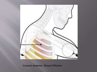
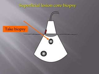
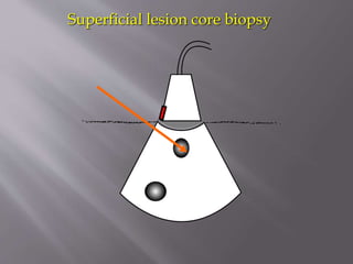
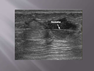
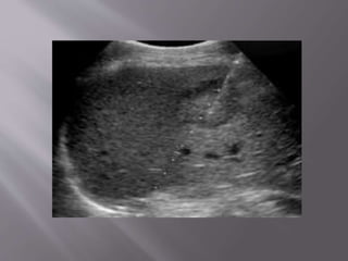
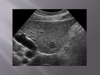
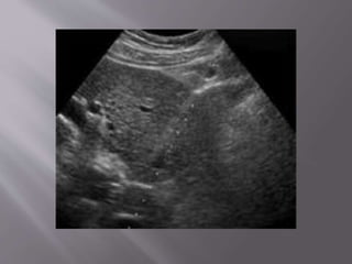
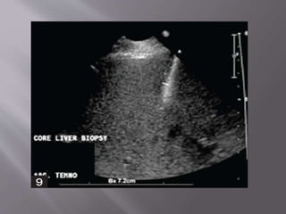
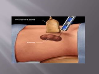
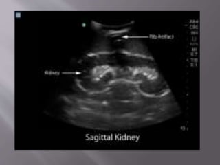
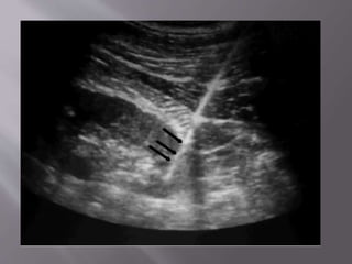
Dr Qurrat-ul-Ain
TMO Radiology


Aspiration



Drainage



Biopsy


Aspirations



Cysts aspiration



Paracentesis (Ascites)



Thoracocentesis (Pleural effusion)


Cysts aspiration:



Cysts are very common.



Usually be diagnosed accurately with
ultrasound.



In most women, they do not usually require
any intervention or follow-up.


Cyst aspirations are done when :

•

Causing significant tenderness

•

The diagnosis of a cyst remains in question
following the ultrasound


POSITION



The patient lie on her back or slightly turned to
one side with the arm placed comfortably

under the head.


The skin is cleaned , numbed with topical
anesthesia. Using ultrasound guidance, a small
needle is advanced into the cyst and suction is
applied to draw the fluid out, causing the lump
to collapse.
The lump (arrow) in this
patient’s right breast was
thought to be a cyst, but some
features are not characteristic
and aspiration was necessary.
Using ultrasound guidance, a fine
needle (white line) is placed so
that its tip (double arrow) is in the
center of the lump (single arrow).
Aspiration is applied by using a
syringe attached to the needle. If
this is a cyst, fluid is drawn into
the syringe as the lesion collapses.
After the aspiration, the
needle (white line) and its
tip (double arrow) are
seen, but the lump is gone.






If is very helpful to get an ultrasound scan of
the ascites before the procedure.
The radiologist will mark the spot for
paracentesis. Two things are important:
What is the distance from the skin to the
fluid? Usually 1 cm.
What is distance to the midpoint of the
collection? Usually 3 cm.
Here we clearly see free fluid in Morrison's pouch that extends
superiorly around the liver
See the needle entering the peritoneal cavity obliquely from just
beneath the indicator marker.


Pleural effusion is an abnormal collection of

fluid in the pleural space. Removal of this fluid
by needle aspiration is called a thoracoentesis.


Patient should be sitting or in the lateral decubitus
position with pleural effusion side up.



The marker on the probe should be pointed
towards the head. Be sure that the transducer is
perpendicular to the chest .


The diaphragm and liver or spleen should be
identified first.



The probe can then be moved towards the head

and from side to side to locate the largest pocket of
fluid between the ribs. Once this is located a mark
is made with indelible ink just above the lower rib.


The distance from the transducer to the pleural
fluid should also be noted.



The probe is then rotated 180 degrees to
visualize the pleural fluid between the ribs to
ensure that there is only fluid visualized ie. no
lung, diaphragm, or liver or spleen.
General Anatomy Pleural Effusion
Thoracocentesis can be both diagnostic and therapeutic .
Using ultrasound to guide this procedure can decrease the
very high complication rate associated with it.
Right Pleural Effusion
Left Pleural Effusion


Procedure allow collections which would

otherwise require open surgery to be drained
via a skin incision only a few mm in size.


Minimally invasive technique



Little procedure related morbidity and equal
applicability to unfit patients,


Any abnormal fluid collection which is
accessible, e.g



Complicated Diverticular abscess



Crohn’s disease related abscess



Appendicular abscess


Localized abscess related to ovary (tubo-ovarian)



Abscess collection after surgery



Hepatic abscess (amebic or post-op)



Renal abscess or retro-peritoneal abscess.



Splenic abscess


The only common contraindications are:



Abscess is not accessible



Patient has a bleeding tendency


Abscess is first delineated &a safe route from
skin to the abscess cavity is identified by
ultrasound.



Prior to the catheter introduction, a diagnostic
needle aspiration may also be done.


The catheter is introduced into the abscess
cavity, either directly using a trocar catheter (as
used for chest intubation (or by modified
Seldinger’s technique using a guide-wire.


Maneuvering of the trocar or guide-wire within
the abdominal cavity should be done strictly
under ultrasound surveillance


Once in position, the catheter is secured and
attached to a drainage bag.



Drainage is recorded daily ,response to the

treatment is assessed by clinical parameters &
u/s.
Indications
Icterus/liver enzyme elevation/elevated bile
acids
Focal nodules or masses anywhere

Renal disease sometimes (i.e. renal dysplasia,
renal masses, lymphosarcoma suspects)
Prostatomegaly
Free abdominal fluid
Cysts

Lymphadenopathy


U/S guided FNA/biopsies generally not done
on:



Adrenal glands



Transitional cell carcinoma suspect masses



Chronic renal failure, glomerulonephritis
Probe orientation
Reference
marker corresponds
to left side of screen
(see
Screen Orientation

Probe
Skin

Superficial “lesion”
to biopsy

Deep “lesion”
to biopsy
Rock and/or slide the probe
to line up the lesion
to a “reachable” position

Deep lesion needs
to be lined up
toward the edge of
the beam

Superficial lesion
can be toward the edge
or in the center
of the beam
Angle to use for a superficial lesion:
Aim needle more perpendicular to beam
Superficial lesion FNA
Superficial lesion core biopsy

Take biopsy
Superficial lesion core biopsy


Percutaneous needle biopsy of the breast
provides reliable diagnosis of both benign and
malignant disease and is a proven alternative
to open surgical biopsy


Ultrasound guidance is an accurate and reliable
biopsy guidance technique and is the method
of choice and suitable for all breast lesions
visible on ultrasound


CNB & FNAB are effective methods for the
diagnosis of most breast lesions



Although CNB has higher sensitivity & positive
predictive value for abnormalities like micro-

calcifications & distortions of architecture.


Focal mass or other lesion of unknown nature –
palpable or non-palpable



Architectural distortion



Micro-calcifications



Cyst aspiration


PROCEDURE:



The long axis of the needle, should be visible along
the long axis of the transducer.



Occasionally, during an FNA biopsy or cyst
aspiration, the transducer can be rotated 90
degrees to visualize the echogenic dot of the
needle within the lesion.


Liver biopsies are performed for both focal and
nonfocal lesions.



The primary indication for parenchymal liver
biopsy is for the diagnosis of hepatic disease.




When imaging guidance is employed, it can
take one of two forms:
US-guided "marking" in which a mark is made

upon the skin during US examination for a
biopsy to be performed later without imaging
guidance or real-time US guidance.


The patient is positioned supine, with the
hands comfortably resting behind the head



A preliminary US scan is performed to identify
the target and mark the skin.


The preliminary scan also ensures that no major
vessels, dilated biliary channels or gall bladder are
in the path of the biopsy needle.


Before the procedure is started, breathing
instructions are practiced with the patient.



performed with the breath held in expiration.



This minimize risk of injury to the pleura or lung.


The skin site is prepped and draped to ensure
asepsis The local area is anesthetized with a
local anesthetic.



The cutting needle is then fired with US
documentation of the site.


Indications:



Biopsy of a focal solid lesion /suspicious cystic
lesion for diagnosis.



Nonfocal biopsy to evaluate for nephropathy
or renal transplant rejection


US has the advantages of real-time needle
placement



No radiation & is therefore well suited for most
nonfocal renal biopsies in thin pts and in biopsies
of some focal solid masses or cystic masses .


The patient is placed in the prone position and
the biopsy is typically taken from the lower

pole of the kidney if there are no specific
locations of interest.


The biopsy needle is guided using ultrasound
to ensure visualization of the needle as it
pierces the kidney parenchyma.


Care is taken not to enter the collecting system
(as it would result in haematuria) or to go near
the renal hilum (to prevent injury to the
vessels).
THANK YOU

Ultrasound guided procedures

  • 1.
  • 2.
  • 3.
  • 4.
     Cysts aspiration:  Cysts arevery common.  Usually be diagnosed accurately with ultrasound.  In most women, they do not usually require any intervention or follow-up.
  • 5.
     Cyst aspirations aredone when : • Causing significant tenderness • The diagnosis of a cyst remains in question following the ultrasound
  • 6.
     POSITION  The patient lieon her back or slightly turned to one side with the arm placed comfortably under the head.
  • 7.
     The skin iscleaned , numbed with topical anesthesia. Using ultrasound guidance, a small needle is advanced into the cyst and suction is applied to draw the fluid out, causing the lump to collapse.
  • 9.
    The lump (arrow)in this patient’s right breast was thought to be a cyst, but some features are not characteristic and aspiration was necessary.
  • 10.
    Using ultrasound guidance,a fine needle (white line) is placed so that its tip (double arrow) is in the center of the lump (single arrow). Aspiration is applied by using a syringe attached to the needle. If this is a cyst, fluid is drawn into the syringe as the lesion collapses.
  • 11.
    After the aspiration,the needle (white line) and its tip (double arrow) are seen, but the lump is gone.
  • 12.
       If is veryhelpful to get an ultrasound scan of the ascites before the procedure. The radiologist will mark the spot for paracentesis. Two things are important: What is the distance from the skin to the fluid? Usually 1 cm. What is distance to the midpoint of the collection? Usually 3 cm.
  • 14.
    Here we clearlysee free fluid in Morrison's pouch that extends superiorly around the liver
  • 15.
    See the needleentering the peritoneal cavity obliquely from just beneath the indicator marker.
  • 16.
     Pleural effusion isan abnormal collection of fluid in the pleural space. Removal of this fluid by needle aspiration is called a thoracoentesis.
  • 17.
     Patient should besitting or in the lateral decubitus position with pleural effusion side up.  The marker on the probe should be pointed towards the head. Be sure that the transducer is perpendicular to the chest .
  • 18.
     The diaphragm andliver or spleen should be identified first.  The probe can then be moved towards the head and from side to side to locate the largest pocket of fluid between the ribs. Once this is located a mark is made with indelible ink just above the lower rib.
  • 19.
     The distance fromthe transducer to the pleural fluid should also be noted.  The probe is then rotated 180 degrees to visualize the pleural fluid between the ribs to ensure that there is only fluid visualized ie. no lung, diaphragm, or liver or spleen.
  • 20.
  • 21.
    Thoracocentesis can beboth diagnostic and therapeutic . Using ultrasound to guide this procedure can decrease the very high complication rate associated with it.
  • 22.
  • 23.
  • 24.
     Procedure allow collectionswhich would otherwise require open surgery to be drained via a skin incision only a few mm in size.
  • 25.
     Minimally invasive technique  Littleprocedure related morbidity and equal applicability to unfit patients,
  • 26.
     Any abnormal fluidcollection which is accessible, e.g  Complicated Diverticular abscess  Crohn’s disease related abscess  Appendicular abscess
  • 27.
     Localized abscess relatedto ovary (tubo-ovarian)  Abscess collection after surgery  Hepatic abscess (amebic or post-op)  Renal abscess or retro-peritoneal abscess.  Splenic abscess
  • 28.
     The only commoncontraindications are:  Abscess is not accessible  Patient has a bleeding tendency
  • 29.
     Abscess is firstdelineated &a safe route from skin to the abscess cavity is identified by ultrasound.  Prior to the catheter introduction, a diagnostic needle aspiration may also be done.
  • 30.
     The catheter isintroduced into the abscess cavity, either directly using a trocar catheter (as used for chest intubation (or by modified Seldinger’s technique using a guide-wire.
  • 31.
     Maneuvering of thetrocar or guide-wire within the abdominal cavity should be done strictly under ultrasound surveillance
  • 32.
     Once in position,the catheter is secured and attached to a drainage bag.  Drainage is recorded daily ,response to the treatment is assessed by clinical parameters & u/s.
  • 35.
    Indications Icterus/liver enzyme elevation/elevatedbile acids Focal nodules or masses anywhere Renal disease sometimes (i.e. renal dysplasia, renal masses, lymphosarcoma suspects) Prostatomegaly Free abdominal fluid Cysts Lymphadenopathy
  • 36.
     U/S guided FNA/biopsiesgenerally not done on:  Adrenal glands  Transitional cell carcinoma suspect masses  Chronic renal failure, glomerulonephritis
  • 37.
    Probe orientation Reference marker corresponds toleft side of screen (see Screen Orientation Probe Skin Superficial “lesion” to biopsy Deep “lesion” to biopsy
  • 38.
    Rock and/or slidethe probe to line up the lesion to a “reachable” position Deep lesion needs to be lined up toward the edge of the beam Superficial lesion can be toward the edge or in the center of the beam
  • 39.
    Angle to usefor a superficial lesion: Aim needle more perpendicular to beam
  • 40.
  • 41.
  • 42.
  • 43.
    Superficial lesion corebiopsy Take biopsy
  • 44.
  • 45.
     Percutaneous needle biopsyof the breast provides reliable diagnosis of both benign and malignant disease and is a proven alternative to open surgical biopsy
  • 46.
     Ultrasound guidance isan accurate and reliable biopsy guidance technique and is the method of choice and suitable for all breast lesions visible on ultrasound
  • 47.
     CNB & FNABare effective methods for the diagnosis of most breast lesions  Although CNB has higher sensitivity & positive predictive value for abnormalities like micro- calcifications & distortions of architecture.
  • 48.
     Focal mass orother lesion of unknown nature – palpable or non-palpable  Architectural distortion  Micro-calcifications  Cyst aspiration
  • 49.
     PROCEDURE:  The long axisof the needle, should be visible along the long axis of the transducer.  Occasionally, during an FNA biopsy or cyst aspiration, the transducer can be rotated 90 degrees to visualize the echogenic dot of the needle within the lesion.
  • 52.
     Liver biopsies areperformed for both focal and nonfocal lesions.  The primary indication for parenchymal liver biopsy is for the diagnosis of hepatic disease.
  • 54.
      When imaging guidanceis employed, it can take one of two forms: US-guided "marking" in which a mark is made upon the skin during US examination for a biopsy to be performed later without imaging guidance or real-time US guidance.
  • 55.
     The patient ispositioned supine, with the hands comfortably resting behind the head  A preliminary US scan is performed to identify the target and mark the skin.
  • 56.
     The preliminary scanalso ensures that no major vessels, dilated biliary channels or gall bladder are in the path of the biopsy needle.
  • 57.
     Before the procedureis started, breathing instructions are practiced with the patient.  performed with the breath held in expiration.  This minimize risk of injury to the pleura or lung.
  • 58.
     The skin siteis prepped and draped to ensure asepsis The local area is anesthetized with a local anesthetic.  The cutting needle is then fired with US documentation of the site.
  • 63.
     Indications:  Biopsy of afocal solid lesion /suspicious cystic lesion for diagnosis.  Nonfocal biopsy to evaluate for nephropathy or renal transplant rejection
  • 64.
     US has theadvantages of real-time needle placement  No radiation & is therefore well suited for most nonfocal renal biopsies in thin pts and in biopsies of some focal solid masses or cystic masses .
  • 65.
     The patient isplaced in the prone position and the biopsy is typically taken from the lower pole of the kidney if there are no specific locations of interest.
  • 66.
     The biopsy needleis guided using ultrasound to ensure visualization of the needle as it pierces the kidney parenchyma.
  • 67.
     Care is takennot to enter the collecting system (as it would result in haematuria) or to go near the renal hilum (to prevent injury to the vessels).
  • 71.