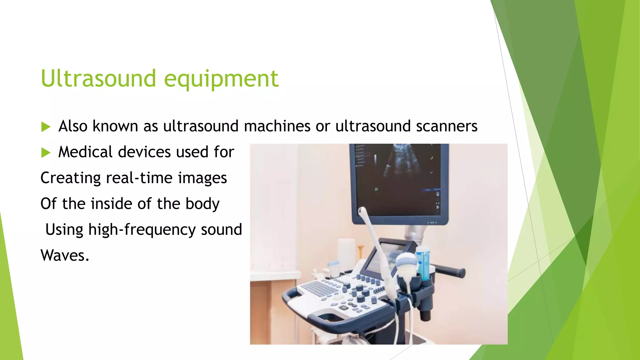
2. Ultrasound equipment
Also known as ultrasound machines or ultrasound scanners
Medical devices used for
Creating real-time images
Of the inside of the body
Using high-frequency sound
Waves.
3. Transducer (Probe):
Monitor:
Control Panel:
Keyboard and User Interface:
Gel Dispenser
Printer
Transducer Ports
Storage and Connectivity
4. Transducer (Probe):
It serves the essential function of emitting ultrasound waves into the
body and receiving the echoes to create images
components of transducer
5. Piezoelectric Crystals:
It’s the functional component of ultrasound
Typically made of materials like lead zirconate titanate (PZT) or
ceramics.
These crystals have the unique property of converting electrical energy
into mechanical vibrations (ultrasound waves) when subjected to an
electrical current and vice versa, converting mechanical vibrations
(echoes) into electrical signals.
An electrical dipole: is a molecular entity containing positive and
negative electric charges that has no net charge.
When mechanically compressed by an externally applied pressure, the
alignment of the dipoles is disturbed from the equilibrium position to
cause an imbalance of the charge distribution.
an external voltage through conductors attached to the surface
electrodes induces the mechanical expansion and contraction of the
transducer element.
6. Damping block: Behind the piezoelectric crystals, there is a backing
material that helps dampen vibrations and reduce the duration of the
emitted ultrasound pulse. This enhances the transducer's ability to
produce high-quality images.
Acoustic Matching Layer: On the front face of the transducer, there is
an acoustic matching layer that helps improve the transmission of
ultrasound waves into the body. This layer has an acoustic impedance
between that of the piezoelectric crystals and human tissue, minimizing
reflection at the crystal-tissue interface.
Lens: Some transducers have a lens or acoustic focusing element in
front of the crystals. The lens helps focus the ultrasound beam to
improve image resolution and penetration depth.
7. 2.Monitor: The ultrasound machine has a built-in monitor or display
screen where the real-time images are displayed. Modern machines often
feature high-resolution color LCD screens for better image quality.
3. Control Panel: The control panel includes buttons and knobs that allow
the operator (sonographer or physician) to adjust various settings, such as
frequency, depth, gain, and focus. It also typically contains preset imaging
modes for specific exams.
4.Keyboard and User Interface: Ultrasound machines have a user-friendly
interface for inputting patient data, annotations, and comments. This
interface allows the operator to control the machine and navigate through
its functions.
8. Housing: The transducer is encased in a protective housing, which is typically
made of plastic or other materials. This housing is designed to be ergonomic for
the operator's comfort and to protect the internal components.
Cable and Connector: The transducer is connected to the ultrasound machine
via a cable. The cable carries the electrical signals to and from the transducer. It
terminates in a connector that plugs into the ultrasound system.
Strain Relief: The cable is often attached to the transducer via a strain relief
mechanism to prevent damage to the cable or connector due to bending or
pulling.
9. 5. Gel Dispenser: Ultrasound gel is essential for ensuring good acoustic contact
between the transducer and the skin. Some machines have integrated gel
dispensers for convenience.
6. Printer: Many ultrasound machines have built-in printers for producing hard
copies of images for patient records or reports.
7. Transducer Ports: Some ultrasound machines have multiple transducer ports,
allowing for easy switching between different transducers during an exam.
8. Storage and Connectivity: Modern ultrasound machines come equipped with
storage options for saving images and patient data. They may also have
connectivity options for transferring images and reports to electronic medical
records (EMRs) or for telemedicine purposes.
10. TYPES OF ULTRASOUND:
1. Diagnostic Ultrasound: This is the most common type of ultrasound
and is used to create images of various parts of the body to diagnose
medical conditions. It includes:
• Abdominal Ultrasound: Used to visualize organs in the abdomen, such
as the liver, gallbladder, kidneys, and pancreas.
• Pelvic Ultrasound: Used to examine the reproductive organs, bladder,
and pelvic structures in both males and females.
• Obstetric Ultrasound: Used during pregnancy to monitor the developing
fetus and assess its health.
• Transvaginal Ultrasound: An internal ultrasound of the female pelvic
organs, often used in gynecology.
11.
12. 2. Doppler Ultrasound: This type of ultrasound measures blood flow in the body's
blood vessels and can help diagnose vascular conditions. There are two main types:
Color Doppler: Provides a color-coded map of blood flow.
Doppler Spectral Analysis: Measures the speed and direction of blood flow
using sound wave frequency changes.
13. 3. Echocardiography: This ultrasound is specifically used to visualize the heart and
its structures. There are different types of echocardiograms, including:
• Transthoracic Echocardiography (TTE): The ultrasound transducer is placed on
the chest to obtain images of the heart.
• Transesophageal Echocardiography (TEE): An ultrasound probe is inserted into
the esophagus to obtain more detailed images of the heart.
14. 4.Musculoskeletal Ultrasound: Used to examine muscles, tendons, ligaments,
joints, and soft tissues for injuries or abnormalities. It's often used in sports
medicine.
15. 5.Breast Ultrasound: Often used alongside mammography to
evaluate breast abnormalities, such as cysts or solid masses.
16. 6. Thyroid Ultrasound: Examines the thyroid gland for nodules,
goiters, or other abnormalities.
7. Endoscopic Ultrasound (EUS): Combines ultrasound with
endoscopy to visualize the gastrointestinal tract and adjacent
organs like the pancreas and bile ducts.
17. 8.Transcranial Doppler Ultrasound: Evaluates blood flow in major
arteries of the brain, often used in neurology to assess conditions
like strokes and vascular disorders.
9.3D and 4D Ultrasound: These techniques create three-
dimensional or real-time moving images, respectively, providing
more detailed information for diagnostic purposes. Commonly used
in obstetrics.
18. 10. Intravascular Ultrasound (IVUS): Used during cardiac
catheterization procedures to visualize the inside of blood vessels,
especially coronary arteries.
11. Contrast-Enhanced Ultrasound (CEUS): Involves injecting
contrast agents to enhance imaging of specific tissues, organs, or
blood vessels.
19. 12.Pediatric Ultrasound: Tailored for imaging infants and
children, often used to evaluate congenital abnormalities or
pediatric-specific conditions.
13.Ophthalmic Ultrasound: Used for eye exams, particularly
for assessing conditions within the eye, such as retinal
detachment.
20. 14.Portable Ultrasound: Compact and often handheld devices
designed for point-of-care applications, emergency medicine, or
field use.
15.Interventional Ultrasound: Utilized for guiding minimally
invasive procedures, such as biopsies, aspirations, or catheter
placements.
21. Indication And Clinical Application Of
Ultrasound
1. Pregnancy and Obstetrics:
• Fetal Monitoring: Ultrasound is used to monitor fetal growth,
development, and well-being during pregnancy.
• Confirming Pregnancy: To confirm the presence of a pregnancy,
determine gestational age, and assess for multiple pregnancies.
• Assessment of Fetal Anomalies: Detecting congenital
abnormalities in the developing fetus.
22. 2.Abdominal Imaging:
• Liver, Gallbladder, and Pancreas: To evaluate the liver for
masses, cirrhosis, or fatty liver, and assess the gallbladder and
pancreas for stones or tumors.
• Kidneys: To diagnose kidney stones, cysts, infections, and
tumors.
• Spleen: To examine the spleen for enlargement or lesions.
• Gastrointestinal Tract: To evaluate the digestive organs, such as
the stomach, intestines, and appendix.
• Bladder: To assess for bladder tumors, urinary retention, or
other abnormalities.
23. 3.Pelvic Imaging:
1. Gynecology: To assess the female reproductive organs,
including the uterus and ovaries, for conditions like fibroids,
ovarian cysts, and endometrial abnormalities.
2. Prostate: To evaluate the prostate gland and assess for
abnormalities, including prostate cancer.
3. Testes: For diagnosing testicular masses or injuries.
4. Cardiac Imaging:
1. Echocardiography: To visualize and assess the heart's
structure and function, including the heart valves, chambers,
and blood flow.
2. Stress Echocardiography: To evaluate heart function during
exercise or stress tests.
24. 5. Vascular Imaging:
1. Vascular Ultrasound: To examine blood vessels for
conditions such as deep vein thrombosis (DVT), arterial
blockages, and aneurysms.
2. Carotid Doppler: To assess blood flow in the carotid
arteries in the neck, often used to evaluate stroke risk.
6. Musculoskeletal Imaging:
1. Orthopedics: To diagnose and monitor conditions
affecting bones, joints, muscles, and tendons, including
fractures, arthritis, and soft tissue injuries.
2. Rheumatology: For evaluating joint inflammation and
conditions like rheumatoid arthritis.
25. 7. Breast Imaging:
1. Breast Ultrasound: As a complement to mammography
to evaluate breast abnormalities, such as cysts, solid
masses, or to guide breast biopsies.
8. Thyroid and Neck Imaging:
1. Thyroid Ultrasound: To assess the thyroid gland for
nodules, goiters, or other abnormalities.
2. Neck: To evaluate lymph nodes and other structures in
the neck.
9. Ophthalmic Ultrasound:
1. To assess conditions within the eye, such as retinal
detachment.
26. 10. Urology and Nephrology:
1. Renal Ultrasound: To diagnose and monitor kidney
conditions, such as kidney stones, cysts, and infections.
2. Bladder: To evaluate bladder function and detect urinary
retention.
11. Interventional Procedures:
1. Ultrasound-Guided Biopsies and Aspirations: Ultrasound is
used to guide the placement of needles for tissue sampling or
fluid drainage.
2. Vascular Access: To assist in placing central venous catheters
or peripherally inserted central catheters (PICCs).
27. 12. Emergency Medicine and Critical Care:
1. Rapid assessment of trauma or critical care patients for
abdominal bleeding, organ injuries, or vascular issues.
13. Pediatrics:
1. Evaluating congenital anomalies, pediatric abdominal issues,
and neonatal brain conditions.
14. Point-of-Care Ultrasound (POCUS):
1. Bedside ultrasound in emergency departments and critical
care settings for rapid diagnostic assessments.
15. Fertility and Reproductive Medicine:
• Monitoring ovarian follicles during assisted reproductive
procedures like in vitro fertilization (IVF).