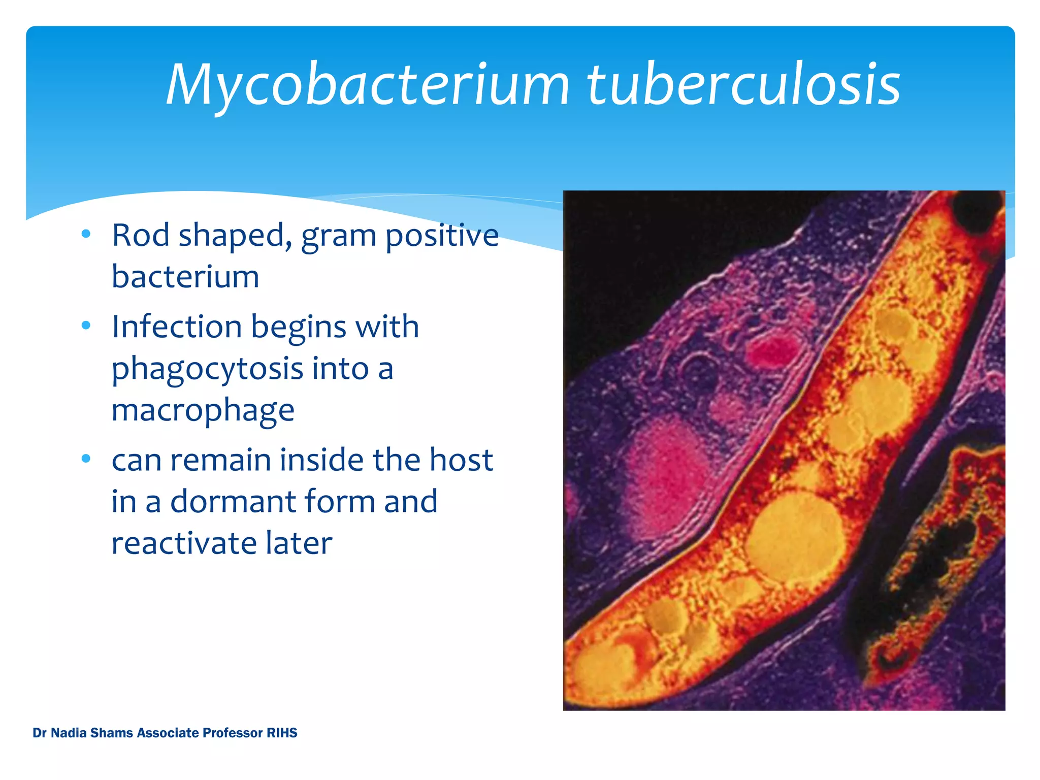
3. World Incidence 2006
World map showing reported cases of tuberculosis per 100,000 citizens. Red = >300,
orange = 200-300; yellow = 100-200; green 50-100; blue = <50 and grey = n/a.
Dr Nadia Shams Associate Professor RIHS
4. #1 on the list of lethal infectious diseases
2 million deaths worldwide annually
Every year 8 million cases reported
Death rate after contracting the disease, if untreated,
is the same as flipping a coin
Statistics
Dr Nadia Shams Associate Professor RIHS
5. Mycobacterium tuberculosis
• Rod shaped, gram positive
bacterium
• Infection begins with
phagocytosis into a
macrophage
• can remain inside the host
in a dormant form and
reactivate later
Dr Nadia Shams Associate Professor RIHS
6. History
TB has been known as
Pthisis, King’s Evil, Pott’s
disease, consumption, and
the White Plague.
Egyptian mummies from
3500 BCE have the
presence of
Mycobacterium
tuberculosis
Dr Nadia Shams Associate Professor RIHS
7. The New World
Infected the New World
before the Europeans
10% deaths in the 19th
century were due to TB
Isolated the infected in
sanitariums, which served
as waiting rooms for death
Dr Nadia Shams Associate Professor RIHS
8. Factors increasing the risk of
Tuberculosis
Children > young < elderly
Immigrants from high prevalence areas
Close contacts of patients who are smear positive
Overcrowding
Immunosuppression
Type 1 Diabetics
CRF
Malnourishment
Dr Nadia Shams Associate Professor RIHS
9. Time table of Tuberculosis
Time from infection manifestations
3-8 weeks Primary complex, +ve tuberculin skin test
3-6 months Meningeal milliary and pleural disease
Upto 3 yrs GIT, bone, joint, lymph node disease
Around 8 yrs Renal tract disease
From 3 yrs onwards Post primary disease due to reactivation/ reinfection
Dr Nadia Shams Associate Professor RIHS
10. Disease progression- Stage 1
Stage 1
Droplet nuclei are inhaled, and are
generated by talking, coughing and
sneezing.
bacteria are taken up by alveolar
macrophages.
The large droplet nuclei reaches
upper respiratory tract, and the small
droplet nuclei reaches air sacs of the
lung (alveoli) where the infection
begins.
Disease onset - when droplet nuclei
reach the alveoli.
Dr Nadia Shams Associate Professor RIHS
11. Begins after 7-21 days after initial infection.
TB multiplies within the inactivated macrophages
until the macrophages burst.
Other macrophages diffuse from peripheral blood,
phagocytose and are inactivated, rendering them
unable to destroy TB.
Disease Progression- Stage 2
Dr Nadia Shams Associate Professor RIHS
12. Lymphocytes/T-cells recognize TB antigen. This results in T-cell
activation and the release of Cytokines, including interferon
(IFN).
The release of IFN causes activation of macrophages, which can
release lytic enzymes and reactive intermediates that facilitates
immune pathology.
Tubercle forms, which contains a semi-solid or “cheesy”
consistency. TB cannot multiply within tubercles due to low PH
and anoxic environment, but TB can persist within these
tubercles for extended periods.
Disease Progression- Stage 3
Dr Nadia Shams Associate Professor RIHS
13. TB uses these macrophages to replicate causing the tubercle to grow.
The growing tubercle may invade a bronchus, causing an infection which
may spread to other parts of the lungs. Tubercle may also invade artery or
other blood supply.
MILIARY TB: Spreading of TB may cause milliary tuberculosis, which can
cause secondary lesions.
SYSTEMIC INVOLVEMENT: Secondary lesions occur in bones, joints, lymph
nodes, genitourinary system and peritoneum.
Disease Progression- Stage 4
Dr Nadia Shams Associate Professor RIHS
14. The caseous centers of the tubercles liquefy.
This liquid is very crucial for the growth of TB, and therefore it
multiplies rapidly (extracellularly).
This later becomes a large antigen load, causing the walls of
nearby bronchi to become necrotic and rupture.
This results in “cavity formation” and allows TB to spread rapidly
into other airways and to other parts of the lung.
Stage 5
Dr Nadia Shams Associate Professor RIHS
15. Pulmonary Versus Extrapulmonary TB
PULMONARY TB
Primary pulmonary TB
Miliary TB
Post Primary Pulmonary
TB
EXTRAPULMONARY TB
Lymphadenitis
GI Tuberculosis
Pericardial Disease
CNS disease
Bone & joint disease
Genitourinary Disease
Dr Nadia Shams Associate Professor RIHS
16. Symptoms of Pulmonary disease
◼ Productive cough
◼ chest pain
◼ night sweats, fatigue, fever
◼ Primary TB: Self limiting febrile illness
◼ Miliary TB: Above symptoms , heptomegaly, Headache, choroid
Tubercles, Millet seed appearance on CXR, anaemia, leukopaenia.
◼ Post Primary TB: Progressive pulmonary Symptoms,
complications, consolidation, collapse, cavitation.
Dr Nadia Shams Associate Professor RIHS
17. Lyphadenitis: Cervical, mediastinal, axillary, Inginal,
supraclavicular. Collar stud abcess and fistula
formation.
GI Tuberculosis: Ileaocaecal disease, Right iliac fossa
mass, tuberculous peritonitis, hepatic dysfunction.
Pericardial disease: Pericardial effusion, constrictive
pericarditis.
Symptoms of Extra Pulmonary
Disease
Dr Nadia Shams Associate Professor RIHS
18. CNS Disease: Tuberculous meningitis/ SOL
Tuberculomas.
Bone and joint disease: Potts disease, Psoas Abscess.
Genitourinary Disease: Sterile Pyuria, Infertility,
endometritis
Symptoms of Extra Pulmonary
Disease
Dr Nadia Shams Associate Professor RIHS
20. TB Tests
ZN (Ziehl Neelsen) staining
Sputum
Gastric Washings
BAL
Transbronchial biopsy
Fluid Examination (CSF, Ascitic, pleural, pericardial, Joint)
Tuberculin Skin Test
Injection of fluid into the skin of the lower arm
48-72 hours later – checked for a reaction
Diagnosis is based on area of the raised skin
Dr Nadia Shams Associate Professor RIHS
21. A= Non Acid fast
B=Acid fast
Dr Nadia Shams Associate Professor RIHS
28. Drug resistant strains of Mycobacterium tuberculosis have
developed
Underdeveloped countries are the most commonly affected by
TB
95% of reported cases come from underdeveloped countries
High HIV rates contribute to the contraction of TB
Reasons for Fear
Dr Nadia Shams Associate Professor RIHS