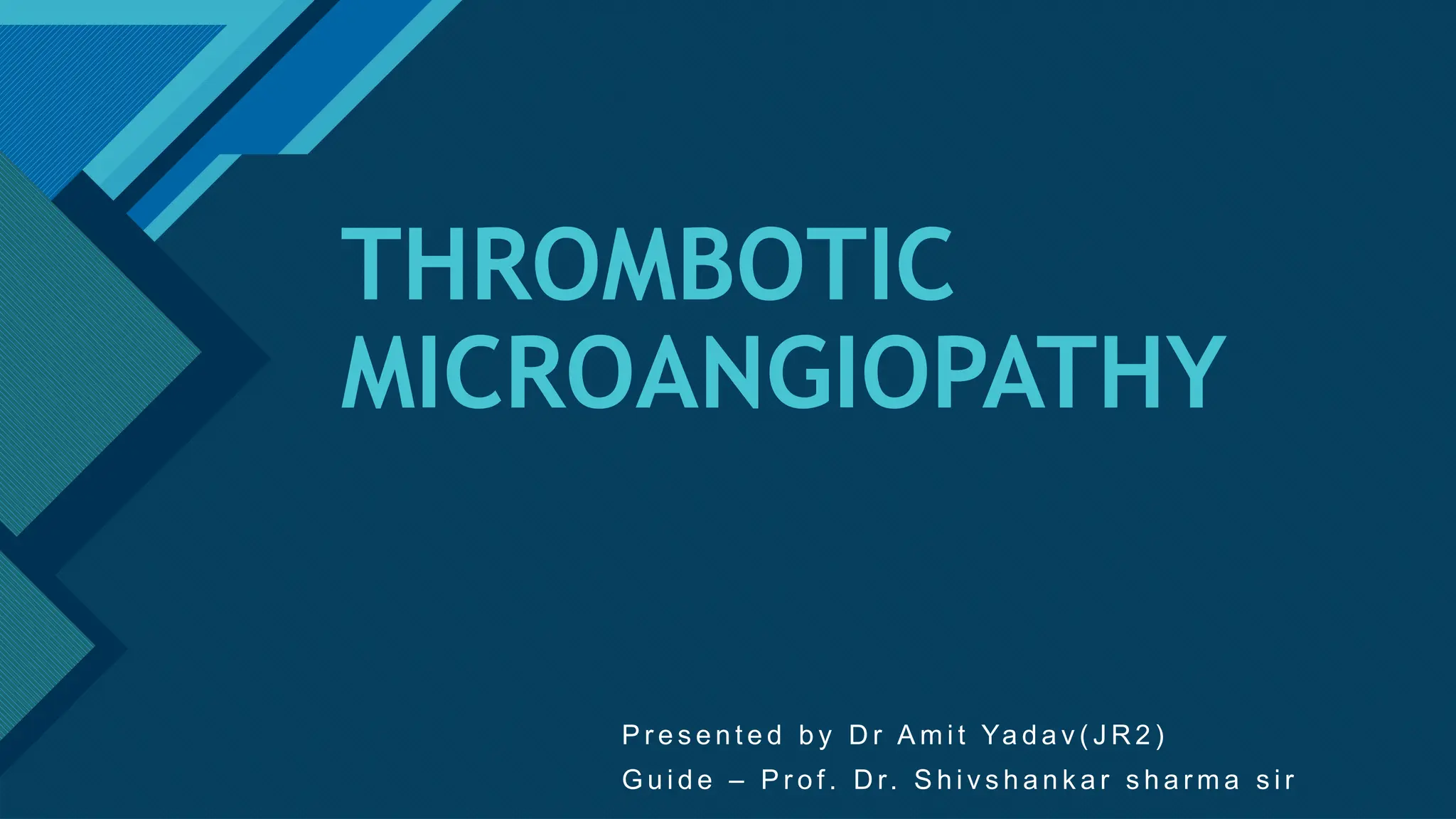
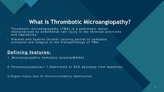
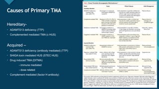
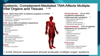
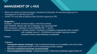
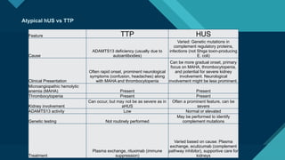
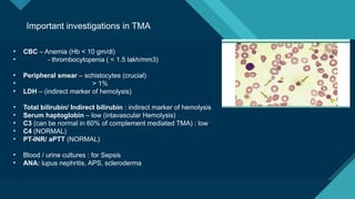
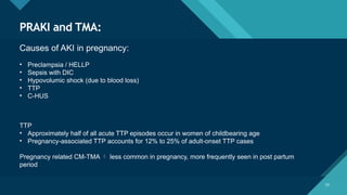
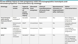
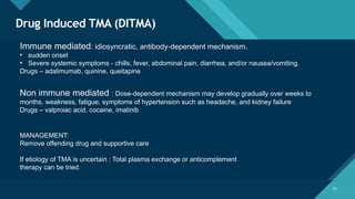
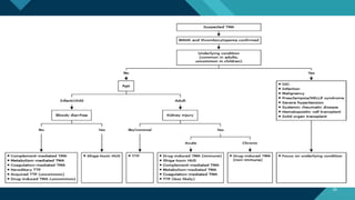
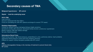
Click to edit Master title style
1
THROMBOTIC
MICROANGIOPATHY
P r e s e n t e d b y D r A m i t Ya d a v ( J R 2 )
G u i d e – P r o f . D r. S h i v s h a n k a r s h a r m a s i r
Click to edit Master title style
2 2
What is Thrombotic Microangiopathy?
• T h r o m b o t i c m i c r o a n g i o p a t h y ( T M A ) i s a p a t h o l o g i c l e s i o n
c h a r a c t e r i z e d b y e n d o t h e l i a l c e l l i n j u r y i n t h e t e r m i n a l a r t e r i o l e s
a n d c a p i l l a r i e s .
• P l a t e l e t a n d h y a l i n e t h r o m b i c a u s i n g p a r t i a l o r c o m p l e t e
o c c l u s i o n a r e i n t e g r a l t o t h e h i s t o p a t h o l o g y o f T M A .
Defining features:
1 . M i c r o a n g i o p a t h i c h e m o l y t i c a n e m i a ( M A H A )
2 . T h r o m b o c y t o p e n i a ( < 1 . 5 l a k h / m m 3 o r 2 5 % d e c r e a s e f r o m b a s e l i n e )
3 . O r g a n i n j u r y d u e t o m i c r o c i r c u l a t o r y o b s t r u c t i o n .
Click to edit Master title style
3 3
What it actually means?
Intraluminal platelet thrombosis
Microangiopathic hemolytic anemia
Consumption of platelets
thrombocytopenia Hemolysis, LDH ↑ , bilirubin ↑
VWF release
Endothelial injury
Click to edit Master title style
4 4
Why is it important?
• Thrombotic microangiopathy (TMA) is frequently suspected but
can be challenging to diagnose definitively due to its complex and
overlapping clinical features with other conditions.
• In pregnancy related AKI it is common.
• Timely diagnosis and management is required to salvage kidney and the patient (24-48
hours)
Click to edit Master title style
5
Causes of Primary TMA
5
Hereditary-
• ADAMTS13 deficiency (TTP)
• Complemented mediated TMA (c HUS)
Acquired –
• ADAMTS13 deficiency (antibody mediated) (TTP)
• SHIGA toxin mediated HUS (STEC HUS)
• Drug induced TMA (DITMA)
- immune mediated
- dose related
• Complement mediated (factor H antibody)
Click to edit Master title style
6
Is Renal Biopsy essential for Diagnosis?
6
NO,
• Histological findings of TMA are not pathognomonic.
• Does not help in establishing the etiology of TMA.
• TMA has a high bleeding risk, due to the presence of high blood
pressure, thrombocytopenia and uremia.
Can be considered in secondary TMA, atypical presentation and Focal TMA.
Click to edit Master title style
7 7
Typical HUS (STEC HUS)
Diarrhea associated (D+)
90% caused by
1) Enterohemorhagic E coli (EHEC)
produces shiga like toxin
2) Shigela dysenteriae type 1: shiga toxin
10% caused by
1) Streptococcus pneumoniae :
Neuroaminidase toxin
Typically seen in children (around 5 years of age )
Click to edit Master title style
8
Typical HUS (cont.)
• Only ~ 15% will develop HUS
• 85% resolves spontaneously
Management:
• Good prognosis
• Aggressive hydration with isotonic fluids
• Antibiotics – avoided in E coli due to preformed toxins release
• Shigellosis – Early antibiotics (azithromycin) indicated to prevent bacteria shredding
Is there a role for plasma exchange?
There is insufficient evidence to support TPE in routine
management of infection-associated HUS; however, it can be
considered in severe cases.
Click to edit Master title style
9
Atypical HUS (c-HUS)
Result of alternate pathway complement
dysregulation.
Congenital(50%) >> acquired (10%)
2/1000000 cases per year
Triad :
1. MAHA
2. Thrombocytopenia
3. Renal dysfunction (severe)
Click to edit Master title style
10
Click to edit Master title style
11
MANAGEMENT OF c-HUS
11
• Before the advent of pharmacologic complement blockade, the standard approach to
management of CM-HUS was TPE.
• Unlike TTP, only 30% of patients with CM-HUS respond to TPE.
Supportive :
Hydration : maintain euvolumic status, avoid fluid overload
Avoid NSAIDS: already high risk of bleeding, may exacerbate AKI
RBC transfusion: indicated in severe ANEMIA (Hb < 7 gm/dl)
Platelets transfusion: routinely avoided, Transfusion of platelets is appropriate when a patient
with a platelet count of < 50,000/mm3 and patient requiring invasive
procedure where critical bleeding is anticipated
Dialysis
Definitive :
• Therapeutic plasma exchange: if anti-complement therapy is not available, and as the initial
approach to complement factor H (CFH) autoantibody-driven disease.
• Terminal complement blockade (recommended)
• Immunosuppressive therapy: cyclophosphamide or Rituximab (antibodies against factor H)
Click to edit Master title style
12
Terminal complement blockade
12
Monoclonal antibodies directed against the C-5 complement component
Should be started preferably in 24-48 hours
Eculizumab
Ravulizumab
Individuals treated with eculizumab or ravulizumab should be vaccinated against encapsulated organisms
Pregnancy : Eculizumab and Ravulizumab can be used
Click to edit Master title style
13
13
Thrombotic Thrombocytopenic purpura
The pathophysiology of TTP involves the accumulation of ultra-large multimers of von Willebrand
factor as a result of the absence or markedly decreased activity of the plasma protease
ADAMTS13.
Acquired (60-90%) >> Congenital
5/1000000 cases per year
Pentad :
1. Microangiopathic hemolytic Anemia
2. Acute renal insufficiency (20-50%)
3. Thrombocytopenia
4. Neurological abnormalities (50-80%)
5. Fever (60-80%)
Click to edit Master title style
14
What is ADAM TS 13? HOW TO INTERPRET ?
ADAMTS13 is a protease which cleaves ultra large von Willebrand factor (VWF) multimers on the
endothelial surface.
The ultra-large VWF multimers that accumulate in the absence of ADAMTS13 are responsible for causing the TMA.
ADAM TS 13 activity-
• ADAMTS13 activity testing can be performed on plasma or serum.
• sample for ADAMTS13 activity measurement should be collected prior to blood product transfusions
and/or initiation of TPE
Click to edit Master title style
15
What is plasmic score?
Devised to predict the likelihood of ADAMTS13 activity
≤10 percent (to help estimate the pretest probability and
support the diagnosis of TTP) in adults with features of
thrombotic microangiopathy (TMA)
While this score cannot substitute for clinical judgment
and cannot be used to definitively confirm or exclude the
diagnosis of TTP, it may be helpful for guiding the
decision of whether to initiate therapy while awaiting the
results of ADAMTS13 testing.
Click to edit Master title style
16
● Severe deficiency (activity <10 percent) –
This confirms TTP in appropriate clinical setting
ADAMTS13 activity <10 percent has also been reported in patients with other causes of MAHA and
thrombocytopenia including sepsis and systemic cancer
●Very low activity (11 to 20 percent) –
Levels of 11 to 20 percent may occur in patients with TTP who have received transfusions or after TPE.
●Low activity (21 to 60 percent) –
Inflammatory disorders (sepsis, malignancy).
●Normal (>60 percent) – look for other etiology of TMA
Interpreting results of ADAM TS 13 Activity:
Click to edit Master title style
17
Management of TTP
17
Supportive measure
Definite measures:
• Therapeutic plasma exchange : (preferably in 24 hours)
• Therapeutic plasma exchange (TPE) remains the mainstay of treatment for TTP .
• It provides ADAMTS13 from donor plasma and removes the autoantibody against ADAMTS13.
• Plasma as the replacement fluid — Plasma is used as the replacement fluid because it provides
ADAMTS13.
• Average cycles needed 7-10
• If TPE unavailable or delayed: FFP transfusions can be given
Click to edit Master title style
18
18
Glucocorticoids — Decrease production of the ADAMTS13 inhibitor (autoantibody).
•Standard Risk:
Prednisone 1 mg/kg/day orally (if alert and awake without neurological
problems or normal troponin).
• If no response within 3-4 days, switch to methylprednisolone 1000 mg/day
intravenously (IV) until improvement.
•High Risk:
Methylprednisolone 1000 mg/day IV Single daily dose for 3 days
continue high dose if patient remains high risk
If improving, switch to prednisone 1 mg/kg/day orally.
Prednisone 1mg/kg/day continues even after plasma exchange (TPE) is stopped.
Tapered over 2-3 weeks
Click to edit Master title style
19
19
Rituximab : 375 mg/m intravenously once a week for four consecutive weeks
Rituximab should be timed immediately after the day's TPE, if possible, because TPE will remove
rituximab from the circulation.
Caplacizumab : Anti von Willebrand factor (VWF) antibody
•Day 1:
•10 mg intravenously (IV)
•Followed by 10 mg subcutaneously (SC) after TPE (total of two doses on day 1)
•Days 2-31: 10 mg SC once daily
•Duration: Continued for 30 days after stopping TPE
•Discontinuation: Treatment is stopped when ADAMTS13 activity rises above 20-30%
Click to edit Master title style
20
Monitoring :
20
•Neurologic symptoms and the serum LDH improve first, platelets start to improve in 2-3 days
•Platelet count is most important: Aim for ≥150,000/μL for 2 days or stable normal/high range for 3
days.
•Close monitoring (daily CBC) crucial in first week to catch relapses.
Early Relapse Detection:
•A sharp drop in platelet count (<150,000/μL) within the first week, even without symptoms, might
indicate relapse.
Click to edit Master title style
21
Atypical hUS vs TTP
Feature TTP HUS
Cause
ADAMTS13 deficiency (usually due to
autoantibodies)
Varied: Genetic mutations in
complement regulatory proteins,
infections (not Shiga toxin-producing
E. coli)
Clinical Presentation
Often rapid onset, prominent neurological
symptoms (confusion, headaches) along
with MAHA and thrombocytopenia
Can be more gradual onset, primary
focus on MAHA, thrombocytopenia,
and potential for severe kidney
involvement. Neurological
involvement might be less prominent.
Microangiopathic hemolytic
anemia (MAHA) Present Present
Thrombocytopenia Present Present
Kidney involvement
Can occur, but may not be as severe as in
aHUS
Often a prominent feature, can be
severe
ADAMTS13 activity Low Normal or elevated
Genetic testing Not routinely performed
May be performed to identify
complement mutations
Treatment
Plasma exchange, rituximab (immune
suppression)
Varied based on cause: Plasma
exchange, eculizumab (complement
pathway inhibitor), supportive care for
kidneys
Click to edit Master title style
22
Important investigations in TMA
• CBC – Anemia (Hb < 10 gm/dl)
• - thrombocytopenia ( < 1.5 lakh/mm3)
• Peripheral smear – schistocytes (crucial)
• > 1%
• LDH – (indirect marker of hemolysis)
• Total bilirubin/ Indirect bilirubin : indirect marker of hemolysis
• Serum haptoglobin – low (intavascular Hemolysis)
• C3 (can be normal in 60% of complement mediated TMA) : low
• C4 (NORMAL)
• PT-INR/ aPTT (NORMAL)
• Blood / urine cultures : for Sepsis
• ANA: lupus nephritis, APS, scleroderma
Click to edit Master title style
23
23
PRAKI and TMA:
Causes of AKI in pregnancy:
• Preclampsia / HELLP
• Sepsis with DIC
• Hypovolumic shock (due to blood loss)
• TTP
• C-HUS
TTP
• Approximately half of all acute TTP episodes occur in women of childbearing age
• Pregnancy-associated TTP accounts for 12% to 25% of adult-onset TTP cases
Pregnancy related CM-TMA  less common in pregnancy, more frequently seen in post partum
period
Click to edit Master title style
24
24
Click to edit Master title style
25
Drug Induced TMA (DITMA)
25
Immune mediated: idiosyncratic, antibody-dependent mechanism.
• sudden onset
• Severe systemic symptoms - chills, fever, abdominal pain, diarrhea, and/or nausea/vomiting.
Drugs – adalimumab, quinine, queitapine
Non immune mediated : Dose-dependent mechanism may develop gradually over weeks to
months. weakness, fatigue, symptoms of hypertension such as headache, and kidney failure
Drugs – valproiac acid, cocaine, imatinib
MANAGEMENT:
Remove offending drug and supportive care
If etiology of TMA is uncertain : Total plasma exchange or anticomplement
therapy can be tried.
Click to edit Master title style
26
26
Click to edit Master title style
27
27
Malignant hypertension  BP control
Sepsis  treat the underlying cause
HIV & TMA
Less common
Occurs in advanced HIV (low CD4+)
Focus on controlling HIV with ART (plasma exchange for severe TTP cases)
Radiation Nephropathy
Caused by radiation therapy damaging kidneys (highly sensitive)
Symptoms months after exposure: decreased kidney function, proteinuria, hypertension
Biopsy confirms TMA with cellular damage
No cure, RAAS blockade
Scleroderma Renal Crisis
Rapid kidney decline, high blood pressure, protein/blood in urine, pulmonary edema, heart problems
Aggressive blood pressure control with ACE inhibitors (first-line)
Dialysis for support, kidney transplant possible after BP control
APS
Lifelong anticoagulation therapy is the mainstay of treatment to prevent blood clots.
BP control
Secondary causes of TMA
Click to edit Master title style
28
Focal TMA
28
Isolated Renal TMA:
Myeloproliferative Neoplasm (MPN)-Related Glomerulopathy:
•MPN: A group of blood cancers (CML, PV, ET, etc.) that can cause late kidney complications (median 7.2 years after MPN
diagnosis).
• Symptoms: Renal impairment and high levels of protein in urine (nephrotic-range proteinuria).
• Treatment: Limited options  renin-angiotensin system blockers and corticosteroids.
POEMS Syndrome:
•Consider POEMS in patients with:
• Peripheral neuropathy (weakness, numbness)
• Abnormal protein levels (IL-6, VEGF, lambda light chains)
• Organomegaly (enlarged organs)
• Mild to moderate kidney impairment with proteinuria (rarely progresses to kidney failure)
Click to edit Master title style
29
TAKE HOME MESSAGE
29
• TMAs are important particularly in Pregnancy related AKI
• Suspect TMA in patients presenting with anemia, thrombocytopenia and organ dysfunction
• TTP if fairly common and easily treatable if thought of early
• One should not wait for renal biopsy to diagnose TTP
• TPE can turn critical situations around
• Diagnostic query- You will suspect TMA in 1000 patients but will come to a diagnosis in approx. 10 patients.
Click to edit Master title style
30
“ Thank you.
30

Thrombotic microangiopathy TMA, TTP.pptx

  • 1.
    Click to editMaster title style 1 THROMBOTIC MICROANGIOPATHY P r e s e n t e d b y D r A m i t Ya d a v ( J R 2 ) G u i d e – P r o f . D r. S h i v s h a n k a r s h a r m a s i r
  • 2.
    Click to editMaster title style 2 2 What is Thrombotic Microangiopathy? • T h r o m b o t i c m i c r o a n g i o p a t h y ( T M A ) i s a p a t h o l o g i c l e s i o n c h a r a c t e r i z e d b y e n d o t h e l i a l c e l l i n j u r y i n t h e t e r m i n a l a r t e r i o l e s a n d c a p i l l a r i e s . • P l a t e l e t a n d h y a l i n e t h r o m b i c a u s i n g p a r t i a l o r c o m p l e t e o c c l u s i o n a r e i n t e g r a l t o t h e h i s t o p a t h o l o g y o f T M A . Defining features: 1 . M i c r o a n g i o p a t h i c h e m o l y t i c a n e m i a ( M A H A ) 2 . T h r o m b o c y t o p e n i a ( < 1 . 5 l a k h / m m 3 o r 2 5 % d e c r e a s e f r o m b a s e l i n e ) 3 . O r g a n i n j u r y d u e t o m i c r o c i r c u l a t o r y o b s t r u c t i o n .
  • 3.
    Click to editMaster title style 3 3 What it actually means? Intraluminal platelet thrombosis Microangiopathic hemolytic anemia Consumption of platelets thrombocytopenia Hemolysis, LDH ↑ , bilirubin ↑ VWF release Endothelial injury
  • 4.
    Click to editMaster title style 4 4 Why is it important? • Thrombotic microangiopathy (TMA) is frequently suspected but can be challenging to diagnose definitively due to its complex and overlapping clinical features with other conditions. • In pregnancy related AKI it is common. • Timely diagnosis and management is required to salvage kidney and the patient (24-48 hours)
  • 5.
    Click to editMaster title style 5 Causes of Primary TMA 5 Hereditary- • ADAMTS13 deficiency (TTP) • Complemented mediated TMA (c HUS) Acquired – • ADAMTS13 deficiency (antibody mediated) (TTP) • SHIGA toxin mediated HUS (STEC HUS) • Drug induced TMA (DITMA) - immune mediated - dose related • Complement mediated (factor H antibody)
  • 6.
    Click to editMaster title style 6 Is Renal Biopsy essential for Diagnosis? 6 NO, • Histological findings of TMA are not pathognomonic. • Does not help in establishing the etiology of TMA. • TMA has a high bleeding risk, due to the presence of high blood pressure, thrombocytopenia and uremia. Can be considered in secondary TMA, atypical presentation and Focal TMA.
  • 7.
    Click to editMaster title style 7 7 Typical HUS (STEC HUS) Diarrhea associated (D+) 90% caused by 1) Enterohemorhagic E coli (EHEC) produces shiga like toxin 2) Shigela dysenteriae type 1: shiga toxin 10% caused by 1) Streptococcus pneumoniae : Neuroaminidase toxin Typically seen in children (around 5 years of age )
  • 8.
    Click to editMaster title style 8 Typical HUS (cont.) • Only ~ 15% will develop HUS • 85% resolves spontaneously Management: • Good prognosis • Aggressive hydration with isotonic fluids • Antibiotics – avoided in E coli due to preformed toxins release • Shigellosis – Early antibiotics (azithromycin) indicated to prevent bacteria shredding Is there a role for plasma exchange? There is insufficient evidence to support TPE in routine management of infection-associated HUS; however, it can be considered in severe cases.
  • 9.
    Click to editMaster title style 9 Atypical HUS (c-HUS) Result of alternate pathway complement dysregulation. Congenital(50%) >> acquired (10%) 2/1000000 cases per year Triad : 1. MAHA 2. Thrombocytopenia 3. Renal dysfunction (severe)
  • 10.
    Click to editMaster title style 10
  • 11.
    Click to editMaster title style 11 MANAGEMENT OF c-HUS 11 • Before the advent of pharmacologic complement blockade, the standard approach to management of CM-HUS was TPE. • Unlike TTP, only 30% of patients with CM-HUS respond to TPE. Supportive : Hydration : maintain euvolumic status, avoid fluid overload Avoid NSAIDS: already high risk of bleeding, may exacerbate AKI RBC transfusion: indicated in severe ANEMIA (Hb < 7 gm/dl) Platelets transfusion: routinely avoided, Transfusion of platelets is appropriate when a patient with a platelet count of < 50,000/mm3 and patient requiring invasive procedure where critical bleeding is anticipated Dialysis Definitive : • Therapeutic plasma exchange: if anti-complement therapy is not available, and as the initial approach to complement factor H (CFH) autoantibody-driven disease. • Terminal complement blockade (recommended) • Immunosuppressive therapy: cyclophosphamide or Rituximab (antibodies against factor H)
  • 12.
    Click to editMaster title style 12 Terminal complement blockade 12 Monoclonal antibodies directed against the C-5 complement component Should be started preferably in 24-48 hours Eculizumab Ravulizumab Individuals treated with eculizumab or ravulizumab should be vaccinated against encapsulated organisms Pregnancy : Eculizumab and Ravulizumab can be used
  • 13.
    Click to editMaster title style 13 13 Thrombotic Thrombocytopenic purpura The pathophysiology of TTP involves the accumulation of ultra-large multimers of von Willebrand factor as a result of the absence or markedly decreased activity of the plasma protease ADAMTS13. Acquired (60-90%) >> Congenital 5/1000000 cases per year Pentad : 1. Microangiopathic hemolytic Anemia 2. Acute renal insufficiency (20-50%) 3. Thrombocytopenia 4. Neurological abnormalities (50-80%) 5. Fever (60-80%)
  • 14.
    Click to editMaster title style 14 What is ADAM TS 13? HOW TO INTERPRET ? ADAMTS13 is a protease which cleaves ultra large von Willebrand factor (VWF) multimers on the endothelial surface. The ultra-large VWF multimers that accumulate in the absence of ADAMTS13 are responsible for causing the TMA. ADAM TS 13 activity- • ADAMTS13 activity testing can be performed on plasma or serum. • sample for ADAMTS13 activity measurement should be collected prior to blood product transfusions and/or initiation of TPE
  • 15.
    Click to editMaster title style 15 What is plasmic score? Devised to predict the likelihood of ADAMTS13 activity ≤10 percent (to help estimate the pretest probability and support the diagnosis of TTP) in adults with features of thrombotic microangiopathy (TMA) While this score cannot substitute for clinical judgment and cannot be used to definitively confirm or exclude the diagnosis of TTP, it may be helpful for guiding the decision of whether to initiate therapy while awaiting the results of ADAMTS13 testing.
  • 16.
    Click to editMaster title style 16 ● Severe deficiency (activity <10 percent) – This confirms TTP in appropriate clinical setting ADAMTS13 activity <10 percent has also been reported in patients with other causes of MAHA and thrombocytopenia including sepsis and systemic cancer ●Very low activity (11 to 20 percent) – Levels of 11 to 20 percent may occur in patients with TTP who have received transfusions or after TPE. ●Low activity (21 to 60 percent) – Inflammatory disorders (sepsis, malignancy). ●Normal (>60 percent) – look for other etiology of TMA Interpreting results of ADAM TS 13 Activity:
  • 17.
    Click to editMaster title style 17 Management of TTP 17 Supportive measure Definite measures: • Therapeutic plasma exchange : (preferably in 24 hours) • Therapeutic plasma exchange (TPE) remains the mainstay of treatment for TTP . • It provides ADAMTS13 from donor plasma and removes the autoantibody against ADAMTS13. • Plasma as the replacement fluid — Plasma is used as the replacement fluid because it provides ADAMTS13. • Average cycles needed 7-10 • If TPE unavailable or delayed: FFP transfusions can be given
  • 18.
    Click to editMaster title style 18 18 Glucocorticoids — Decrease production of the ADAMTS13 inhibitor (autoantibody). •Standard Risk: Prednisone 1 mg/kg/day orally (if alert and awake without neurological problems or normal troponin). • If no response within 3-4 days, switch to methylprednisolone 1000 mg/day intravenously (IV) until improvement. •High Risk: Methylprednisolone 1000 mg/day IV Single daily dose for 3 days continue high dose if patient remains high risk If improving, switch to prednisone 1 mg/kg/day orally. Prednisone 1mg/kg/day continues even after plasma exchange (TPE) is stopped. Tapered over 2-3 weeks
  • 19.
    Click to editMaster title style 19 19 Rituximab : 375 mg/m intravenously once a week for four consecutive weeks Rituximab should be timed immediately after the day's TPE, if possible, because TPE will remove rituximab from the circulation. Caplacizumab : Anti von Willebrand factor (VWF) antibody •Day 1: •10 mg intravenously (IV) •Followed by 10 mg subcutaneously (SC) after TPE (total of two doses on day 1) •Days 2-31: 10 mg SC once daily •Duration: Continued for 30 days after stopping TPE •Discontinuation: Treatment is stopped when ADAMTS13 activity rises above 20-30%
  • 20.
    Click to editMaster title style 20 Monitoring : 20 •Neurologic symptoms and the serum LDH improve first, platelets start to improve in 2-3 days •Platelet count is most important: Aim for ≥150,000/μL for 2 days or stable normal/high range for 3 days. •Close monitoring (daily CBC) crucial in first week to catch relapses. Early Relapse Detection: •A sharp drop in platelet count (<150,000/μL) within the first week, even without symptoms, might indicate relapse.
  • 21.
    Click to editMaster title style 21 Atypical hUS vs TTP Feature TTP HUS Cause ADAMTS13 deficiency (usually due to autoantibodies) Varied: Genetic mutations in complement regulatory proteins, infections (not Shiga toxin-producing E. coli) Clinical Presentation Often rapid onset, prominent neurological symptoms (confusion, headaches) along with MAHA and thrombocytopenia Can be more gradual onset, primary focus on MAHA, thrombocytopenia, and potential for severe kidney involvement. Neurological involvement might be less prominent. Microangiopathic hemolytic anemia (MAHA) Present Present Thrombocytopenia Present Present Kidney involvement Can occur, but may not be as severe as in aHUS Often a prominent feature, can be severe ADAMTS13 activity Low Normal or elevated Genetic testing Not routinely performed May be performed to identify complement mutations Treatment Plasma exchange, rituximab (immune suppression) Varied based on cause: Plasma exchange, eculizumab (complement pathway inhibitor), supportive care for kidneys
  • 22.
    Click to editMaster title style 22 Important investigations in TMA • CBC – Anemia (Hb < 10 gm/dl) • - thrombocytopenia ( < 1.5 lakh/mm3) • Peripheral smear – schistocytes (crucial) • > 1% • LDH – (indirect marker of hemolysis) • Total bilirubin/ Indirect bilirubin : indirect marker of hemolysis • Serum haptoglobin – low (intavascular Hemolysis) • C3 (can be normal in 60% of complement mediated TMA) : low • C4 (NORMAL) • PT-INR/ aPTT (NORMAL) • Blood / urine cultures : for Sepsis • ANA: lupus nephritis, APS, scleroderma
  • 23.
    Click to editMaster title style 23 23 PRAKI and TMA: Causes of AKI in pregnancy: • Preclampsia / HELLP • Sepsis with DIC • Hypovolumic shock (due to blood loss) • TTP • C-HUS TTP • Approximately half of all acute TTP episodes occur in women of childbearing age • Pregnancy-associated TTP accounts for 12% to 25% of adult-onset TTP cases Pregnancy related CM-TMA  less common in pregnancy, more frequently seen in post partum period
  • 24.
    Click to editMaster title style 24 24
  • 25.
    Click to editMaster title style 25 Drug Induced TMA (DITMA) 25 Immune mediated: idiosyncratic, antibody-dependent mechanism. • sudden onset • Severe systemic symptoms - chills, fever, abdominal pain, diarrhea, and/or nausea/vomiting. Drugs – adalimumab, quinine, queitapine Non immune mediated : Dose-dependent mechanism may develop gradually over weeks to months. weakness, fatigue, symptoms of hypertension such as headache, and kidney failure Drugs – valproiac acid, cocaine, imatinib MANAGEMENT: Remove offending drug and supportive care If etiology of TMA is uncertain : Total plasma exchange or anticomplement therapy can be tried.
  • 26.
    Click to editMaster title style 26 26
  • 27.
    Click to editMaster title style 27 27 Malignant hypertension  BP control Sepsis  treat the underlying cause HIV & TMA Less common Occurs in advanced HIV (low CD4+) Focus on controlling HIV with ART (plasma exchange for severe TTP cases) Radiation Nephropathy Caused by radiation therapy damaging kidneys (highly sensitive) Symptoms months after exposure: decreased kidney function, proteinuria, hypertension Biopsy confirms TMA with cellular damage No cure, RAAS blockade Scleroderma Renal Crisis Rapid kidney decline, high blood pressure, protein/blood in urine, pulmonary edema, heart problems Aggressive blood pressure control with ACE inhibitors (first-line) Dialysis for support, kidney transplant possible after BP control APS Lifelong anticoagulation therapy is the mainstay of treatment to prevent blood clots. BP control Secondary causes of TMA
  • 28.
    Click to editMaster title style 28 Focal TMA 28 Isolated Renal TMA: Myeloproliferative Neoplasm (MPN)-Related Glomerulopathy: •MPN: A group of blood cancers (CML, PV, ET, etc.) that can cause late kidney complications (median 7.2 years after MPN diagnosis). • Symptoms: Renal impairment and high levels of protein in urine (nephrotic-range proteinuria). • Treatment: Limited options  renin-angiotensin system blockers and corticosteroids. POEMS Syndrome: •Consider POEMS in patients with: • Peripheral neuropathy (weakness, numbness) • Abnormal protein levels (IL-6, VEGF, lambda light chains) • Organomegaly (enlarged organs) • Mild to moderate kidney impairment with proteinuria (rarely progresses to kidney failure)
  • 29.
    Click to editMaster title style 29 TAKE HOME MESSAGE 29 • TMAs are important particularly in Pregnancy related AKI • Suspect TMA in patients presenting with anemia, thrombocytopenia and organ dysfunction • TTP if fairly common and easily treatable if thought of early • One should not wait for renal biopsy to diagnose TTP • TPE can turn critical situations around • Diagnostic query- You will suspect TMA in 1000 patients but will come to a diagnosis in approx. 10 patients.
  • 30.
    Click to editMaster title style 30 “ Thank you. 30

Editor's Notes

  • #7 Shiga toxin binds to the glycolipid surface receptor globotriaosylceramide (Gb3), which is richly expressed on cells of the renal microvasculature. Upon binding, the toxin enters the cells, inducing inflammatory cytokines (interleukin 8 [IL-8], monocyte chemotactic protein 1 [MCP-1], and stromal cell–derived factor 1 [SDF-1]) and chemokine receptors (CXCR4 and CXCR7); this action results in platelet aggregation and the microangiopathic process. Streptococcus pneumoniae can also cause HUS. Certain strains produce a neuraminidase that cleaves the N-acetylneuraminic acid moieties normally covering the ThomsenFriedenreich antigen on platelets and endothelial cells. Exposure of this cryptic antigen to preformed IgM results in severe MAHA
  • #8 other infections (rare)- *Campylobacter jejuni*, *Klebsiella pneumoniae* - Viral: Influenza, human immunodeficiency virus, Epstein-Barr virus, cytomegalovirus, BK virus, parvovirus B19, SARS-CoV-2 - Fungal: Histoplasmosis (reference: Am J Kidney Dis. 2022 Dec 9;S0272-6386(22)01056-3)
  • #12 Eculizumab – IV 900mg one per week for 4 weeks IV 1200mg one week later maintenance: 1200 mg once every two weeks Ravulizumab – Standard adult dosing for ravulizumab is 2700 mg intravenous loading dose (3000 mg if weight ≥100 kg) followed by a maintenance dose of 3300 mg (3600 mg if weight ≥100 kg), given every eight weeks, starting two weeks after the loading dose.
  • #13 ADAMTS13: A Disintegrin And Metalloproteinase with a ThromboSpondin type 1 Often, an additional inflammatory trigger (such as infection, surgery, pancreatitis, or pregnancy) is required to initiate clinical TTP. This may be mediated by human neutrophil peptides that inhibit cleavage of von Willebrand factor by ADAMTS13. Congenital TTP (cTTP): Upshaw schulman syndrome: Usually starts within first week of life. ADAMTS 13 mutation alone is not enough to cause TTP, it requires an additional trigger – infection, surgery, pancreatitis, pregnancy. Acquired TTP (iTTP) : 60-90% of all TTP Female : male = 3:1 If untreated – 90% mortality
  • #14 (A Disintegrin And Metalloprotease with a Thrombospondin type 1 motif, member 13), Although these results alone should never change the decision to initiate or withhold treatment, they can significantly contribute to the confidence in the clinical diagnosis. Importantly, these values should not be used in isolation, and definitive therapy with TPE should not be delayed while determining ADAMTS13 activity levels in patients with a reasonable clinical suspicion for TTP, because such delays may be fatal.
  • #15 A PLASMIC score of 5 or higher provided a sensitivity of 99 percent (95% CI 0.91-1.00) and a specificity of 57 percent (95% CI 0.41-0.72). A score of 6 or higher lowered the sensitivity to 85 percent and raised the specificity to 89 percent. These results suggest that, for this potentially fatal disease where withholding therapy can be life-threatening, all patients with a PLASMIC score of 5 or higher should be empirically treated for TTP unless there is an obvious alternative explanation for the individual's clinical presentation
  • #16 Almost all commercial assays use fluorescence-based detection such as fluorescence resonance energy transfer (FRET) high bilirubin levels (eg, >20 mg/dL) may interfere with fluorescence in FRET-based assays [54,64,65]. High levels of free hemoglobin caused by intravascular hemolysis can also interfere with FRET-based assays [66]. Reported ADAMTS13 activity can be artificially high if the anti-ADAMTS13 autoantibodies dissociate from ADAMTS13 during the in vitro incubation required for the assay [57]
  • #17 fresh frozen Plasma (FFP) infusion is not an adequate substitute for TPE in the treatment of immune TTP, and TPE should not be delayed to allow for plasma infusion or because plasma infusion has been administered. Plasma infusion does not remove the inhibitor (autoantibody) to ADAMTS13, and the volume of plasma (and amount of ADAMTS13) that can be delivered is significantly less than in TPE.
  • #19 Supporting evidence Randomized trials – The HERCULES trial randomly assigned 145 patients (mostly adults) with a clinical diagnosis of immune TTP to receive caplacizumab or placebo daily until 30 days beyond the last TPE procedure • [58]. All patients received daily TPE and glucocorticoids, and slightly less than one-half received rituximab. Caplacizumab treatment resulted in: Fewer deaths (1 with caplacizumab versus 3 with placebo [1 versus 4 percent]). - Faster normalization of the platelet count, which correlated with fewer days of TPE (mean, 5.8 versus 9.4). - Fewer exacerbations (12 versus 38 percent). Exacerbations occurred up to 25 days after stopping TPE. - Shorter hospitalization (mean, 9.9 versus 14.4 days) and fewer days in the intensive care unit (mean, 3.4 versus 9.7).
  • #22 Normal schistocytes <0.5% In Sepsis and Dic sichistocytes 0.5 – 1% > 1% we have to suspect TMA
  • #23 Pre eclampsia: 20 weeks BP >140/90 on 2 occasions 4 hours apart + IUGR or ORGAN Damage - RENAL involvement: AKI, proteinuria > 300mg/g (UACR) - neurological (seizures) - hematological (thrombocytopenia, MAHA) Hellp 0.2 – 0.9 % of pregnancy ,10-20% of pre-eclampsia 3rd trimester > post partum (30%) > in 27 weeks (10%) ACOG Diagnostic criteria: LDH > 600 : AST/ALT > 2 times UNL : thrombocytopenia < 1 lakh/mm3 AFLP 3rd Trimester Due to LCHAD (long chain hydroxy acyl co enzyme A dehydrogenase) Presents like Acute hepatitis (malaise, jaundice)  fulminant liver failure Coagulopathy + hepatic encephalopathy
  • #25 Immune mediated - Patients can often recall the exact time and place when these symptoms began. Neurologic findings may range from mild confusion to coma. Gastrointestinal symptoms - nausea, vomiting, diarrhea, or abdominal pain Our avoidance of TPE when there is a high level of confidence in the diagnosis of DITMA is supported by a lack of high-quality evidence for a benefit of TPE in DITMA and is consistent with recommendations from the American Society for Apheresis (ASFA), which publishes an evidence-based categorization of the usefulness of TPE on review of available evidence and considers quinine and gemcitabine to be category IV (TPE ineffective or harmful)