Thrombotic thrombocytopenic purpura in pregnancy as grave as it comes jiacm jan 2014
•
1 like•479 views

case report

Recommended
Recommended
More Related Content
What's hot
What's hot (20)
Similar to Thrombotic thrombocytopenic purpura in pregnancy as grave as it comes jiacm jan 2014
Similar to Thrombotic thrombocytopenic purpura in pregnancy as grave as it comes jiacm jan 2014 (20)
More from Sachin Adukia
More from Sachin Adukia (20)
Recently uploaded
Recently uploaded (20)
Thrombotic thrombocytopenic purpura in pregnancy as grave as it comes jiacm jan 2014
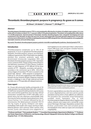
- 1. C A S E R E P O R T JIACM 2014; 15(1): 56-9 Thrombotic thrombocytopenic purpura in pregnancy: As grave as it comes AG Diwan*, SA Adukia**, S Kannan***, GN Wagh**** Abstract Thrombocytopenic thrombotic purpura (TTP) is a microangiopathy affecting the circulation of multiple organ systems.It is a rare entity with an incidence of about 2 to 11 cases per million in the general population.Thrombotic microangiopathies affect about 1 in 25,000 pregnancies. Classical pentad of haemolytic anaemia, thrombocytopenia, fever, alongwith neurological and renal involvement is rare.Herewith,we report the case of an 18-year-old primigravida of 26 weeks gestation diagnosed asTTP,who had astormycoursewithposteriorreversibleencephalopathysyndrome(PRES)alongwithfever,thrombocytopenia,microangiopathic haemolytic anaemia, and acute kidney injury. Key words: Thrombotic thrombocytopenic purpura, posterior reversible encephalopathy syndrome, classical pentad of TTP. *ProfessorandHead,**Post-graduateStudent,***ConsultantHaematologist,DepartmentofMedicine,****Professor and Head, Department of Obstetrics and Gynaecology, Bharati Vidyapeeth University Medical College and Bharati Hospital,Pune - 411 043,Maharashtra. Introduction Thrombocytopenia complicates up to 10% of all pregnancies. It occurs in gestational thrombocytopenia, idiopathic thrombocytopenic purpura, pre-eclampsia, malignant hypertension,HELLP (haemolysis,low platelets, elevated liver enzymes) syndrome, sepsis with disseminated intravascular coagulation (DIC) and thromboticthrombocytopenicpurpura(TTP)-haemolytic uremicsyndrome(HUS). Alargeoverlapofclinicalfeatures and laboratory parameters in these disease conditions makes an early diagnosis of TTP difficult. Of the three varieties of TTP – idiopathic, secondary, and genetic – secondary is the commonest (up to 60%). And of the various triggers of secondaryTTP, none is as potent as the gravid state. Women – either pregnant or postpartum – makeup10-25%ofcases,suggestinganinter-relationship between the two.However,despite a diagnosis,treatment remains a challenge in view of high maternal and foetal mortality seen inTTP,especially with delayed treatment. Case report An 18-year-old, previously healthy, primigravida of 26 weeks gestation was admitted late night with complaints of mild fronto-temporal throbbing headache with scanty clear vomit and low-grade fever since two days.Clinical examination revealed fever of 99°F, mild pallor, and a gravid uterus, but was otherwise unremarkable. Early morning she had an episode of generalised tonic clonic seizure lasting 2 to 3 minutes followed by loss of consciousness. Her blood pressure remained 170/110 mm Hg inspite of giving nifedipine and alpha- methyldopa. Both pupils were equal, reacting to light; bilateral plantars were extensor. Other systems were normal. Scanty cola-coloured urine suggestive of haemoglobinuria was noted upon Foley’s catherisation. Urgent MRI brain with venogram revealed features suggestive of PRES (Fig.1). Pre-term vaginal delivery was done the same day in view of severe oligohydramnios and intra-uterine growth retardation during which a fresh stillbirth was delivered. The procedure was uneventful. Serial haemograms revealed severe haemolysis (Fig. 2) with thrombocytopenia.Negativebloodculture,negativeurine culture, reduced urine output and a normal DIC screen (serum fibrinogen, fibrinogen degradation products like D-dimer; prothrombin time and activated partial thromboplastin time) ruled out sepsis and consumptive coagulopathy. Fig.1:MRIBrainshowingbilateralhighsignalsinfrontal,temporal,parietal and occipital lobes,lentiform nucleus,caudate nuclei (arrows) suggestive of PRES (posterior reversible encephalopathy syndrome).
- 2. Typical pentad including microangiopathic haemolytic anaemia (evidenced by schistocytes),thrombocytopenia, fever, renal impairment (oliguria, haemoglobinuria) and neurological involvement (seizures due to PRES) supported the diagnosis of TTP. Elevated lactate dehydrogenase(LDH)levelswerecorroborativeevidence. She underwent 5 sittings of plasmapheresis (therapeutic plasma exchange) with fresh frozen plasma (FFP). Table I shows serial investigations from admission up to discharge. Once clinically stable, she was discharged on day 12. Discussion TTP or Moschcowitz syndrome, first described by Dr Eli Moschcowitz in 1925, occurs due to deficiency of ADAMTS13.Thus,ultra-large multimers of vonWillebrand factor(ULVWF)releasedfromendotheliumarenotcleaved appropriately,andcausespontaneousplateletaggregates inconditionsofhighshear,suchasinthemicrovasculature of the brain,heart,and kidneys1 . Classified as idiopathic, secondary, and familial, TTP (secondaryvariety)hasastrongrelationtopregnancy.The reason for this is that pregnancy is associated with increasing concentrations of procoagulant factors, decreasing fibrinolytic activity, loss of endothelial cell thrombomodulin,and decreasing activity of ADAMTS-13. All of these abnormalities worsen through the course of pregnancy until delivery and immediately post-partum2 . Classic pentad is rare. However, the triad of Coombs’- negative microangiopathic schistocytic haemolytic anaemia,consumptivethrombocytopeniacausingsevere haemorrhagic diathesis, and fluctuating neurological symptomscanbeobservedinupto75%ofpatients3 . Due Fig.2:Peripheralbloodsmearshowingseverehaemolysisandschistocytes (arrows). to the high mortality of untreated TTP, a presumptive diagnosis of TTP is made even when only microangiopathic haemolytic anaemia and thrombocytopenia is present. Table II4 suggests a rational clinico-pathological approach to ruling-out entities with remarkably similar presentations as TTP. Despitethis,thedistinctionbetweenTTPandHELLPisvery difficult. Delivery generally leads to a rapid resolution of pre-eclampsia and HELLP syndrome; however, if no improvement is seen after 48 to 72 hours of delivery, possibility of thrombotic microangiopathies should be considered.Thedifferentiatingfeaturesbetweenthethree are as shown in Table II5 . Posterior reversible encephalopathy syndrome (PRES) is the predominant brain neuroimaging abnormality in patientswithTTP. Thereisnoassociationbetweendegree of hypertension, haematocrit or platelet count, D-dimer, fibrinogen,lactatedehydrogenase,ortotalbilirubinlevels and occurrence of PRES. The presenting feature can be benign – like headache, vomiting; or severe – like confusion,seizures,visual abnormalities,and motor signs. However, this pathology is completely reversible if the underlying cause is treated early.PRES inTTP is associated with worse renal function. Plasmapheresis (or therapeutic plasma exchange – TPE) has reduced mortality in TTP from over 90% to 10 - 20%. Earlier initiation correlates with a better prognosis. TPE allows removal of autoantibody, and repletes ADAMTS- 13. Large volume plasma infusions are indicated if there is to be a delay in TPE. Daily exchange should continue for minimum 2 days after complete remission,defined as normal platelet count (> 1,50,000/dl). More intensive exchange, such as twice daily TPE, may be required in resistant cases especially with neurological or cardiac events4 .Immunosuppressive therapy with rituximab may be used singularly or as an adjunctive to plasmapheresis. Recently, intravenous immunoglobulins have been successfully used alone to treat a case of TTP with PRES with other co-morbidities6 . Maternal mortality in TTP is related to widespread microvascular thromboses and multiple organ dysfunction. Placental infarction leads to foetal intra-uterine growth retardation and/or mortality. Women with previous history who wish to conceive should be counselled and closely monitored for platelet count, haemoglobin, LDH, and peripheral smear throughout the course of pregnancy. Plasma therapy should be started at the earliest evidence of a relapse of TTP. Prophylactic plasma infusion in a pregnant woman with a history of relapsing TTP may be considered7 . No association of TTP in subsequent pregnancies in women presenting withTTP in an earlier pregnancy can be made. Thus assurance is required for anxious women. Journal, Indian Academy of Clinical Medicine Vol. 15, No. 1 January-March, 2014 57
- 3. 58 Journal, Indian Academy of Clinical Medicine Vol. 15, No. 1 January-March, 2014 Table I: Showing serial investigations from admission up to discharge. Investigation Day 1 Day 3 Day 5 Day 12 Haemoglobin 9.9 gm/dl 6.6 gm/dl 7.6 gm/dl 10.8 gm/dl Mean corpuscular volume 80.6 fL 76.1 fL 97.7 fL 92.6 fL Total leucocytic count 22,800 cells/cmm 18,400 cells/cmm 28,300 cells/cmm 11,300 cells/cmm Differential count Neutrophils 87% Neutrophils 75% Neutrophils 72% Neutrophils 72% Lymphocytes 7% Lymphocytes 19% Lymphocytes 24% Lymphocytes 24% Platelets 50,000 cells/cmm 10,000 cells/cmm 30,000 cells/cmm 2,93,000 cells/cmm Peripheral smear Normocytic, normochromic Microcytic,hypochromic picture; Occasional anisopoikilocytosis, Normocytic normochromic picture with no evidence of anisopoikilocytosis with schistocytes, microcytes, macrocytes, picture with occasional haemolysis and evidence of severe haemolysis polychromatic cells and helmet anisopoikilocytosis cells,occasional spherocytes, schistocytes Reticulocyte count 1.5 % Urea 35 mg/dl 45 mg/dl 30 mg/dL Creatinine 0.9 mg/dl 1.1 mg/dl 0.8 mg/dL Bilirubin - Direct 0.5 mg/dl 0.78 mg/dl 0.73 mg/dl - Indirect 1.1 mg/dl 1.67 mg/dl 1.93 mg/dl Aspartate transaminase 484 IU/L 455 IU/L 69 IU/L 38 IU/L Alanine transaminase 311 IU/L 268 IU/L 76 IU/L 41 IU/L Prothrombin time 19.5sec /1.5 215.6sec /1.18 13.1sec /0.97 APTT activated partial 27.9 sec 29.9 sec 21.5 sec thromboplastin time Fibrinogen 344.80 mg/dl (Normal) D-dimer Negative Negative Lactate dehydrogenase 4,687 IU/L 1,187 IU/L 700 IU/L 550 IU/L ANA Negative Antiphospholipid antibody Negative (IgG/IgM) Blood culture No growth Urine routine Haemoglobinuria RBCs 6 - 8/hpf Urine culture No growth Chest X-ray Normal Normal USG abdomen Single live intra-uterine pregnancy of 25 weeks 6 days with Coarse hepatic echotexture with moderate to gross ascites and oligohydramnios and asymmetric intra-uterine growth retardation. bilateral pleural effusion.Bulky uterus. MRI brain with venogram Venogram normal.Bilateral high signals are seen in frontal,temporal,parietal and occipital lobes,lentiform nucleus,caudate nuclei,cerebellar hemispheres and brainstem,suggestive of PRES. Table II:Typical features in pregnancy-associated micro-angiopathies. Entity MAHA Thrombocytopenia Coagulopathy High BP Abdominal Renal Neurological symptoms impairment symptoms PET + + +/- +++ +/- +/- ++ HELLP + ++ +/- + +++ + +/- TTP ++ +++ - +/- + ++ +++ HUS + ++ +/- ++ + +++ +/- SLE + + +/- + +/- ++ + PET = Pre-eclampsia;HELLP = Haemolysis,elevated liver enzymes,and low platelets;TTP =Thrombotic thrombocytopenic purpura;HUS = Haemolytic uraemic syndrome;SLE = Systemic lupus erythematosus; MAHA = Micro-angiopathic haemolytic anaemia;BP = Blood pressure.
- 4. Journal, Indian Academy of Clinical Medicine Vol. 15, No. 1 January-March, 2014 59 Table III: Differentiating features between TTP, HUS, and HELLP. Feature TTP HUS HELLP Neurological features +++ ± ± Fever +++ ± - Hypertension ± ± ± Renal dysfunction ± +++ ± Purpura with bleeding ++ - - Platelets Markedly reduced Moderately reduced Reduced PT/APTT Normal Normal Prolonged Fibrinogen Normal Normal Reduced BUN/Creatinine Increased Increased Increased Liver enzymes ± ± + LDH Markedly increased Moderately increased Can be increased TTP=Thromboticthrombocytopenicpurpura;HUS=Haemolyticuraemicsyndrome;HELLP= Haemolysis,elevatedliverenzymesandlowplatelets;PT=Prothrombintime;APTT=Activated partial thromboplastin time;BUN = Blood urea nitrogen;LDH = Lactate dehydrogenase. References 1. Moake JL. von Willebrand factor, ADAMTS-13, and thrombotic thrombocytopenic purpura. Semin Hematol 2004; 41(1): 4-14. 2. George JN. The association of pregnancy with thrombotic thrombocytopenic purpura–hemolytic uremic syndrome.Current Opinion in Hematology 2003;10: 339-44 3. Proia A, Paesano R, Torcia F et al. Thrombotic thrombocytopenic purpuraandpregnancy:acasereportandareviewoftheliterature. Ann Hematol 2002; 81: 210-4. 4. Scully M,Hunt BJ,Benjamin S,Liesner Retal and on behalf of British Committee for Standards in Haematology. Guidelines on the diagnosis and management of Thrombotic thrombocytopenic purpura and other thrombotic microangiopathies. British Journal of Haematology 2012; 158(3): 323-35. 5. Patnaik MM, Deshpande AK, Nagar VS et al. Thrombotic microangiopathies presenting as an obstetric emergency. JAPI 2004; 52: 152-3. 6. Khobragade AK, Chogle AR, Ram RP et al. Reversible Posterior Leukoencephalopathy Syndrome in a case of Adult Onset Still’s disease with concurrent Thrombotic thrombocytopenic purpura: Response to High Dose Immunoglobulin Infusions. JAPI 2012; 60: 59-62. 7. Abdulla F, Zeheb MA, Awidi A. Thrombotic thrombocytopenic purpura associated with pregnancy in two sisters. Postgrad Med J 1993; 69: 229-31. “I wish to say what I think and feel today, with the proviso that tomorrow probably I shall contradict it all.” – Ralph Waldo Emerson.