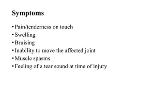
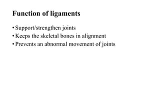
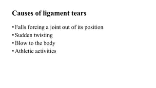
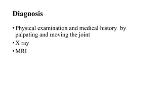
SlideShare a Scribd company logo
Traumatology 11
Department: OTM
Year : 2nd year
Semester : 1
Learning outcomes,
By the end of this lesson you should be able to:
Manage injuries of the upper limbs
Manage injuries of the lower limbs
Manage injuries of the head, chest and spine
Manage joint and soft tissue injuries
Definition
Definition- Traumatology is the study of diagnosis and
treatment of severe, acute physical injuries e.g. from
accidents, gunshot wounds sustained by individuals
requiring immediate medical attention
Cont., of definition
Or Traumatology is the evaluation and treatment of
psychological trauma in individuals affected by severe
mental or emotional stress or physical injury
Fractures of the upper extremity
• Shoulder girdle fractures; Fractures and dislocations
involving the shoulder girdle are common
Cause/etiology
• High energy trauma
Assessment; Follow advanced trauma life
support(ATLS)guide lines to identify associated life
threatening injuries
Physical examination
• Must include thorough neurovascular assessment of
upper limb
Caution- Pay attention to axillary nerve/brachial plexus
prone to shoulder girdle injuries
Cont.,
• Frequently traumatized region of the skeleton
• Complex structure comprised of several articulations,
numerous ligaments, tendons and muscles suspending
the upper extremity to the thorax
• Allows a tremendous range of motion
Fractures of the scapula (shoulder blade)
• Caused by a blunt trauma
• Include fractures of body, spine, acromion, scapula
neck and coracoid process fractures
• Commonly affects ipsilateral shoulder girdle, upper
extremity lung and chest wall
• Pulmonary injuries include haempneumothorax or
pulmonary contusion
Cont.,
• Acromion fractures are caused by significant blunt
force to shoulder directed anteriorly
• Glenoid neck fractures occur due to falls with an
outstretched arm, blunt force
Associated injuries
• Humerus fractures
• Shoulder dislocation
Cause:
• Direct trauma/violence
Incidence:
• Uncommon
• Less than 1% of all fractures
Location; 50% involve body and spine
Signs/symptoms; Pain and ecchymosis
Mortality rate; 2.5% associated mortality
Associated conditions
• Rip fractures 52%
• Ipsilateral clavicle fracture 25%
• Spine fractures 29%
• Brachial plexus injuries 75%
Medical:
• Pulmonary (lung) injury
• Pneumothorax (32%)
• Pulmonary contusion (41%)
• Head injury (34%)
• Vascular injury (11%)
Investigation
• X-rays AP/LAT, scapula Y, and lateral views
• CT scan
Treatment
• Sling for 2 weeks followed by early range of motion
exercises
Operative treatment- ORIF (open reduction internal
fixation)
Complications
• Infection in open fractures
• Neurovascular deficit
• Loss of function of gleno humeral joint
Clavicle fractures
• Incidence- make up 4% of all fractures
• Demographics- often seen in young active patients
• Associated injuries- ipsilateral scapula fracture and
scapula thoracic dissociation, rib fracture,
pneumothorax and neurovascular injury
Mechanism of injury
• Direct blow to lateral aspect of shoulder
• Fall on an outstretched arm
• Direct trauma
Pathoanatomy
• In displaced fractures, the sternocleidomastoid muscle
pulls the medial fragment posterior superiorly
• The pectoralis and weight of the arm pulls the lateral
fragment inferior medially
• Open fractures button hole through the platysma
Clinical features
• Physical deformity
• Shoulder pain- worse on movement
• Swelling
• Tenderness
• Bruising
• Gritty sensation felt over broken bones
Investigation
• X rays
• CT scan
Treatment
• Arm sling immobilization with gentle range of motion
exercises at 2-4 weeks and strengthening at 6-10
weeks. No attempt to reduction should be made
• Pain medications (NSAIDS)
• Surgery in cases when pieces of bone move far out of
place to realign the collar bone
Complications
• Neurovascular injury
• Pneumothorax
• Mal union
• Nonunion
• Post traumatic arthritis
• Re fractures
• Scarring
• Hardware prominence
Radius fractures
• Epidemiology- Occur in up to 20% of all elbow
injuries
• Incidence- Most common elbow fractures
Associated injuries
• 30% have associated soft tissue or skeletal injuries
• Distal radioulnar joint injury
• Interosseous membrane disruption
• Coronoid fractures
• Elbow dislocation
• Carpal fractures
Clinical features
• Pain /tenderness along the lateral aspect of elbow
• Limited elbow/forearm motion particularly
supination/pronation
Physical examination
• Reduced range of motion
• Instability
• Imaging- X-ray AP/LAT, CTscan
Treatment
• Non-operative; Short period of immobilization
followed by early range of motion in isolated
minimally displaced fractures
• Operative- ORIF, Radial head replacement
Complications
• Displacement of fracture
• Posterior interosseous nerve palsy
• Loss of fixation
• Loss of forearm rotation
• Elbow stiffness
• Radiocapitellar joint arthritis
• infection
Radius/Ulna shaft fractures(both bone
forearm fractures)
• Epidemiology- More common in men than women
• ratio of open to closed fractures is higher than for any
other bone except tibia
Mechanism- Direct trauma (often while protecting ones
head)
• Indirect trauma- motor vehicle accidents
• Falls from heights and athletic competition
Associated conditions
• Elbow injuries
• Compartment syndrome
Signs/symptoms- gross deformity
• Pain
• Swelling
• Loss of forearm and hand function
Physical examination
• Inspection- in open injuries check for tense forearm
compartments
• Neurovascular exam-assess radial/ulna nerve pulses
- Document median, radial and ulna nerve function
• Investigation- x rays AP/LAT views
Treatment
• Non operative- Functional brace with good
interosseous mold in non displaced or distal two thirds
ulna shaft fractures
• Operative- Plates/screws
• external fixators
• IMnailing
Complications
• Refracture
• Neurovascular damage
• Malunion
• Nonunion
• Compartment syndrome
• Infection
• synostosis
Fracture Humerus:
•Incidence; 3.5% of all fractures
•Bimodal age of distribution; High energy in
young patients
•Low energy in elderly with osteoporotic bones
•Symptoms; pain and extremity weakness
•Physical exam; Examine overall limb alignment
•Examine and document status of radial nerve pre/post
reduction
Investigation;
• X-rays AP/LAT
• MRI
• C T scan
Treatment;
Non operative- Cooptation splint followed by
functional brace
Operative treatment- ( indicated in)
• open fractures
• Vascular injury requiring repair
• Brachial plexus injury
• Ipsilateral forearm fracture (floating elbow)
• Compartment syndrome
Operative treatment;
• Plates/screws
• IM nails
Complications;
• Radial nerve palsy
• Malunion
• Nonunion
• Infection in open fractures
Proximal Humerus fracture;
Incidence- 4-6% of all fractures
• Third most common fracture pattern seen in elderly
Demographics- 2:1 female to male ratio
• Increasing age correlates with increasing risk of
fractures in women
Mechanism- low energy falls
• Elderly with osteoporotic bones
• High energy trauma in young individuals
Associated conditions;
• Nerve injury- axillary nerve palsy most common
• Fracture dislocations most commonly associated with
nerve injuries
Symptoms- Pain
• Swelling
• Decreased motion
Physical exam;
• On inspection- there is an extensive ecchymosis of
chest, arm and forearm
Diagnosis- x rays AP/LAT
• Scapula Y views
• Axillary views
Treatment;
Non operative- Sling immobilization followed by
progressive rehabilitation (exercises) early range of
motion within 14 days, stretching program
Operative treatment-
• IM nails
• Plates/screws
• arthroplasty
Complications;
• Nerve injury
• Malunion
• Nonunion
• Post traumatic arthritis
• infection
Distal Humerus fractures
• Incidence- Distal intercondylar fractures are the most
fracture patterns
• Common in young males and older females
Mechanism
• Low energy falls in elderly
• High energy impact in young population
Associated injuries
• Elbow dislocation
• Terrible triad injury
• Floating elbow
• Volkmann contracture (as a result of a missed forearm
compartment syndrome)
Symptoms;
• Elbow pain
• swelling
Physical examination;
• Cross instability often present
Neurovascular exam
• Check function of radial, ulna and medial nerves
• Check for distal pulses
• Monitor carefully for forearm compartment syndrome
Diagnosis;
• X rays AP/LAT views
• CT scan
• MRI
Treatment- Non operative- Cast immobilization in non
displaced fractures
Operative- (CRPP) closed reduction percutaneous
pinning and total elbow arthroplasty
Complications
• Ulna nerve palsy
• Elbow stiffness
• Nonunion
• Malunion
• Degenerative joint disease
Fracture Carpals;
Definition- a fracture of one or more carpal bones of
the wrist
- Treatment varies depending on element involved
Incidence- accounts for 18% of all hand fractures
- Over 80% of carpal fractures involve the scaphoid
- Triquetrum and others are less involved
Scaphoid fracture
• Break of scaphoid in the wrist
• General symptoms include pain at base of thumb
• Worsens on use of hand
Mechanism of injury:
• Fall on an outstretched hand
Epidemiology:
• Common in young males
• Less common in children /older adults due to weak
radius and accounts for 50-80% carpal injuries
Signs/symptoms
• Focal tenderness in volar prominence at the distal
wrist for distal pole fractures
• Anatomic snuff box for waist or mid body fractures
Diagnosis
• X-rays
• MRI
• Bone
DDX:
• Distal radius fracture
• Dislocation
• Wrist sprain
Prevention:
• Wrist guards during activities
Treatment: Non- operative
• If not displaced apply a thumb Spica
Cont.,
Operative:
• Percutaneous screw fixation- screwing the scaphoid
bone back together
Complications:
• Avascular necrosis
• Non union
Metacarpal fractures:
Incidence:- comprise between 18-44% of all hand
fractures
- Non thumb metacarpals account for around 88% of all
metacarpal fractures with the 5th finger most
commonly involved
- Majority of metacarpal fractures are isolated injuries
which are simple, closed and stable
Mechanism of injury:
• Punching when the fist is in a clenched position
• Direct trauma to the dorsum of the hand
Signs/symptoms
• Pain /tenderness
• Deformity
• Inability to move the finger
• Swelling
• bruising
Cont.,
• Shortened finger
• Scissoring of the injured finger over neighbor when
making a partial fist
Diagnosis:
• X rays
• MRI
• Ultrasound
• Blood culture
Treatment:
• Antibiotics for 4-6 weeks if osteomyelitis is present
• Incision /drainage
• Primarily non-operative sedation or local anesthesia
followed by closed reduction of the fracture
• Application of a forearm based splint and secure with
a loose compressive wrap for 3 weeks
Complications;
• Rare but most common is mal union
• Rotational deformity
• Loss of function
• stiffness
Fracture phalanges:
• Usually caused by direct trauma
• Twisting injury
• Crush injuries to the distal phalanx resulting to nail
trauma and open fractures
Signs/symptoms:
• Swelling on the fracture site
• Tenderness at fracture site
• Bruising
Cont.,
• Inability to move the injured finger
• Deformity of the finger
Diagnosis:
• X rays
• Physical examination
• Patient history
• ultrasound
Treatment:
Non- operative- is the general choice of treatment for
distal phalanx fractures because of its small size
- Conservative treatment with splints is used for
displaced fractures
Operative treatment- surgical treatment of displaced
proximal phalanx fracture after reduction is common
because of instability and rotation trouble
- Use of Krishna wires by percutaneous pinning or open
reduction or very small screws/plates
DDX:
• Metacarpal fracture
• Mallet finger
• Jersey finger
• Volar plate
• Avulsion injury
Complication:
• Loss of motion
• Nonunion
• Mal-union
• infection
11:Fractures of the lower extremity
Fractures of the pelvic girdle;
Incidence:
• High energy blunt trauma
• High mortality rate: 15-25% for closed fractures/50% for
open fractures
• In pediatrics, reradiate cartilage is open, the iliac wing is
weaker than the elastic pelvic ligaments resulting in bone
failure before pelvic ring disruption
Associated injuries
• Chest injury for up to 63%
• Long bone fractures in 50%
• Sexual dysfunction up to 50%
• Head and abdominal injury in 40%
• Spine fractures in 25%
Clinical symptoms
• Pain /inability to bear weight
• Test stability by placing gentle rotational force on each
iliac crest- low sensitivity for detecting instability
- perform only once
• Look for abnormal lower extremity positioning
-external rotation of one or both extremities
-limb length discrepancy
• Rectal exam to evaluate sphincter tone/perirectal
sensation
Diagnosis:
• X ray- AP pelvis, inlet view, outlet view
Tiles classification:
• A- Stable
• B- Rotationally unstable, vertically stable
• C- Rotationally and vertically unstable
Young barges classification:
• Anterior posterior compression
• Lateral compression
Cont.
• Vertical shear
Treatment: resuscitation
• Fluids- blood/crystalloids
• Pelvic binder/sheet
• External fixation
• Angiography/embolization
Non operative treatment
• Indicated for mechanically stable pelvic ring injuries
including anterior impaction fracture of sacrum and
oblique ramus fracture with less than 1cm of posterior
ring displacement
Operative treatment: (ORIF) in cases like-
Cont.,
• Sacroiliac joint displacement more than 1cm
• Displacement or rotation of hemi pelvis
• Open fracture
Diverting colostomy- indicated when there is an open
pelvis fracture especially with extensive perineal injury
or rectal involvement
Fracture head femur:
• Incidence- rare fracture pattern associated with hip
dislocation
• Location /size of fracture fragment/degree of
comminution depend on the position of the hip at the
time of dislocation
Mechanism- impaction, avulsion or shear forces
involved -unrestrained passenger(knee against
dashboard) -falls from height
-sports injury
-industrial accidents
Associated injuries
• 5-15% of posterior hip dislocations are associated with
a femoral head fracture
• Anterior hip dislocations usually associated with
impaction/indentation fractures of the femoral head
• Femoral neck fracture
• Acetabular fracture
• Sciatic nerve injury
• Femoral head avascular necrosis
• Ipsilateral knee ligamentous instability
Clinical presentation
• History from patient e.g. fall from a height
• Frontal impact with knee striking dashboard
Symptoms-localized hip pain, unable to bear weight
Physical exam- on inspection- shortened lower limb,
posterior dislocation and anterior dislocation
Neurovascular- may have signs of sciatic nerve injury
Diagnosis
• X rays- AP pelvis, lateral hip and judet views both on
pre/post reduction
• CT scan after reduction to evaluate- concentric
reduction, loose bodies in joint, acetabular fracture and
femoral or neck fracture
Findings; femoral head fracture, intra articular
fragments, posterior pelvic ring injury, impaction and
acetabular fracture
Treatment
Non operative (indications)- acute dislocation, reduce
hip dislocation within 6hrs
Technique- obtain post reduction (CT continuous
traction) -perform serial radiographs to document
maintained reduction
Operative (ORIF) indications:
• Presence of loose bodies in the joint
• Associated neck/acetabular fracture
• Poly trauma
Cont.,
• Irreducible fracture dislocation
• Arthroplasty- significantly displaced fractures,
osteoporotic and comminuted fractures
Complications:
• Heterotopic ossifications
• Avascular, decreased range of motion (ROM)
• Sciatic nerve injury
• Degenerative joint disease (DJD)
Femoral neck fractures
Epidemiology:
• Increasingly common due to aging population
• Women more than men
• Whites more than blacks
• Most expensive fracture to treat on per person basis
Mechanism:
• High energy in young patients
• Low energy falls in older patients
Pathophysiology:
Healing potential- femoral neck is intra capsular, bathed in
synovial fluid, lacks periosteal layer, callus formation is
limited which affects healing
Associated injuries;
• Femoral shaft fractures 6.9%
Symptoms:
• Slight pain in the groin or referred pain along the medial
side of the thigh/knee in impacted/stress fractures
• Pain in the entire hip region in displaced fractures
Physical examination:
• No obvious clinical deformity
• Minor discomfort with active or passive hip range of
motion, muscle spasms at extremes of motion
• Pain with percussion over greater trochanter
• In displaced fractures, the leg is in external rotation,
abduction with shortening
Diagnosis
• X rays
• CT scan
• MRI
• Duplex scanning
Treatment:
Non operative- indicated in non-ambulatory patients who
have minimal pain and at high risk of surgical intervention
Operative treatment:
• ORIF indicated in displaced fractures in young or
physiologically young patients less than 55 yrs. of age
• Hemi arthroplasty- indicated in patients with metabolic
diseases and the elderly
• Total hip arthroplasty- in controversial, in older active
patients and in patients with progressively hip osteoarthritis
Complications:
• Osteonecrosis
• Nonunion
• dislocation
Femoral shaft fractures:
incidence;
• Traumatic- (high energy)-most common in young
population as a result of high speed motor vehicle
accidents
• Low energy- more common in the elderly often as a
result of fall on standing
Associated conditions
• Ipsilateral femoral neck fractures (26% often
incidence)
• Bilateral femur fractures – significant risk of
pulmonary complications with increased rate of
mortality as compared to unilateral fractures
Clinical presentation
• Pain in thigh
• In inspection swollen tense thigh
• Tenderness around the thigh
• Difficult examination of ipsilateral femoral neck
secondary to pain on from the fracture
Diagnosis:
• X rays- AP/LAT views for entire femur and ipsilateral
hip and knee
• Ct scan to rule out associated femoral neck fracture
Treatment- (non operative) long leg cast in un
displaced fractures with multiple medical comorbidities
operative
• IM nails
• Plates/screws
• Exofix
Complications:
• Mal union
• Neurovascular damage (femora)
• Non union
• Infection
• Quadriceps weakness
Distal femur fractures:
Defined as fractures from articular surface 5cm above
metaphyseal flare
Mechanism- in young patients high energy with
significant displacement
In older patients low energy in osteoporotic bone with
less displacement
Physical exam in vascular evaluation potential for
injury to popliteal artery in significant displacement
• Angiography is done if there is no pulse after
alignment/reduction
Diagnosis:
• X rays AP/LAT
• CT scan to establish intra articular involvement
• Angiography indicated when there is diminished distal
pulses with gross alignment restored
Treatment:
Non operative- in non displaced , non ambulatory and those
with significant comorbidities
• Long leg cast
• Continuous traction
• Hinged knee brace with immediate ROM non weight
bearing for 6 weeks
Operative
• Plates/screws
• Screws only
• IM nails
Complications:
• Symptomatic hardware
• Malunion
• Nonunion
• Infection
• Joint stiffness
Patella fracture
• Incidence- Patella fractures account for 1% of all skeletal
injuries
• Male to female 2:1
• Most fractures occur in 20-50 yrs. Old
Patella sleeve fractures- seen in pediatric population(8- 10
yrs.) - high index of suspicion required
Bipartite patella- may be mistaken for patella fractures and
affects 8% of population
- Characteristic superior lateral position/50% bilateral cases
Classification
• Can be described based on fracture pattern
• Non displaced
• Transverse
• Pole or sleeve (upper or lower)
• Vertical
• Marginal
• Osteo chondral
• comminuted
Physical exam
• Palpable patella defect
• Significant hem arthrosis
• Unable to perform a straight leg raise indicating failure
of extensor mechanism and retinaculum disrupted
Diagnosis
• X-rays AP/LAT to check for displacement of patella
especially on the lateral view/degree of retinacula
disruption
• MRI- in cases of difficult leg raise
Treatment:
Non operative: knee immobilization in extension
(brace or cylinder cast) and full weight bearing)
indicated in
•Intact extensor mechanism (patient is able to
perform straight leg raise)
•In non-displaced or minimally displaced fractures
Cont.,
• In vertical fracture patterns
• Early active ROM with hinged knee brace
• Early weight bearing in full extension
• Progress in flexion after 2-3 weeks
Operative
• TBW Tension Band Wiring
• Lag screws
• Circlage wires and patellectomy
Complications:
• Weakness and anterior knee pain
• Symptomatic hardware ( most common)
• Malunion
• Nonunion
• Osteonecrosis(proximal fragment) due to excessive
initial fracture displacement
• Infection
• Stiffness of the knee
Tibia fractures (proximal third)
• Defined as fractures of the proximal tibia shaft that are
associated with high rates of malunion, valgus and soft
tissue compromise
Incidence- 5-10% of all tibia shaft fractures
Mechanism- (low energy) occur as a result of
rotational injury and a direct trauma
In high energy- occurs as a result of direct trauma
Clinical presentation:
Symptoms- pain and inability to bear weight
Physical examination- on inspection/palpation-
contusions, blisters, open wounds and compartments
Diagnosis;
• X rays AP/LAT of affected tibia ipsilateral knee and
ipsilateral ankle
• CT scan- indicated for intra-articular fracture
extension
Treatment:
Non operative- closed reduction/cast immobilization in
long leg cast for low energy fractures with acceptable
alignment
Technique- place in long leg cast and convert to
functional brace at 4 weeks
- cast in 10-20 degrees of flexion
Outcomes- rotational control is difficult to achieve by
closed methods
- Intact fibula may lead to Varus deformity with weight
bearing
Cont.,
Operative treatment:
• External fixation
• Intra medullary nailing
• Plates/screws ( percutaneous locking plate)
Complication:
• Mal union
• Non union
• infection
Tibia plafond fractures
Incidence ( pillon fractures) account for more than 10%
of lower extremity injuries
- Incidence increasing as survival rates after motor
vehicle accidents and falls from heights
Characteristics;
• Articular impaction/comminution
• Metaphyseal bone comminution
• Soft tissue injury associated with musculoskeletal
injuries 75% have associated fibula fractures
Cont.,
• 3 fragments typical with intact ankle ligaments i.e.
medial malleolar (deltoid ligament)
• Posterior lateral /Volkmann fragment(posterior inferior
tibiofibular ligament
Symptoms
• Ankle pain
• Inability to bear weight
• deformity
Cont.,
Physical exam- examine soft tissue integrity
Inspection- swelling, abrasion, ecchymosis, blisters and
open wounds
ROM/stability examine stability and ligament of the
ankle joint
Neurovascular- check distal pulses and proximal tibia
pulses, look for neurologic compromise and signs of
compartment syndrome
Diagnosis
• X rays AP/LAT, mortise views of ankle and full length
tibia/fibula and foot x rays performed for fracture
extension
• CT scan – delineate articular involvement/surgical
planning
Treatment- (non operative) immobilization with a
long keg cast for 6 weeks
cont.
Indications for immobilization
• Stable fracture patterns without articular surface
displacement
• Critically ill or non ambulatory patients
• Significant risk of skin problems (diabetes, vascular
disease/neuropathy
Technique:
• Long cast for 6 weeks followed by fracture brace
/ROM exercises
• Alternative treatment is with early range of motion
Outcomes;
• Intra articular fragments are likely to reduce with
manipulation of displaced fractures
• Loss of reduction is common
• Inability to monitor soft tissue injuries is a major
disadvantage
Operative treatment:
• Temporizing spanning external fixation across ankle
joint
• ORIF
• Exofix :definitively
• IM nails
Complications:
• Wound slough- wound flap for post operative
breakdown
• Dehiscence- wait for soft tissue edema to subside
before ORIF (1-2 weeks)
• Varus mal union
• Non union
• Post traumatic arthritis- most commonly begins 1-2
years post injury
Tibia plateau fractures:
• Incidence- peri articular injuries of the proximal tibia
frequently associated with soft tissue injuries
• Bimodal distribution- males in 40s (high energy)
• Females in 70s (falls)
Frequency- lateral /biconylar/medial
Mechanism-
• Varus/valgus load-or without axial load
• High energy- frequently associated with soft tissue
injuries while low energy is associated with fractures
Associated conditions:
• Meniscal tears- lateral meniscal tear- most common
than medial
• ACL-Anterior Cruciate Ligament injuries
• Compartment syndrome
• Vascular injury
Examination:
• History- high energy trauma in young patients
- low energy falls in elderly
Physical exam-( inspection)-look for open injuries
-(palpation)-consider compartment
syndrome when compartments are firm and not compressible
Varus/valgus stress testing- often difficult to perform given
pain
Neurovascular exam- any difficulties in pulse exam
between extremities should be further investigated
Diagnosis:
• X rays
• CT scan
• MRI
Treatment (non operative)-hinged knee brace 8-12
weeks /immediate ROM exercises
Indications- Minimally displaced split or depressed
fractures
-low energy fractures stable to Varus/valgus
alignment and also in non ambulatory patients
Operative treatment
• Plates/screws
• External fixators
• Screws only
Complications:
• Post traumatic arthritis
• Infections
• Compartment syndrome
• Vascular injuries
Cont.
• Knee instability
Fibula fractures:
• Definition- A fibula fracture is used to describe a break in
the fibular bone
Mechanism:
• A forceful impact e.g. landing after a high jump/impact to
the outer aspect of the leg
• Rolling /spraining of the ankle joint puts stress on the fibula
which leads to fracture
• Direct injury to the leg
• Gun shot wound
• Direct blow/blunt trauma e.g. sports/motor vehicle accident
Function of fibula
• Supports the tibia bone and helps stabilize the ankle
joint and the lower leg muscles
• Connects to the tibia via a interosseous membrane
Epidemiology:
• quite rare
• Occurs commonly in the young athletes
Physical examination:
Examine- skin for any laceration, abrasion/bruising and
any signs of trauma e.g. range of motion
Palpation- tenderness over the fibula
Symptoms:
• Pain (acute/sharp)
• Swelling
• Inability to bear weight
Diagnosis:
• X rays
Treatment:(non operative)
• Immobilization with a long leg cast/splint for several
weeks then pop walking boot with weight bearing
DDX- osteoid osteoma, Ewing sarcoma, osteosarcoma,
osteomyelitis, muscle spasm, ankle sprain, tendon
rupture, compartment syndrome and nerve entrapment
Cont.,
Operative treatment
• ORIF if the fracture is located on the upper half of the
bone
Complications:
• Damage to superficial peroneal nerve
• Arterial damage and compartment syndrome in
isolated fibula fractures
• Non union
Tarsal fractures:
• Is a break of any of the tarsal bones in the foot
• 7 in number- calcaneus, talus, cuboid, navicular,
medial, middle and lateral cuneiforms
Mechanism:
• Track and field athletes
• Associated with long term morbidity
• Stress fracture
Clinical features:
• Aching pain in the dorsal mid foot which radiates
along the medial arch. Pain increases with activity eg
running/jumping
• Loss of function of the foot
• Tenderness
• deformity
Treatment:
Non-operative- boot cast for 6 weeks /non weight for a
fractured bone /walking boot with crutches in hairline
fractures to reduce weight
Operative- done in too unstable fractures( but not
recommended)
Navicular
• most common
Symptoms:
• Poorly localized ache in the mid foot which gets worse
with exercise
• Pain radiating inside the foot Arche
• Tenderness on press on top of the foot
• On and off pain upon rest
Assignment –( read more on navicular fractures)
Cuboid stress fracture
Occurrence-(rare) occurs on calcaneus compression
and the 4th/5th metatarsal bones
- Diagnosis is always missed on x ray on initial stages
but clear on MRI
- Treatment- non weight rest for 4-6 weeks followed by
gradual return to full fitness
3 Cuneiform stress fractures
• Exceptionally rare tarsal fractures
• Caused by overuse
Treatment
• Non/light weight bearing rest
• Operation to fix the middle cuneiform if displaced
Calcaneus fracture
• A stress fracture of the calcaneus is a small break in
the heal bone
• The calcaneus is essential for walking and provides
support and stability to the foot
Signs/symptoms
• Sudden pain in the heel and inability to bear weight on
that foot
• Swelling in the heel area
• Bruising of the heel and ankle
Diagnosis:
• X ray then repeat in 2 weeks
• Bone scan
• MRI
Treatment- immobilization with a cast, splint or brace
is applied to hold the bones of the foot in proper
position while they heal for 6-8 weeks or possibly
longer
Complication of tarsal fractures:
• Chronic pain
• Limb arthritis
• Delayed union
• Non union
• deformity
Metatarsal fractures:
• Occur when one of the long bones of the mid foot is
cracked or broken due to sudden injury (an acute
fracture) or due to repeated stress (stress fracture)
Incidence
• Most common
• They are five in number
• Long slim which run the length of the foot to the base
of the toes
5th metatarsal fracture
• Most common to fractures
• Breaks at various points along its length
• Other metatarsals may break depending on mechanism
• 1st/2nd/5th are commonly injured in sport/footballers
• Fractures may be acute , caused immediately by injury
or may occur over a longer period of time
• Fractures may be open/closed/displaced/not displaced
Causes:
• direct injury to the foot(stepping on /kicking
foot/dropping something on foot or falling on foot
• Twisting of foot on the ankle joint can cause fractures
on base of 5th metatarsal
• Twisting of foot from landing from a jump( e.g. ballet
dancers)
Causes of stress fractures
• Repeated stress to the bone (overuse)
• Matching /running long distances carrying heavy packs
(match fractures)
• Practicing in athletes during training exercises
• Running on routine basis
• Wearing poor foot wear for running that does not suit the
foot
• Abnormalities of foot structure e.g. rheumatoid
arthritis/thinning of bones (osteoporosis)
• Loss of nerve sensation in neurological patients(diabetes)
Signs/symptoms
• Pain (pin point)
• Tenderness
• Bleeding from broken bones causing bruising and
swelling
• Inability to use foot
Diagnosis
• X rays
• CT scan
• MRI
• Ultrasound
• Bone scan
Treatment
• Pain killers (NSAIDS)
• Ice for 10-30 minutes immediately after injury to
reduce blood flow to the area. Not done directly to the
skin as it causes an ice burn (use an ice park) and press
gently onto the injured part
• Rest/elevate to reduce swelling
• Stop the causative activity by resting
• Immobilize with a below knee cast
• Surgery to realign moved bones/physiotherapy care
Prevention
• Exercise slowly and gradually
• Rest/recovery time needs to be build into any training
schedule
• Wear proper fitting footwear
• Be aware of stress fracture symptoms to stop
worsening
Complications
• Chronic metatarsal fractures
• Fracture of 1st metatarsal can lead to later arthritis of
big toe joint
• Continued pain and healing problems
• Foot deformity
• Non union
Assignment read on fractures of the phallanges
111. Injuries of the head and spine:
• Vertebral bones injuries- injuries involve
fractures/dislocations
• Fractures include any break of the vertebrae
• A dislocation is when the vertebral bones do not line
up correctly or are out of place
• A fracture /dislocation may cause damage to the spinal
cord
Types of fractures /dislocations that cause
spinal/head injury:
• Compression fracture: occurs due to hyper flexion
(front to back)
-It forces part of the spinal column forward and
downward
• Burst fracture: a serious force of compression
fracture where the bone is shattered from the injury
-bone fragments may pierce the spinal cord
-occurs due to downward or upward force along the
spine
Cont.
• Subluxation: weakening of vertebral joints by an abnormal
movement of the bones
- It is a partial dislocation of vertebrae caused by injury of
muscles/ligaments in the spine which may also cause a
spinal cord injury
• Dislocation: caused by torn/badly stretched ligaments from
an injury
-it causes too much movements of the vertebrae
-The vertebrae may lock over each other on one or both
sides
Cont.
-Traction or surgery reduction is needed for reduction
-use of a brace/halovest /surgery is used to fuse the
vertebrae for alignment/line up
• Fracture dislocation: occurs when there is a
fracture/a dislocation of the vertebrae
-It causes injury to the soft tissues/ligament injury
-It may also cause injury to the spinal cord
Dislocation of the inter -vertebral bones
• Joints in the back part of vertebrae are weakened by an
abnormal movement
• Dislocation of vertebrae occurs due to injury to the
muscles/ligaments in the spine causing injury to he
spinal cord
Causes;
• Car accidents
• Sports collisions/injuries
• falls
Treatment
• Reduction- trying gentle maneuvers to help put bones
back to position
• Immobilization- with a spinal splint /sling after
reduction for several weeks
• Surgery
• rehabilitation
Complications
• Dural tears
• Misdirected instrumentation
• Excessive bleeding necessitating transfusions/fluids
replacement
Raptures of vertebral discs
• Occurs when the spinal column tear and the discs
protrude outward, press or pinch the nearby spinal
nerves
• A ruptured disc is also called herniated or slipped disc
• Ruptured discs causes severe low back pain and
sometimes a shooting pain down the back of the legs
known as ( sciatica)
Cause
• Inflammation of the spinal nerves
• Every day activities/work
• Strains/sprains of muscles/tendons/ligaments
• Aging
• Sports
• Car accidents
• falls
Symptoms
• Severe low back pain/leg (usually 1 leg)
• Tingling in part of the leg or foot
• Weakness in the leg
Diagnosis
• On symptoms (sciatica)
• CT scan
• MRI
• Patient history
Treatment of ruptured discs
• Heat and cold- apply cold packs to the painful area to
numb the nerves that cause pain
- Heating pads/hot paths later reduce tightness/spasms
in the muscles of the lower back for free movement
• Pain relievers- NSAIDS
(ibuprofen/advil/motrin/aspirin/acetaminophen/naprox
en
• Stay active- do a normal daily activity- extended bed
rest is harmful
cont.
• Gentle stretches/exercises helps one return to normal
activity
• Complementary care- spinal manipulation, massage
and acupuncture help relieve pain and discomfort
while healing
- It should be done by a professional person with license
Surgical treatment
• Considered if pain/sciatica persists for 3 months or
more
• Discectomy- removal of a ruptured disk parts to relief
compression of the spinal nerve roots
(Assignment- discuss the complications)
Ligamentous tears
• A ligament- is a tough band of fibrous tissue that
connects bone to bone or bone to cartilage
• they can stretch and tear resulting to sprains
• Ligament tear occur due to extreme force to a joint
such as a fall/high impact event
Commonly affected areas-
Ankle/knee/wrist/thumb/neck or back ligaments
Symptoms
• Pain/tenderness on touch
• Swelling
• Bruising
• Inability to move the affected joint
• Muscle spasms
• Feeling of a tear sound at time of injury
Function of ligaments
• Support/strengthen joints
• Keeps the skeletal bones in alignment
• Prevents an abnormal movement of joints
Causes of ligament tears
• Falls forcing a joint out of its position
• Sudden twisting
• Blow to the body
• Athletic activities
Diagnosis
• Physical examination and medical history by
palpating and moving the joint
• X ray
• MRI
Grading of sprains
• Grade1- a mild sprain damaging the ligament but does
not cause significant tearing
• Grade2- moderate sprain that includes a partial tear of
the ligament. This may show an abnormal looseness
• Grade3- This is a severe sprain with a complete tear
of the ligament resulting to joint instability and loss of
function
Treatment
• (RICE- ACRONYM) Rest/Ice/Compress/Elevate
• R- rest the joint by stopping further activity that stress
the joint
• I- ice/cold contact provides short term pain
relief/limits swelling
• C- compress(wrap) the injured area with an elastic
bandage to reduce pain/swelling
• E- elevate to control blood flow to the area to reduce
swelling (elevate above the level of the heart)
Complications
• Fractures
• Infections
• Vascular/neurologic complications following
injury/surgery
• Compartment syndrome
• Complex regional pain syndrome
• Deep venous thrombosis
• Loss of motion /persistent laxity issues
Injuries to the spinal cord (SCI)
• Involves damage to the spinal cord that causes
temporary/permanent changes in its function
• Affects people between 16-30 yrs. of age
• Most commonly caused by an external trauma
• Affects men more than women
Causes
• Tumors
• Blood loss
Cont.
• Stenosis
Types of spinal cord injuries:
• A complete spinal cord injury- causes a permanent
damage to the area of the spinal cord that is affected
• Paraplegia(lower part of the body) occurs
• An incomplete spinal cord injury- is a partial
damage to the spinal cord
Levels of spinal cord injuries
• Cervical
• Thoracic
• Lumbar
• sacral
Signs/symptoms
• Muscle weakness
• Reduced sensation to touch or pin/needles
• Leaking of urine/retention
• Abnormal and painful sensation
• Leaking of stool
• Shortness of breath
Diagnosis
• X ray
• CT scan
• MRI
Treatment
• 1st aid immobilization to align the spine
• Traction to realign the vertebrae
• Surgery- in patients with neurological deterioration to
prevent future pain/deformity
• Follow-up to monitor for secondary complications
such as pneumonia, pressure, ulcers and deep vein
thrombosis
Complications
• Autonomic dysreflexia- lesions at T6 or higher which
can cause stroke/seizures due to increased blood
pressure
• Deep vein thrombosis- slow blood flow inside a vein
• Syringomyelia- formation of fluid filled cavity on the
spinal cord causing pressure on surrounding areas
Cont.
• Spasticity- involuntary increase in muscle tone- very
stiff muscles with no movement
• Heterotopic ossification- small areas of bone crop up
in non-bony areas
• Pressure sores- sitting/lying too long in one position
• Pain- around the joints
Joint and soft tissue injuries
• Dislocation-injury to a joint in which the bone ends
are forced to move from their normal positions or a
displacement of one or more bones at a joint
Treatment
• Rest the dislocated joint /don’t repeat the action that
caused the injury /try to avoid painful movements
• Apply ice and heat to reduce swelling
• Maintain range of motion
• NSAIDS drugs (ibuprofen)
Causes of dislocation
• Trauma
• Car accidents
• Falls
• Sports (foot ball)
• Regular activities when the muscles and tendons
surrounding the joint are weak
Prevention of dislocations
• Being cautious on stairs to help avoid falls
• Wearing protective gear during contact sports
• Staying physically active to keep muscles /tendons
around the joint strong
• Maintaining a healthy weight to avoid increased
pressure on the bones
Complications
• Tearing of muscles/ligaments/tendons that reinforce
the injured joint
• Nerve or blood vessel damage in /around the joint
• Susceptibility to re injury in severe dislocation or
repeated dislocation
Strains
• An act of straining or the condition of being strained/
an excessive extension/effort of muscles or ligaments
Causes
• Sporting injuries
• Falls/sudden movement
• Attempt to lift heavy objects
• Repeated coughing strains the ribcage muscles
Treatment
• (Pain relievers, ice, splinting)
• Rest the affected muscle and apply ice and heat
• NSAIDS to reduce pain and swelling
• Compression with an elastic bandage to provide
support and decrease swelling
• Elevate to reduce swelling
Complications
• Joint dislocation
• Pain
• Recurring swelling
• Ruptured muscle
• Cartilage injuries
Sprains
• Stretching or tearing of ligaments and fibrous tissue
that connects bones to joints
Causes
• Stretched /torn ligaments (fibers connecting bone to
bone)
• Injury e.g. twisting of joints
Treatment
• Pain relievers, ice or splinting with an elastic bandage
to support and reduce swelling
• Rest the strained muscle
• Apply ice
• NSAIDS (acetaminophen (Tylenol)
• Heat the muscle with a warm bath after pain decreases
• Stretching and light exercises to bring blood to the
injured area
Complication of sprain
• Joint dislocation
• Pain
• Recurring swelling
• Ruptured muscle
• Cartilage injuries
Subluxation
• An incomplete or partial dislocation or abnormal
spacing of vertebrae or intervertebral units or organ
Causes
• Accidents
• Bad posture
• Sitting for long hours
• Alcohol or drug use
• Improper lifting and emotional stress
Signs/symptoms
• Dizziness or balance problems
• Reduced range of motion or spinal mobility
• Spinal muscle tightness/weakness or spasms
• Pain/numbness or tingling sensation in extremities
• Joint pain/soreness or tenderness
Treatment
• Closed reduction by maneuvers to relocate joint to
position
• Surgery when there is a recurrent dislocation
• Use of braces/splints
• Medications (NSAIDS)
• Rehabilitation
Complication
• Damage to blood vessels and nerves
• Ligament /muscle tears
• Loss of movement/flexibility
Ligament/tendon injuries
• These are injuries to the soft tissues that connect
muscles/joints
Symptoms
• Pain
• Feeling of pop sound when tissue is torn
Treatment
• Brace/splint
• NSAIDS/or surgery in severe cases
Causes
• Twisting of a joint –traumatic injury
• Overuse activities
Neurovascular injuries
• Is the damage to the major blood vessels supplying the
brain, brainstem and upper spinal cord including the
vertebral, basilar and carotid arteries
• These vessels are located both extra and intra cranially
and injuries can occur in either or both of these
locations
Causes
• Minor/severe blunt or penetrating trauma to the head
and neck
Clinical features
• Decreased sensation
• Loss of sensation
• Dysesthesia
• Numbness
• Tingling or pins and needles
Diagnosis
• Assessment of distal pulses, capillary refill, skin color
and temperature
• CT scan for skull base fractures
Complications
• Ilia inguinal nerve compression
• Meralgia paresthesia
• Obturator nerve compression
• Sciatica and also piriformis syndrome
Contusions
• A contusion is a bruise caused by a direct blow or an
impact such as a fall
• Are common in sport injuries
• Happens when blood vessels get torn and leak under
the skin or the surrounding area
• Is any collection of blood outside a blood vessel
Cause
• Trauma e.g. cut or blow to an area of the body
Signs/symptoms
• Stiffness/swelling in bone contusions
• Tenderness
• Trouble bending
• Pain
Diagnosis
• MRI in bone contusions
Complications
• Damage to the internal organs
• Fractures
• Dislocations
• Torn muscles
• sprains
Lacerations
• A wound produced by tearing of soft body tissue
• A laceration wound is often contaminated with
bacteria and debris from the object caused by a cut
Cause
• Cut by sharp objects
Symptoms
• Bruising
• Bleeding
Cont.
• Swelling
• Skin discoloration
• pain
Treatment
• Application of antibiotic ointment and cover wound
with a sterile bandage
• Daily cleaning of wound with soap/water
Complication
• Infection
• bleeding
Muscle tears
• A muscle tear is stretching and tearing of muscle fibres
Cause
• Fatigue
• Overuse
Commonly affected muscles
• Lower back muscles
• Neck muscles
• Shoulder muscles
• hamstring
Signs of muscle tear
• Pain
• Swelling
• Muscle spasms
• Limited range of motion
• Redness
• bruising
Diagnosis
• Physical examination and ask patient patients history
• X rays
Treatment
• Pain relievers
• Ice
• splinting
Complication
• Compartment syndrome
• Swelling
• Rupture of muscle in long immobilization
END
THANK YOU
TRAUMATOLOGY 11 copy 2 (1).pptx

More Related Content

What's hot

median nerve injuries.pptx
median nerve injuries.pptxmedian nerve injuries.pptx
median nerve injuries.pptx
Saurabh Agrawal
 
Hand injuries by Dr.SUNIL C
Hand injuries by Dr.SUNIL CHand injuries by Dr.SUNIL C
Hand injuries by Dr.SUNIL C
Sunil Chandrashekar
 
supracondylar fracrture of humerus in children
supracondylar fracrture of humerus in childrensupracondylar fracrture of humerus in children
supracondylar fracrture of humerus in children
Hardik Pawar
 
radial nerve palsy
radial nerve palsy radial nerve palsy
radial nerve palsy
Sumer Yadav
 
Aneurysm coiling
Aneurysm coiling Aneurysm coiling
Aneurysm coiling
Sai Sudarsan
 
Gun shot injury
Gun shot injuryGun shot injury
Gun shot injury
Jamil Kifayatullah
 
Femur fracture
Femur fractureFemur fracture
Femur fracture
muhammad bilal
 
management of closed fracture
management of closed fracture management of closed fracture
management of closed fracture
ekhlass ramadan
 
Cubitus valgus varus
Cubitus valgus varusCubitus valgus varus
ortho-trauma-presentation
ortho-trauma-presentation ortho-trauma-presentation
ortho-trauma-presentation
drhakim90
 
Suprascapular Nerve Palsy - Lennard Funk 2015
Suprascapular Nerve Palsy - Lennard Funk 2015Suprascapular Nerve Palsy - Lennard Funk 2015
Suprascapular Nerve Palsy - Lennard Funk 2015
Lennard Funk
 
Ankle fractures final
Ankle fractures finalAnkle fractures final
Ankle fractures final
Ankur Mittal
 
Median nerve injuries
Median nerve injuriesMedian nerve injuries
Median nerve injuries
Mahak Jain
 
peripheral nerve injury concepts and management
peripheral nerve injury concepts and management peripheral nerve injury concepts and management
peripheral nerve injury concepts and management
yasim786
 
Thoracic outlet syndrome
Thoracic outlet syndromeThoracic outlet syndrome
Thoracic outlet syndrome
chetan narra
 
Orthopaedic trauma
Orthopaedic trauma Orthopaedic trauma
Orthopaedic trauma
Oryza Satria
 
Hand injuries
Hand injuries Hand injuries
Hand injuries
MONTHER ALKHAWLANY
 
The distal radioulnar joint and tfcc
The distal radioulnar joint and tfccThe distal radioulnar joint and tfcc
The distal radioulnar joint and tfcc
Rafael Salazar II, MHS, OTR/L
 
Οξεία Αστάθεια της Άπω Κερκιδωλενικής 016/ Acute DRUJ Instability 016
Οξεία Αστάθεια της Άπω Κερκιδωλενικής 016/ Acute DRUJ Instability 016Οξεία Αστάθεια της Άπω Κερκιδωλενικής 016/ Acute DRUJ Instability 016
Οξεία Αστάθεια της Άπω Κερκιδωλενικής 016/ Acute DRUJ Instability 016
Nikos Darlis
 
Facial palsy
Facial palsyFacial palsy
Facial palsy
Ahmed Shawky
 

What's hot (20)

median nerve injuries.pptx
median nerve injuries.pptxmedian nerve injuries.pptx
median nerve injuries.pptx
 
Hand injuries by Dr.SUNIL C
Hand injuries by Dr.SUNIL CHand injuries by Dr.SUNIL C
Hand injuries by Dr.SUNIL C
 
supracondylar fracrture of humerus in children
supracondylar fracrture of humerus in childrensupracondylar fracrture of humerus in children
supracondylar fracrture of humerus in children
 
radial nerve palsy
radial nerve palsy radial nerve palsy
radial nerve palsy
 
Aneurysm coiling
Aneurysm coiling Aneurysm coiling
Aneurysm coiling
 
Gun shot injury
Gun shot injuryGun shot injury
Gun shot injury
 
Femur fracture
Femur fractureFemur fracture
Femur fracture
 
management of closed fracture
management of closed fracture management of closed fracture
management of closed fracture
 
Cubitus valgus varus
Cubitus valgus varusCubitus valgus varus
Cubitus valgus varus
 
ortho-trauma-presentation
ortho-trauma-presentation ortho-trauma-presentation
ortho-trauma-presentation
 
Suprascapular Nerve Palsy - Lennard Funk 2015
Suprascapular Nerve Palsy - Lennard Funk 2015Suprascapular Nerve Palsy - Lennard Funk 2015
Suprascapular Nerve Palsy - Lennard Funk 2015
 
Ankle fractures final
Ankle fractures finalAnkle fractures final
Ankle fractures final
 
Median nerve injuries
Median nerve injuriesMedian nerve injuries
Median nerve injuries
 
peripheral nerve injury concepts and management
peripheral nerve injury concepts and management peripheral nerve injury concepts and management
peripheral nerve injury concepts and management
 
Thoracic outlet syndrome
Thoracic outlet syndromeThoracic outlet syndrome
Thoracic outlet syndrome
 
Orthopaedic trauma
Orthopaedic trauma Orthopaedic trauma
Orthopaedic trauma
 
Hand injuries
Hand injuries Hand injuries
Hand injuries
 
The distal radioulnar joint and tfcc
The distal radioulnar joint and tfccThe distal radioulnar joint and tfcc
The distal radioulnar joint and tfcc
 
Οξεία Αστάθεια της Άπω Κερκιδωλενικής 016/ Acute DRUJ Instability 016
Οξεία Αστάθεια της Άπω Κερκιδωλενικής 016/ Acute DRUJ Instability 016Οξεία Αστάθεια της Άπω Κερκιδωλενικής 016/ Acute DRUJ Instability 016
Οξεία Αστάθεια της Άπω Κερκιδωλενικής 016/ Acute DRUJ Instability 016
 
Facial palsy
Facial palsyFacial palsy
Facial palsy
 

Similar to TRAUMATOLOGY 11 copy 2 (1).pptx

Acetabular fracture new
Acetabular fracture newAcetabular fracture new
Acetabular fracture new
rohit raj
 
Spinal cord injury
Spinal cord injurySpinal cord injury
Spinal cord injury
Zahid Khan
 
Spinal emergencies role of imaging-dr.arvind
Spinal emergencies   role of imaging-dr.arvindSpinal emergencies   role of imaging-dr.arvind
Spinal emergencies role of imaging-dr.arvind
Teleradiology Solutions
 
THORACOLUMBAR SPINE INJURIES
THORACOLUMBAR SPINE INJURIESTHORACOLUMBAR SPINE INJURIES
THORACOLUMBAR SPINE INJURIES
Suman Subedi
 
Thoracolumbar-spine-fracture-.ppt
Thoracolumbar-spine-fracture-.pptThoracolumbar-spine-fracture-.ppt
Thoracolumbar-spine-fracture-.ppt
yiminli12
 
Thoracolumbar-spine-fracture-.ppt
Thoracolumbar-spine-fracture-.pptThoracolumbar-spine-fracture-.ppt
Thoracolumbar-spine-fracture-.ppt
DR KHALID FIYAZ M
 
Spine and extermity injury.pptx
Spine and extermity injury.pptxSpine and extermity injury.pptx
Spine and extermity injury.pptx
kalilinux24
 
4. CNS Spinal cord injury.pptx
4. CNS Spinal cord injury.pptx4. CNS Spinal cord injury.pptx
4. CNS Spinal cord injury.pptx
NimonaAAyele
 
4. CNS Spinal cord injury.pptx
4. CNS Spinal cord injury.pptx4. CNS Spinal cord injury.pptx
4. CNS Spinal cord injury.pptx
ShambelDebele
 
imaging in skeletal trauma.ppt
imaging in skeletal trauma.pptimaging in skeletal trauma.ppt
imaging in skeletal trauma.ppt
himani sharma
 
Introduction to Upper limb trauma
Introduction to Upper limb traumaIntroduction to Upper limb trauma
Introduction to Upper limb trauma
Prateek Goel
 
BRACHIAL PLEXUS INJURY EDITED.pptx
BRACHIAL PLEXUS INJURY EDITED.pptxBRACHIAL PLEXUS INJURY EDITED.pptx
BRACHIAL PLEXUS INJURY EDITED.pptx
UzairieAnwar2
 
Shoulder fx & dislocation
Shoulder fx & dislocationShoulder fx & dislocation
Shoulder fx & dislocation
ayousefelahi
 
L01 EAU-MSB-M.pptx osteoarthritis osteoporosis
L01 EAU-MSB-M.pptx osteoarthritis osteoporosisL01 EAU-MSB-M.pptx osteoarthritis osteoporosis
L01 EAU-MSB-M.pptx osteoarthritis osteoporosis
SugulleXaabsey
 
Spine injuries in our daily lifestyle.ppt
Spine injuries in our daily lifestyle.pptSpine injuries in our daily lifestyle.ppt
Spine injuries in our daily lifestyle.ppt
MartinMalyawere1
 
Spine injuries in our daily lifestyle.ppt
Spine injuries in our daily lifestyle.pptSpine injuries in our daily lifestyle.ppt
Spine injuries in our daily lifestyle.ppt
MartinMalyawere1
 
Ortho absite
Ortho absiteOrtho absite
Ortho absite
KevinClimaco
 
Cervical fractures
Cervical fracturesCervical fractures
Cervical fractures
Tarek ElHewala
 
Spinal cord injury
Spinal cord injurySpinal cord injury
Spinal cord injury
Shweta Sharma
 
Evaluation of Spinal Injury & Emergency Management
Evaluation of Spinal Injury & Emergency ManagementEvaluation of Spinal Injury & Emergency Management
Evaluation of Spinal Injury & Emergency Management
Atif Shahzad
 

Similar to TRAUMATOLOGY 11 copy 2 (1).pptx (20)

Acetabular fracture new
Acetabular fracture newAcetabular fracture new
Acetabular fracture new
 
Spinal cord injury
Spinal cord injurySpinal cord injury
Spinal cord injury
 
Spinal emergencies role of imaging-dr.arvind
Spinal emergencies   role of imaging-dr.arvindSpinal emergencies   role of imaging-dr.arvind
Spinal emergencies role of imaging-dr.arvind
 
THORACOLUMBAR SPINE INJURIES
THORACOLUMBAR SPINE INJURIESTHORACOLUMBAR SPINE INJURIES
THORACOLUMBAR SPINE INJURIES
 
Thoracolumbar-spine-fracture-.ppt
Thoracolumbar-spine-fracture-.pptThoracolumbar-spine-fracture-.ppt
Thoracolumbar-spine-fracture-.ppt
 
Thoracolumbar-spine-fracture-.ppt
Thoracolumbar-spine-fracture-.pptThoracolumbar-spine-fracture-.ppt
Thoracolumbar-spine-fracture-.ppt
 
Spine and extermity injury.pptx
Spine and extermity injury.pptxSpine and extermity injury.pptx
Spine and extermity injury.pptx
 
4. CNS Spinal cord injury.pptx
4. CNS Spinal cord injury.pptx4. CNS Spinal cord injury.pptx
4. CNS Spinal cord injury.pptx
 
4. CNS Spinal cord injury.pptx
4. CNS Spinal cord injury.pptx4. CNS Spinal cord injury.pptx
4. CNS Spinal cord injury.pptx
 
imaging in skeletal trauma.ppt
imaging in skeletal trauma.pptimaging in skeletal trauma.ppt
imaging in skeletal trauma.ppt
 
Introduction to Upper limb trauma
Introduction to Upper limb traumaIntroduction to Upper limb trauma
Introduction to Upper limb trauma
 
BRACHIAL PLEXUS INJURY EDITED.pptx
BRACHIAL PLEXUS INJURY EDITED.pptxBRACHIAL PLEXUS INJURY EDITED.pptx
BRACHIAL PLEXUS INJURY EDITED.pptx
 
Shoulder fx & dislocation
Shoulder fx & dislocationShoulder fx & dislocation
Shoulder fx & dislocation
 
L01 EAU-MSB-M.pptx osteoarthritis osteoporosis
L01 EAU-MSB-M.pptx osteoarthritis osteoporosisL01 EAU-MSB-M.pptx osteoarthritis osteoporosis
L01 EAU-MSB-M.pptx osteoarthritis osteoporosis
 
Spine injuries in our daily lifestyle.ppt
Spine injuries in our daily lifestyle.pptSpine injuries in our daily lifestyle.ppt
Spine injuries in our daily lifestyle.ppt
 
Spine injuries in our daily lifestyle.ppt
Spine injuries in our daily lifestyle.pptSpine injuries in our daily lifestyle.ppt
Spine injuries in our daily lifestyle.ppt
 
Ortho absite
Ortho absiteOrtho absite
Ortho absite
 
Cervical fractures
Cervical fracturesCervical fractures
Cervical fractures
 
Spinal cord injury
Spinal cord injurySpinal cord injury
Spinal cord injury
 
Evaluation of Spinal Injury & Emergency Management
Evaluation of Spinal Injury & Emergency ManagementEvaluation of Spinal Injury & Emergency Management
Evaluation of Spinal Injury & Emergency Management
 

More from KeyaArere

PHARMACOLGY I-Lecturer 2a notes h-1.pptx
PHARMACOLGY I-Lecturer 2a notes h-1.pptxPHARMACOLGY I-Lecturer 2a notes h-1.pptx
PHARMACOLGY I-Lecturer 2a notes h-1.pptx
KeyaArere
 
5. PHARMACOLOGY II (1).pptx Analgesic and nsaids
5. PHARMACOLOGY II (1).pptx Analgesic and nsaids5. PHARMACOLOGY II (1).pptx Analgesic and nsaids
5. PHARMACOLOGY II (1).pptx Analgesic and nsaids
KeyaArere
 
ORTHOPEDIC CONDITIONS-2-1 infections.pptx
ORTHOPEDIC  CONDITIONS-2-1 infections.pptxORTHOPEDIC  CONDITIONS-2-1 infections.pptx
ORTHOPEDIC CONDITIONS-2-1 infections.pptx
KeyaArere
 
UE 9 Radial Head and Neck Fractures. pdf
UE 9 Radial Head and Neck Fractures. pdfUE 9 Radial Head and Neck Fractures. pdf
UE 9 Radial Head and Neck Fractures. pdf
KeyaArere
 
Amino glycosides and streptomycin pharmac
Amino glycosides and streptomycin pharmacAmino glycosides and streptomycin pharmac
Amino glycosides and streptomycin pharmac
KeyaArere
 
REHABILITATION.pptx
REHABILITATION.pptxREHABILITATION.pptx
REHABILITATION.pptx
KeyaArere
 
8. PHARMACOLOGY II-1.pdf
8. PHARMACOLOGY II-1.pdf8. PHARMACOLOGY II-1.pdf
8. PHARMACOLOGY II-1.pdf
KeyaArere
 
scrubbinggowningandglovingtechnique-lennah.pdf
scrubbinggowningandglovingtechnique-lennah.pdfscrubbinggowningandglovingtechnique-lennah.pdf
scrubbinggowningandglovingtechnique-lennah.pdf
KeyaArere
 
Mobility Aids and Positioning-1.pdf
Mobility Aids and Positioning-1.pdfMobility Aids and Positioning-1.pdf
Mobility Aids and Positioning-1.pdf
KeyaArere
 
Spine Orthotics-1.pdf
Spine Orthotics-1.pdfSpine Orthotics-1.pdf
Spine Orthotics-1.pdf
KeyaArere
 
Spine Biomechanics-1.pdf
Spine Biomechanics-1.pdfSpine Biomechanics-1.pdf
Spine Biomechanics-1.pdf
KeyaArere
 
THE DIGESTIVE SYSTEM [Autosaved].pptx
THE DIGESTIVE SYSTEM [Autosaved].pptxTHE DIGESTIVE SYSTEM [Autosaved].pptx
THE DIGESTIVE SYSTEM [Autosaved].pptx
KeyaArere
 
ORGANIZATION OF HEALTH CARE SERVICES-1.ppt
ORGANIZATION OF HEALTH CARE SERVICES-1.pptORGANIZATION OF HEALTH CARE SERVICES-1.ppt
ORGANIZATION OF HEALTH CARE SERVICES-1.ppt
KeyaArere
 
OFA 2021.pptx
OFA 2021.pptxOFA 2021.pptx
OFA 2021.pptx
KeyaArere
 
Adverse Reactions.ppt
Adverse Reactions.pptAdverse Reactions.ppt
Adverse Reactions.ppt
KeyaArere
 
EOral-Obesity.ppt
EOral-Obesity.pptEOral-Obesity.ppt
EOral-Obesity.ppt
KeyaArere
 
Drug interactions.ppt
Drug interactions.pptDrug interactions.ppt
Drug interactions.ppt
KeyaArere
 
diploma HEALTH SYSTEM MANAGEMENT.pptx
diploma HEALTH SYSTEM MANAGEMENT.pptxdiploma HEALTH SYSTEM MANAGEMENT.pptx
diploma HEALTH SYSTEM MANAGEMENT.pptx
KeyaArere
 
Parmacodynamics.pptx
Parmacodynamics.pptxParmacodynamics.pptx
Parmacodynamics.pptx
KeyaArere
 
DIPLOMA OTM HSM1.ppt
DIPLOMA OTM HSM1.pptDIPLOMA OTM HSM1.ppt
DIPLOMA OTM HSM1.ppt
KeyaArere
 

More from KeyaArere (20)

PHARMACOLGY I-Lecturer 2a notes h-1.pptx
PHARMACOLGY I-Lecturer 2a notes h-1.pptxPHARMACOLGY I-Lecturer 2a notes h-1.pptx
PHARMACOLGY I-Lecturer 2a notes h-1.pptx
 
5. PHARMACOLOGY II (1).pptx Analgesic and nsaids
5. PHARMACOLOGY II (1).pptx Analgesic and nsaids5. PHARMACOLOGY II (1).pptx Analgesic and nsaids
5. PHARMACOLOGY II (1).pptx Analgesic and nsaids
 
ORTHOPEDIC CONDITIONS-2-1 infections.pptx
ORTHOPEDIC  CONDITIONS-2-1 infections.pptxORTHOPEDIC  CONDITIONS-2-1 infections.pptx
ORTHOPEDIC CONDITIONS-2-1 infections.pptx
 
UE 9 Radial Head and Neck Fractures. pdf
UE 9 Radial Head and Neck Fractures. pdfUE 9 Radial Head and Neck Fractures. pdf
UE 9 Radial Head and Neck Fractures. pdf
 
Amino glycosides and streptomycin pharmac
Amino glycosides and streptomycin pharmacAmino glycosides and streptomycin pharmac
Amino glycosides and streptomycin pharmac
 
REHABILITATION.pptx
REHABILITATION.pptxREHABILITATION.pptx
REHABILITATION.pptx
 
8. PHARMACOLOGY II-1.pdf
8. PHARMACOLOGY II-1.pdf8. PHARMACOLOGY II-1.pdf
8. PHARMACOLOGY II-1.pdf
 
scrubbinggowningandglovingtechnique-lennah.pdf
scrubbinggowningandglovingtechnique-lennah.pdfscrubbinggowningandglovingtechnique-lennah.pdf
scrubbinggowningandglovingtechnique-lennah.pdf
 
Mobility Aids and Positioning-1.pdf
Mobility Aids and Positioning-1.pdfMobility Aids and Positioning-1.pdf
Mobility Aids and Positioning-1.pdf
 
Spine Orthotics-1.pdf
Spine Orthotics-1.pdfSpine Orthotics-1.pdf
Spine Orthotics-1.pdf
 
Spine Biomechanics-1.pdf
Spine Biomechanics-1.pdfSpine Biomechanics-1.pdf
Spine Biomechanics-1.pdf
 
THE DIGESTIVE SYSTEM [Autosaved].pptx
THE DIGESTIVE SYSTEM [Autosaved].pptxTHE DIGESTIVE SYSTEM [Autosaved].pptx
THE DIGESTIVE SYSTEM [Autosaved].pptx
 
ORGANIZATION OF HEALTH CARE SERVICES-1.ppt
ORGANIZATION OF HEALTH CARE SERVICES-1.pptORGANIZATION OF HEALTH CARE SERVICES-1.ppt
ORGANIZATION OF HEALTH CARE SERVICES-1.ppt
 
OFA 2021.pptx
OFA 2021.pptxOFA 2021.pptx
OFA 2021.pptx
 
Adverse Reactions.ppt
Adverse Reactions.pptAdverse Reactions.ppt
Adverse Reactions.ppt
 
EOral-Obesity.ppt
EOral-Obesity.pptEOral-Obesity.ppt
EOral-Obesity.ppt
 
Drug interactions.ppt
Drug interactions.pptDrug interactions.ppt
Drug interactions.ppt
 
diploma HEALTH SYSTEM MANAGEMENT.pptx
diploma HEALTH SYSTEM MANAGEMENT.pptxdiploma HEALTH SYSTEM MANAGEMENT.pptx
diploma HEALTH SYSTEM MANAGEMENT.pptx
 
Parmacodynamics.pptx
Parmacodynamics.pptxParmacodynamics.pptx
Parmacodynamics.pptx
 
DIPLOMA OTM HSM1.ppt
DIPLOMA OTM HSM1.pptDIPLOMA OTM HSM1.ppt
DIPLOMA OTM HSM1.ppt
 

Recently uploaded

The simplified electron and muon model, Oscillating Spacetime: The Foundation...
The simplified electron and muon model, Oscillating Spacetime: The Foundation...The simplified electron and muon model, Oscillating Spacetime: The Foundation...
The simplified electron and muon model, Oscillating Spacetime: The Foundation...
RitikBhardwaj56
 
BBR 2024 Summer Sessions Interview Training
BBR  2024 Summer Sessions Interview TrainingBBR  2024 Summer Sessions Interview Training
BBR 2024 Summer Sessions Interview Training
Katrina Pritchard
 
How to Build a Module in Odoo 17 Using the Scaffold Method
How to Build a Module in Odoo 17 Using the Scaffold MethodHow to Build a Module in Odoo 17 Using the Scaffold Method
How to Build a Module in Odoo 17 Using the Scaffold Method
Celine George
 
Azure Interview Questions and Answers PDF By ScholarHat
Azure Interview Questions and Answers PDF By ScholarHatAzure Interview Questions and Answers PDF By ScholarHat
Azure Interview Questions and Answers PDF By ScholarHat
Scholarhat
 
PCOS corelations and management through Ayurveda.
PCOS corelations and management through Ayurveda.PCOS corelations and management through Ayurveda.
PCOS corelations and management through Ayurveda.
Dr. Shivangi Singh Parihar
 
What is Digital Literacy? A guest blog from Andy McLaughlin, University of Ab...
What is Digital Literacy? A guest blog from Andy McLaughlin, University of Ab...What is Digital Literacy? A guest blog from Andy McLaughlin, University of Ab...
What is Digital Literacy? A guest blog from Andy McLaughlin, University of Ab...
GeorgeMilliken2
 
ANATOMY AND BIOMECHANICS OF HIP JOINT.pdf
ANATOMY AND BIOMECHANICS OF HIP JOINT.pdfANATOMY AND BIOMECHANICS OF HIP JOINT.pdf
ANATOMY AND BIOMECHANICS OF HIP JOINT.pdf
Priyankaranawat4
 
How to Make a Field Mandatory in Odoo 17
How to Make a Field Mandatory in Odoo 17How to Make a Field Mandatory in Odoo 17
How to Make a Field Mandatory in Odoo 17
Celine George
 
How to Setup Warehouse & Location in Odoo 17 Inventory
How to Setup Warehouse & Location in Odoo 17 InventoryHow to Setup Warehouse & Location in Odoo 17 Inventory
How to Setup Warehouse & Location in Odoo 17 Inventory
Celine George
 
PIMS Job Advertisement 2024.pdf Islamabad
PIMS Job Advertisement 2024.pdf IslamabadPIMS Job Advertisement 2024.pdf Islamabad
PIMS Job Advertisement 2024.pdf Islamabad
AyyanKhan40
 
Pollock and Snow "DEIA in the Scholarly Landscape, Session One: Setting Expec...
Pollock and Snow "DEIA in the Scholarly Landscape, Session One: Setting Expec...Pollock and Snow "DEIA in the Scholarly Landscape, Session One: Setting Expec...
Pollock and Snow "DEIA in the Scholarly Landscape, Session One: Setting Expec...
National Information Standards Organization (NISO)
 
Main Java[All of the Base Concepts}.docx
Main Java[All of the Base Concepts}.docxMain Java[All of the Base Concepts}.docx
Main Java[All of the Base Concepts}.docx
adhitya5119
 
MARY JANE WILSON, A “BOA MÃE” .
MARY JANE WILSON, A “BOA MÃE”           .MARY JANE WILSON, A “BOA MÃE”           .
MARY JANE WILSON, A “BOA MÃE” .
Colégio Santa Teresinha
 
The History of Stoke Newington Street Names
The History of Stoke Newington Street NamesThe History of Stoke Newington Street Names
The History of Stoke Newington Street Names
History of Stoke Newington
 
South African Journal of Science: Writing with integrity workshop (2024)
South African Journal of Science: Writing with integrity workshop (2024)South African Journal of Science: Writing with integrity workshop (2024)
South African Journal of Science: Writing with integrity workshop (2024)
Academy of Science of South Africa
 
How to Fix the Import Error in the Odoo 17
How to Fix the Import Error in the Odoo 17How to Fix the Import Error in the Odoo 17
How to Fix the Import Error in the Odoo 17
Celine George
 
How to Add Chatter in the odoo 17 ERP Module
How to Add Chatter in the odoo 17 ERP ModuleHow to Add Chatter in the odoo 17 ERP Module
How to Add Chatter in the odoo 17 ERP Module
Celine George
 
DRUGS AND ITS classification slide share
DRUGS AND ITS classification slide shareDRUGS AND ITS classification slide share
DRUGS AND ITS classification slide share
taiba qazi
 
Community pharmacy- Social and preventive pharmacy UNIT 5
Community pharmacy- Social and preventive pharmacy UNIT 5Community pharmacy- Social and preventive pharmacy UNIT 5
Community pharmacy- Social and preventive pharmacy UNIT 5
sayalidalavi006
 
Walmart Business+ and Spark Good for Nonprofits.pdf
Walmart Business+ and Spark Good for Nonprofits.pdfWalmart Business+ and Spark Good for Nonprofits.pdf
Walmart Business+ and Spark Good for Nonprofits.pdf
TechSoup
 

Recently uploaded (20)

The simplified electron and muon model, Oscillating Spacetime: The Foundation...
The simplified electron and muon model, Oscillating Spacetime: The Foundation...The simplified electron and muon model, Oscillating Spacetime: The Foundation...
The simplified electron and muon model, Oscillating Spacetime: The Foundation...
 
BBR 2024 Summer Sessions Interview Training
BBR  2024 Summer Sessions Interview TrainingBBR  2024 Summer Sessions Interview Training
BBR 2024 Summer Sessions Interview Training
 
How to Build a Module in Odoo 17 Using the Scaffold Method
How to Build a Module in Odoo 17 Using the Scaffold MethodHow to Build a Module in Odoo 17 Using the Scaffold Method
How to Build a Module in Odoo 17 Using the Scaffold Method
 
Azure Interview Questions and Answers PDF By ScholarHat
Azure Interview Questions and Answers PDF By ScholarHatAzure Interview Questions and Answers PDF By ScholarHat
Azure Interview Questions and Answers PDF By ScholarHat
 
PCOS corelations and management through Ayurveda.
PCOS corelations and management through Ayurveda.PCOS corelations and management through Ayurveda.
PCOS corelations and management through Ayurveda.
 
What is Digital Literacy? A guest blog from Andy McLaughlin, University of Ab...
What is Digital Literacy? A guest blog from Andy McLaughlin, University of Ab...What is Digital Literacy? A guest blog from Andy McLaughlin, University of Ab...
What is Digital Literacy? A guest blog from Andy McLaughlin, University of Ab...
 
ANATOMY AND BIOMECHANICS OF HIP JOINT.pdf
ANATOMY AND BIOMECHANICS OF HIP JOINT.pdfANATOMY AND BIOMECHANICS OF HIP JOINT.pdf
ANATOMY AND BIOMECHANICS OF HIP JOINT.pdf
 
How to Make a Field Mandatory in Odoo 17
How to Make a Field Mandatory in Odoo 17How to Make a Field Mandatory in Odoo 17
How to Make a Field Mandatory in Odoo 17
 
How to Setup Warehouse & Location in Odoo 17 Inventory
How to Setup Warehouse & Location in Odoo 17 InventoryHow to Setup Warehouse & Location in Odoo 17 Inventory
How to Setup Warehouse & Location in Odoo 17 Inventory
 
PIMS Job Advertisement 2024.pdf Islamabad
PIMS Job Advertisement 2024.pdf IslamabadPIMS Job Advertisement 2024.pdf Islamabad
PIMS Job Advertisement 2024.pdf Islamabad
 
Pollock and Snow "DEIA in the Scholarly Landscape, Session One: Setting Expec...
Pollock and Snow "DEIA in the Scholarly Landscape, Session One: Setting Expec...Pollock and Snow "DEIA in the Scholarly Landscape, Session One: Setting Expec...
Pollock and Snow "DEIA in the Scholarly Landscape, Session One: Setting Expec...
 
Main Java[All of the Base Concepts}.docx
Main Java[All of the Base Concepts}.docxMain Java[All of the Base Concepts}.docx
Main Java[All of the Base Concepts}.docx
 
MARY JANE WILSON, A “BOA MÃE” .
MARY JANE WILSON, A “BOA MÃE”           .MARY JANE WILSON, A “BOA MÃE”           .
MARY JANE WILSON, A “BOA MÃE” .
 
The History of Stoke Newington Street Names
The History of Stoke Newington Street NamesThe History of Stoke Newington Street Names
The History of Stoke Newington Street Names
 
South African Journal of Science: Writing with integrity workshop (2024)
South African Journal of Science: Writing with integrity workshop (2024)South African Journal of Science: Writing with integrity workshop (2024)
South African Journal of Science: Writing with integrity workshop (2024)
 
How to Fix the Import Error in the Odoo 17
How to Fix the Import Error in the Odoo 17How to Fix the Import Error in the Odoo 17
How to Fix the Import Error in the Odoo 17
 
How to Add Chatter in the odoo 17 ERP Module
How to Add Chatter in the odoo 17 ERP ModuleHow to Add Chatter in the odoo 17 ERP Module
How to Add Chatter in the odoo 17 ERP Module
 
DRUGS AND ITS classification slide share
DRUGS AND ITS classification slide shareDRUGS AND ITS classification slide share
DRUGS AND ITS classification slide share
 
Community pharmacy- Social and preventive pharmacy UNIT 5
Community pharmacy- Social and preventive pharmacy UNIT 5Community pharmacy- Social and preventive pharmacy UNIT 5
Community pharmacy- Social and preventive pharmacy UNIT 5
 
Walmart Business+ and Spark Good for Nonprofits.pdf
Walmart Business+ and Spark Good for Nonprofits.pdfWalmart Business+ and Spark Good for Nonprofits.pdf
Walmart Business+ and Spark Good for Nonprofits.pdf
 

TRAUMATOLOGY 11 copy 2 (1).pptx

  • 1. Traumatology 11 Department: OTM Year : 2nd year Semester : 1
  • 2. Learning outcomes, By the end of this lesson you should be able to: Manage injuries of the upper limbs Manage injuries of the lower limbs Manage injuries of the head, chest and spine Manage joint and soft tissue injuries
  • 3. Definition Definition- Traumatology is the study of diagnosis and treatment of severe, acute physical injuries e.g. from accidents, gunshot wounds sustained by individuals requiring immediate medical attention
  • 4. Cont., of definition Or Traumatology is the evaluation and treatment of psychological trauma in individuals affected by severe mental or emotional stress or physical injury
  • 5. Fractures of the upper extremity • Shoulder girdle fractures; Fractures and dislocations involving the shoulder girdle are common
  • 6. Cause/etiology • High energy trauma Assessment; Follow advanced trauma life support(ATLS)guide lines to identify associated life threatening injuries
  • 7. Physical examination • Must include thorough neurovascular assessment of upper limb Caution- Pay attention to axillary nerve/brachial plexus prone to shoulder girdle injuries
  • 8. Cont., • Frequently traumatized region of the skeleton • Complex structure comprised of several articulations, numerous ligaments, tendons and muscles suspending the upper extremity to the thorax • Allows a tremendous range of motion
  • 9. Fractures of the scapula (shoulder blade) • Caused by a blunt trauma • Include fractures of body, spine, acromion, scapula neck and coracoid process fractures • Commonly affects ipsilateral shoulder girdle, upper extremity lung and chest wall • Pulmonary injuries include haempneumothorax or pulmonary contusion
  • 10. Cont., • Acromion fractures are caused by significant blunt force to shoulder directed anteriorly • Glenoid neck fractures occur due to falls with an outstretched arm, blunt force
  • 11. Associated injuries • Humerus fractures • Shoulder dislocation
  • 13. Incidence: • Uncommon • Less than 1% of all fractures Location; 50% involve body and spine Signs/symptoms; Pain and ecchymosis Mortality rate; 2.5% associated mortality
  • 14. Associated conditions • Rip fractures 52% • Ipsilateral clavicle fracture 25% • Spine fractures 29% • Brachial plexus injuries 75%
  • 15. Medical: • Pulmonary (lung) injury • Pneumothorax (32%) • Pulmonary contusion (41%) • Head injury (34%) • Vascular injury (11%)
  • 16. Investigation • X-rays AP/LAT, scapula Y, and lateral views • CT scan
  • 17. Treatment • Sling for 2 weeks followed by early range of motion exercises Operative treatment- ORIF (open reduction internal fixation)
  • 18. Complications • Infection in open fractures • Neurovascular deficit • Loss of function of gleno humeral joint
  • 19. Clavicle fractures • Incidence- make up 4% of all fractures • Demographics- often seen in young active patients • Associated injuries- ipsilateral scapula fracture and scapula thoracic dissociation, rib fracture, pneumothorax and neurovascular injury
  • 20. Mechanism of injury • Direct blow to lateral aspect of shoulder • Fall on an outstretched arm • Direct trauma
  • 21. Pathoanatomy • In displaced fractures, the sternocleidomastoid muscle pulls the medial fragment posterior superiorly • The pectoralis and weight of the arm pulls the lateral fragment inferior medially • Open fractures button hole through the platysma
  • 22. Clinical features • Physical deformity • Shoulder pain- worse on movement • Swelling • Tenderness • Bruising • Gritty sensation felt over broken bones
  • 24. Treatment • Arm sling immobilization with gentle range of motion exercises at 2-4 weeks and strengthening at 6-10 weeks. No attempt to reduction should be made • Pain medications (NSAIDS) • Surgery in cases when pieces of bone move far out of place to realign the collar bone
  • 25. Complications • Neurovascular injury • Pneumothorax • Mal union • Nonunion • Post traumatic arthritis • Re fractures • Scarring • Hardware prominence
  • 26. Radius fractures • Epidemiology- Occur in up to 20% of all elbow injuries • Incidence- Most common elbow fractures
  • 27. Associated injuries • 30% have associated soft tissue or skeletal injuries • Distal radioulnar joint injury • Interosseous membrane disruption • Coronoid fractures • Elbow dislocation • Carpal fractures
  • 28. Clinical features • Pain /tenderness along the lateral aspect of elbow • Limited elbow/forearm motion particularly supination/pronation
  • 29. Physical examination • Reduced range of motion • Instability • Imaging- X-ray AP/LAT, CTscan
  • 30. Treatment • Non-operative; Short period of immobilization followed by early range of motion in isolated minimally displaced fractures • Operative- ORIF, Radial head replacement
  • 31. Complications • Displacement of fracture • Posterior interosseous nerve palsy • Loss of fixation • Loss of forearm rotation • Elbow stiffness • Radiocapitellar joint arthritis • infection
  • 32. Radius/Ulna shaft fractures(both bone forearm fractures) • Epidemiology- More common in men than women • ratio of open to closed fractures is higher than for any other bone except tibia Mechanism- Direct trauma (often while protecting ones head) • Indirect trauma- motor vehicle accidents • Falls from heights and athletic competition
  • 33. Associated conditions • Elbow injuries • Compartment syndrome Signs/symptoms- gross deformity • Pain • Swelling • Loss of forearm and hand function
  • 34. Physical examination • Inspection- in open injuries check for tense forearm compartments • Neurovascular exam-assess radial/ulna nerve pulses - Document median, radial and ulna nerve function • Investigation- x rays AP/LAT views
  • 35. Treatment • Non operative- Functional brace with good interosseous mold in non displaced or distal two thirds ulna shaft fractures • Operative- Plates/screws • external fixators • IMnailing
  • 36. Complications • Refracture • Neurovascular damage • Malunion • Nonunion • Compartment syndrome • Infection • synostosis
  • 37. Fracture Humerus: •Incidence; 3.5% of all fractures •Bimodal age of distribution; High energy in young patients •Low energy in elderly with osteoporotic bones •Symptoms; pain and extremity weakness •Physical exam; Examine overall limb alignment •Examine and document status of radial nerve pre/post reduction
  • 39. Treatment; Non operative- Cooptation splint followed by functional brace Operative treatment- ( indicated in) • open fractures • Vascular injury requiring repair • Brachial plexus injury • Ipsilateral forearm fracture (floating elbow) • Compartment syndrome
  • 41. Complications; • Radial nerve palsy • Malunion • Nonunion • Infection in open fractures
  • 42. Proximal Humerus fracture; Incidence- 4-6% of all fractures • Third most common fracture pattern seen in elderly Demographics- 2:1 female to male ratio • Increasing age correlates with increasing risk of fractures in women Mechanism- low energy falls • Elderly with osteoporotic bones • High energy trauma in young individuals
  • 43. Associated conditions; • Nerve injury- axillary nerve palsy most common • Fracture dislocations most commonly associated with nerve injuries Symptoms- Pain • Swelling • Decreased motion
  • 44. Physical exam; • On inspection- there is an extensive ecchymosis of chest, arm and forearm Diagnosis- x rays AP/LAT • Scapula Y views • Axillary views
  • 45. Treatment; Non operative- Sling immobilization followed by progressive rehabilitation (exercises) early range of motion within 14 days, stretching program Operative treatment- • IM nails • Plates/screws • arthroplasty
  • 46. Complications; • Nerve injury • Malunion • Nonunion • Post traumatic arthritis • infection
  • 47. Distal Humerus fractures • Incidence- Distal intercondylar fractures are the most fracture patterns • Common in young males and older females Mechanism • Low energy falls in elderly • High energy impact in young population
  • 48. Associated injuries • Elbow dislocation • Terrible triad injury • Floating elbow • Volkmann contracture (as a result of a missed forearm compartment syndrome) Symptoms; • Elbow pain • swelling
  • 49. Physical examination; • Cross instability often present Neurovascular exam • Check function of radial, ulna and medial nerves • Check for distal pulses • Monitor carefully for forearm compartment syndrome
  • 50. Diagnosis; • X rays AP/LAT views • CT scan • MRI Treatment- Non operative- Cast immobilization in non displaced fractures Operative- (CRPP) closed reduction percutaneous pinning and total elbow arthroplasty
  • 51. Complications • Ulna nerve palsy • Elbow stiffness • Nonunion • Malunion • Degenerative joint disease
  • 52. Fracture Carpals; Definition- a fracture of one or more carpal bones of the wrist - Treatment varies depending on element involved Incidence- accounts for 18% of all hand fractures - Over 80% of carpal fractures involve the scaphoid - Triquetrum and others are less involved
  • 53. Scaphoid fracture • Break of scaphoid in the wrist • General symptoms include pain at base of thumb • Worsens on use of hand Mechanism of injury: • Fall on an outstretched hand Epidemiology: • Common in young males • Less common in children /older adults due to weak radius and accounts for 50-80% carpal injuries
  • 54. Signs/symptoms • Focal tenderness in volar prominence at the distal wrist for distal pole fractures • Anatomic snuff box for waist or mid body fractures Diagnosis • X-rays • MRI • Bone
  • 55. DDX: • Distal radius fracture • Dislocation • Wrist sprain Prevention: • Wrist guards during activities Treatment: Non- operative • If not displaced apply a thumb Spica
  • 56. Cont., Operative: • Percutaneous screw fixation- screwing the scaphoid bone back together Complications: • Avascular necrosis • Non union
  • 57. Metacarpal fractures: Incidence:- comprise between 18-44% of all hand fractures - Non thumb metacarpals account for around 88% of all metacarpal fractures with the 5th finger most commonly involved - Majority of metacarpal fractures are isolated injuries which are simple, closed and stable
  • 58. Mechanism of injury: • Punching when the fist is in a clenched position • Direct trauma to the dorsum of the hand Signs/symptoms • Pain /tenderness • Deformity • Inability to move the finger • Swelling • bruising
  • 59. Cont., • Shortened finger • Scissoring of the injured finger over neighbor when making a partial fist Diagnosis: • X rays • MRI • Ultrasound • Blood culture
  • 60. Treatment: • Antibiotics for 4-6 weeks if osteomyelitis is present • Incision /drainage • Primarily non-operative sedation or local anesthesia followed by closed reduction of the fracture • Application of a forearm based splint and secure with a loose compressive wrap for 3 weeks
  • 61. Complications; • Rare but most common is mal union • Rotational deformity • Loss of function • stiffness
  • 62. Fracture phalanges: • Usually caused by direct trauma • Twisting injury • Crush injuries to the distal phalanx resulting to nail trauma and open fractures Signs/symptoms: • Swelling on the fracture site • Tenderness at fracture site • Bruising
  • 63. Cont., • Inability to move the injured finger • Deformity of the finger Diagnosis: • X rays • Physical examination • Patient history • ultrasound
  • 64. Treatment: Non- operative- is the general choice of treatment for distal phalanx fractures because of its small size - Conservative treatment with splints is used for displaced fractures Operative treatment- surgical treatment of displaced proximal phalanx fracture after reduction is common because of instability and rotation trouble - Use of Krishna wires by percutaneous pinning or open reduction or very small screws/plates
  • 65. DDX: • Metacarpal fracture • Mallet finger • Jersey finger • Volar plate • Avulsion injury
  • 66. Complication: • Loss of motion • Nonunion • Mal-union • infection
  • 67. 11:Fractures of the lower extremity Fractures of the pelvic girdle; Incidence: • High energy blunt trauma • High mortality rate: 15-25% for closed fractures/50% for open fractures • In pediatrics, reradiate cartilage is open, the iliac wing is weaker than the elastic pelvic ligaments resulting in bone failure before pelvic ring disruption
  • 68. Associated injuries • Chest injury for up to 63% • Long bone fractures in 50% • Sexual dysfunction up to 50% • Head and abdominal injury in 40% • Spine fractures in 25%
  • 69. Clinical symptoms • Pain /inability to bear weight • Test stability by placing gentle rotational force on each iliac crest- low sensitivity for detecting instability - perform only once • Look for abnormal lower extremity positioning -external rotation of one or both extremities -limb length discrepancy • Rectal exam to evaluate sphincter tone/perirectal sensation
  • 70. Diagnosis: • X ray- AP pelvis, inlet view, outlet view Tiles classification: • A- Stable • B- Rotationally unstable, vertically stable • C- Rotationally and vertically unstable Young barges classification: • Anterior posterior compression • Lateral compression
  • 71. Cont. • Vertical shear Treatment: resuscitation • Fluids- blood/crystalloids • Pelvic binder/sheet • External fixation • Angiography/embolization
  • 72. Non operative treatment • Indicated for mechanically stable pelvic ring injuries including anterior impaction fracture of sacrum and oblique ramus fracture with less than 1cm of posterior ring displacement Operative treatment: (ORIF) in cases like-
  • 73. Cont., • Sacroiliac joint displacement more than 1cm • Sacroiliac joint displacement more than 1cm • Displacement or rotation of hemi pelvis • Open fracture Diverting colostomy- indicated when there is an open pelvis fracture especially with extensive perineal injury or rectal involvement
  • 74. Fracture head femur: • Incidence- rare fracture pattern associated with hip dislocation • Location /size of fracture fragment/degree of comminution depend on the position of the hip at the time of dislocation Mechanism- impaction, avulsion or shear forces involved -unrestrained passenger(knee against dashboard) -falls from height -sports injury -industrial accidents
  • 75. Associated injuries • 5-15% of posterior hip dislocations are associated with a femoral head fracture • Anterior hip dislocations usually associated with impaction/indentation fractures of the femoral head • Femoral neck fracture • Acetabular fracture • Sciatic nerve injury • Femoral head avascular necrosis • Ipsilateral knee ligamentous instability
  • 76. Clinical presentation • History from patient e.g. fall from a height • Frontal impact with knee striking dashboard Symptoms-localized hip pain, unable to bear weight Physical exam- on inspection- shortened lower limb, posterior dislocation and anterior dislocation Neurovascular- may have signs of sciatic nerve injury
  • 77. Diagnosis • X rays- AP pelvis, lateral hip and judet views both on pre/post reduction • CT scan after reduction to evaluate- concentric reduction, loose bodies in joint, acetabular fracture and femoral or neck fracture Findings; femoral head fracture, intra articular fragments, posterior pelvic ring injury, impaction and acetabular fracture
  • 78. Treatment Non operative (indications)- acute dislocation, reduce hip dislocation within 6hrs Technique- obtain post reduction (CT continuous traction) -perform serial radiographs to document maintained reduction Operative (ORIF) indications: • Presence of loose bodies in the joint • Associated neck/acetabular fracture • Poly trauma
  • 79. Cont., • Irreducible fracture dislocation • Arthroplasty- significantly displaced fractures, osteoporotic and comminuted fractures Complications: • Heterotopic ossifications • Avascular, decreased range of motion (ROM) • Sciatic nerve injury • Degenerative joint disease (DJD)
  • 80. Femoral neck fractures Epidemiology: • Increasingly common due to aging population • Women more than men • Whites more than blacks • Most expensive fracture to treat on per person basis Mechanism: • High energy in young patients • Low energy falls in older patients
  • 81. Pathophysiology: Healing potential- femoral neck is intra capsular, bathed in synovial fluid, lacks periosteal layer, callus formation is limited which affects healing Associated injuries; • Femoral shaft fractures 6.9% Symptoms: • Slight pain in the groin or referred pain along the medial side of the thigh/knee in impacted/stress fractures • Pain in the entire hip region in displaced fractures
  • 82. Physical examination: • No obvious clinical deformity • Minor discomfort with active or passive hip range of motion, muscle spasms at extremes of motion • Pain with percussion over greater trochanter • In displaced fractures, the leg is in external rotation, abduction with shortening
  • 83. Diagnosis • X rays • CT scan • MRI • Duplex scanning
  • 84. Treatment: Non operative- indicated in non-ambulatory patients who have minimal pain and at high risk of surgical intervention Operative treatment: • ORIF indicated in displaced fractures in young or physiologically young patients less than 55 yrs. of age • Hemi arthroplasty- indicated in patients with metabolic diseases and the elderly • Total hip arthroplasty- in controversial, in older active patients and in patients with progressively hip osteoarthritis
  • 86. Femoral shaft fractures: incidence; • Traumatic- (high energy)-most common in young population as a result of high speed motor vehicle accidents • Low energy- more common in the elderly often as a result of fall on standing
  • 87. Associated conditions • Ipsilateral femoral neck fractures (26% often incidence) • Bilateral femur fractures – significant risk of pulmonary complications with increased rate of mortality as compared to unilateral fractures
  • 88. Clinical presentation • Pain in thigh • In inspection swollen tense thigh • Tenderness around the thigh • Difficult examination of ipsilateral femoral neck secondary to pain on from the fracture
  • 89. Diagnosis: • X rays- AP/LAT views for entire femur and ipsilateral hip and knee • Ct scan to rule out associated femoral neck fracture Treatment- (non operative) long leg cast in un displaced fractures with multiple medical comorbidities operative • IM nails • Plates/screws • Exofix
  • 90. Complications: • Mal union • Neurovascular damage (femora) • Non union • Infection • Quadriceps weakness
  • 91. Distal femur fractures: Defined as fractures from articular surface 5cm above metaphyseal flare Mechanism- in young patients high energy with significant displacement In older patients low energy in osteoporotic bone with less displacement Physical exam in vascular evaluation potential for injury to popliteal artery in significant displacement • Angiography is done if there is no pulse after alignment/reduction
  • 92. Diagnosis: • X rays AP/LAT • CT scan to establish intra articular involvement • Angiography indicated when there is diminished distal pulses with gross alignment restored
  • 93. Treatment: Non operative- in non displaced , non ambulatory and those with significant comorbidities • Long leg cast • Continuous traction • Hinged knee brace with immediate ROM non weight bearing for 6 weeks Operative • Plates/screws • Screws only • IM nails
  • 94. Complications: • Symptomatic hardware • Malunion • Nonunion • Infection • Joint stiffness
  • 95. Patella fracture • Incidence- Patella fractures account for 1% of all skeletal injuries • Male to female 2:1 • Most fractures occur in 20-50 yrs. Old Patella sleeve fractures- seen in pediatric population(8- 10 yrs.) - high index of suspicion required Bipartite patella- may be mistaken for patella fractures and affects 8% of population - Characteristic superior lateral position/50% bilateral cases
  • 96. Classification • Can be described based on fracture pattern • Non displaced • Transverse • Pole or sleeve (upper or lower) • Vertical • Marginal • Osteo chondral • comminuted
  • 97. Physical exam • Palpable patella defect • Significant hem arthrosis • Unable to perform a straight leg raise indicating failure of extensor mechanism and retinaculum disrupted Diagnosis • X-rays AP/LAT to check for displacement of patella especially on the lateral view/degree of retinacula disruption • MRI- in cases of difficult leg raise
  • 98. Treatment: Non operative: knee immobilization in extension (brace or cylinder cast) and full weight bearing) indicated in •Intact extensor mechanism (patient is able to perform straight leg raise) •In non-displaced or minimally displaced fractures
  • 99. Cont., • In vertical fracture patterns • Early active ROM with hinged knee brace • Early weight bearing in full extension • Progress in flexion after 2-3 weeks Operative • TBW Tension Band Wiring • Lag screws • Circlage wires and patellectomy
  • 100. Complications: • Weakness and anterior knee pain • Symptomatic hardware ( most common) • Malunion • Nonunion • Osteonecrosis(proximal fragment) due to excessive initial fracture displacement • Infection • Stiffness of the knee
  • 101. Tibia fractures (proximal third) • Defined as fractures of the proximal tibia shaft that are associated with high rates of malunion, valgus and soft tissue compromise Incidence- 5-10% of all tibia shaft fractures Mechanism- (low energy) occur as a result of rotational injury and a direct trauma In high energy- occurs as a result of direct trauma
  • 102. Clinical presentation: Symptoms- pain and inability to bear weight Physical examination- on inspection/palpation- contusions, blisters, open wounds and compartments Diagnosis; • X rays AP/LAT of affected tibia ipsilateral knee and ipsilateral ankle • CT scan- indicated for intra-articular fracture extension
  • 103. Treatment: Non operative- closed reduction/cast immobilization in long leg cast for low energy fractures with acceptable alignment Technique- place in long leg cast and convert to functional brace at 4 weeks - cast in 10-20 degrees of flexion Outcomes- rotational control is difficult to achieve by closed methods - Intact fibula may lead to Varus deformity with weight bearing
  • 104. Cont., Operative treatment: • External fixation • Intra medullary nailing • Plates/screws ( percutaneous locking plate) Complication: • Mal union • Non union • infection
  • 105. Tibia plafond fractures Incidence ( pillon fractures) account for more than 10% of lower extremity injuries - Incidence increasing as survival rates after motor vehicle accidents and falls from heights Characteristics; • Articular impaction/comminution • Metaphyseal bone comminution • Soft tissue injury associated with musculoskeletal injuries 75% have associated fibula fractures
  • 106. Cont., • 3 fragments typical with intact ankle ligaments i.e. medial malleolar (deltoid ligament) • Posterior lateral /Volkmann fragment(posterior inferior tibiofibular ligament Symptoms • Ankle pain • Inability to bear weight • deformity
  • 107. Cont., Physical exam- examine soft tissue integrity Inspection- swelling, abrasion, ecchymosis, blisters and open wounds ROM/stability examine stability and ligament of the ankle joint Neurovascular- check distal pulses and proximal tibia pulses, look for neurologic compromise and signs of compartment syndrome
  • 108. Diagnosis • X rays AP/LAT, mortise views of ankle and full length tibia/fibula and foot x rays performed for fracture extension • CT scan – delineate articular involvement/surgical planning Treatment- (non operative) immobilization with a long keg cast for 6 weeks
  • 109. cont. Indications for immobilization • Stable fracture patterns without articular surface displacement • Critically ill or non ambulatory patients • Significant risk of skin problems (diabetes, vascular disease/neuropathy
  • 110. Technique: • Long cast for 6 weeks followed by fracture brace /ROM exercises • Alternative treatment is with early range of motion Outcomes; • Intra articular fragments are likely to reduce with manipulation of displaced fractures • Loss of reduction is common • Inability to monitor soft tissue injuries is a major disadvantage
  • 111. Operative treatment: • Temporizing spanning external fixation across ankle joint • ORIF • Exofix :definitively • IM nails
  • 112. Complications: • Wound slough- wound flap for post operative breakdown • Dehiscence- wait for soft tissue edema to subside before ORIF (1-2 weeks) • Varus mal union • Non union • Post traumatic arthritis- most commonly begins 1-2 years post injury
  • 113. Tibia plateau fractures: • Incidence- peri articular injuries of the proximal tibia frequently associated with soft tissue injuries • Bimodal distribution- males in 40s (high energy) • Females in 70s (falls) Frequency- lateral /biconylar/medial Mechanism- • Varus/valgus load-or without axial load • High energy- frequently associated with soft tissue injuries while low energy is associated with fractures
  • 114. Associated conditions: • Meniscal tears- lateral meniscal tear- most common than medial • ACL-Anterior Cruciate Ligament injuries • Compartment syndrome • Vascular injury
  • 115. Examination: • History- high energy trauma in young patients - low energy falls in elderly Physical exam-( inspection)-look for open injuries -(palpation)-consider compartment syndrome when compartments are firm and not compressible Varus/valgus stress testing- often difficult to perform given pain Neurovascular exam- any difficulties in pulse exam between extremities should be further investigated
  • 116. Diagnosis: • X rays • CT scan • MRI Treatment (non operative)-hinged knee brace 8-12 weeks /immediate ROM exercises Indications- Minimally displaced split or depressed fractures -low energy fractures stable to Varus/valgus alignment and also in non ambulatory patients
  • 117. Operative treatment • Plates/screws • External fixators • Screws only Complications: • Post traumatic arthritis • Infections • Compartment syndrome • Vascular injuries
  • 119. Fibula fractures: • Definition- A fibula fracture is used to describe a break in the fibular bone Mechanism: • A forceful impact e.g. landing after a high jump/impact to the outer aspect of the leg • Rolling /spraining of the ankle joint puts stress on the fibula which leads to fracture • Direct injury to the leg • Gun shot wound • Direct blow/blunt trauma e.g. sports/motor vehicle accident
  • 120. Function of fibula • Supports the tibia bone and helps stabilize the ankle joint and the lower leg muscles • Connects to the tibia via a interosseous membrane Epidemiology: • quite rare • Occurs commonly in the young athletes
  • 121. Physical examination: Examine- skin for any laceration, abrasion/bruising and any signs of trauma e.g. range of motion Palpation- tenderness over the fibula Symptoms: • Pain (acute/sharp) • Swelling • Inability to bear weight
  • 122. Diagnosis: • X rays Treatment:(non operative) • Immobilization with a long leg cast/splint for several weeks then pop walking boot with weight bearing DDX- osteoid osteoma, Ewing sarcoma, osteosarcoma, osteomyelitis, muscle spasm, ankle sprain, tendon rupture, compartment syndrome and nerve entrapment
  • 123. Cont., Operative treatment • ORIF if the fracture is located on the upper half of the bone
  • 124. Complications: • Damage to superficial peroneal nerve • Arterial damage and compartment syndrome in isolated fibula fractures • Non union
  • 125. Tarsal fractures: • Is a break of any of the tarsal bones in the foot • 7 in number- calcaneus, talus, cuboid, navicular, medial, middle and lateral cuneiforms Mechanism: • Track and field athletes • Associated with long term morbidity • Stress fracture
  • 126. Clinical features: • Aching pain in the dorsal mid foot which radiates along the medial arch. Pain increases with activity eg running/jumping • Loss of function of the foot • Tenderness • deformity
  • 127. Treatment: Non-operative- boot cast for 6 weeks /non weight for a fractured bone /walking boot with crutches in hairline fractures to reduce weight Operative- done in too unstable fractures( but not recommended)
  • 128. Navicular • most common Symptoms: • Poorly localized ache in the mid foot which gets worse with exercise • Pain radiating inside the foot Arche • Tenderness on press on top of the foot • On and off pain upon rest Assignment –( read more on navicular fractures)
  • 129. Cuboid stress fracture Occurrence-(rare) occurs on calcaneus compression and the 4th/5th metatarsal bones - Diagnosis is always missed on x ray on initial stages but clear on MRI - Treatment- non weight rest for 4-6 weeks followed by gradual return to full fitness
  • 130. 3 Cuneiform stress fractures • Exceptionally rare tarsal fractures • Caused by overuse Treatment • Non/light weight bearing rest • Operation to fix the middle cuneiform if displaced
  • 131. Calcaneus fracture • A stress fracture of the calcaneus is a small break in the heal bone • The calcaneus is essential for walking and provides support and stability to the foot Signs/symptoms • Sudden pain in the heel and inability to bear weight on that foot • Swelling in the heel area • Bruising of the heel and ankle
  • 132. Diagnosis: • X ray then repeat in 2 weeks • Bone scan • MRI Treatment- immobilization with a cast, splint or brace is applied to hold the bones of the foot in proper position while they heal for 6-8 weeks or possibly longer
  • 133. Complication of tarsal fractures: • Chronic pain • Limb arthritis • Delayed union • Non union • deformity
  • 134. Metatarsal fractures: • Occur when one of the long bones of the mid foot is cracked or broken due to sudden injury (an acute fracture) or due to repeated stress (stress fracture) Incidence • Most common • They are five in number • Long slim which run the length of the foot to the base of the toes
  • 135. 5th metatarsal fracture • Most common to fractures • Breaks at various points along its length • Other metatarsals may break depending on mechanism • 1st/2nd/5th are commonly injured in sport/footballers • Fractures may be acute , caused immediately by injury or may occur over a longer period of time • Fractures may be open/closed/displaced/not displaced
  • 136. Causes: • direct injury to the foot(stepping on /kicking foot/dropping something on foot or falling on foot • Twisting of foot on the ankle joint can cause fractures on base of 5th metatarsal • Twisting of foot from landing from a jump( e.g. ballet dancers)
  • 137. Causes of stress fractures • Repeated stress to the bone (overuse) • Matching /running long distances carrying heavy packs (match fractures) • Practicing in athletes during training exercises • Running on routine basis • Wearing poor foot wear for running that does not suit the foot • Abnormalities of foot structure e.g. rheumatoid arthritis/thinning of bones (osteoporosis) • Loss of nerve sensation in neurological patients(diabetes)
  • 138. Signs/symptoms • Pain (pin point) • Tenderness • Bleeding from broken bones causing bruising and swelling • Inability to use foot
  • 139. Diagnosis • X rays • CT scan • MRI • Ultrasound • Bone scan
  • 140. Treatment • Pain killers (NSAIDS) • Ice for 10-30 minutes immediately after injury to reduce blood flow to the area. Not done directly to the skin as it causes an ice burn (use an ice park) and press gently onto the injured part • Rest/elevate to reduce swelling • Stop the causative activity by resting • Immobilize with a below knee cast • Surgery to realign moved bones/physiotherapy care
  • 141. Prevention • Exercise slowly and gradually • Rest/recovery time needs to be build into any training schedule • Wear proper fitting footwear • Be aware of stress fracture symptoms to stop worsening
  • 142. Complications • Chronic metatarsal fractures • Fracture of 1st metatarsal can lead to later arthritis of big toe joint • Continued pain and healing problems • Foot deformity • Non union Assignment read on fractures of the phallanges
  • 143. 111. Injuries of the head and spine: • Vertebral bones injuries- injuries involve fractures/dislocations • Fractures include any break of the vertebrae • A dislocation is when the vertebral bones do not line up correctly or are out of place • A fracture /dislocation may cause damage to the spinal cord
  • 144. Types of fractures /dislocations that cause spinal/head injury: • Compression fracture: occurs due to hyper flexion (front to back) -It forces part of the spinal column forward and downward • Burst fracture: a serious force of compression fracture where the bone is shattered from the injury -bone fragments may pierce the spinal cord -occurs due to downward or upward force along the spine
  • 145. Cont. • Subluxation: weakening of vertebral joints by an abnormal movement of the bones - It is a partial dislocation of vertebrae caused by injury of muscles/ligaments in the spine which may also cause a spinal cord injury • Dislocation: caused by torn/badly stretched ligaments from an injury -it causes too much movements of the vertebrae -The vertebrae may lock over each other on one or both sides
  • 146. Cont. -Traction or surgery reduction is needed for reduction -use of a brace/halovest /surgery is used to fuse the vertebrae for alignment/line up • Fracture dislocation: occurs when there is a fracture/a dislocation of the vertebrae -It causes injury to the soft tissues/ligament injury -It may also cause injury to the spinal cord
  • 147. Dislocation of the inter -vertebral bones • Joints in the back part of vertebrae are weakened by an abnormal movement • Dislocation of vertebrae occurs due to injury to the muscles/ligaments in the spine causing injury to he spinal cord Causes; • Car accidents • Sports collisions/injuries • falls
  • 148. Treatment • Reduction- trying gentle maneuvers to help put bones back to position • Immobilization- with a spinal splint /sling after reduction for several weeks • Surgery • rehabilitation
  • 149. Complications • Dural tears • Misdirected instrumentation • Excessive bleeding necessitating transfusions/fluids replacement
  • 150. Raptures of vertebral discs • Occurs when the spinal column tear and the discs protrude outward, press or pinch the nearby spinal nerves • A ruptured disc is also called herniated or slipped disc • Ruptured discs causes severe low back pain and sometimes a shooting pain down the back of the legs known as ( sciatica)
  • 151. Cause • Inflammation of the spinal nerves • Every day activities/work • Strains/sprains of muscles/tendons/ligaments • Aging • Sports • Car accidents • falls
  • 152. Symptoms • Severe low back pain/leg (usually 1 leg) • Tingling in part of the leg or foot • Weakness in the leg Diagnosis • On symptoms (sciatica) • CT scan • MRI • Patient history
  • 153. Treatment of ruptured discs • Heat and cold- apply cold packs to the painful area to numb the nerves that cause pain - Heating pads/hot paths later reduce tightness/spasms in the muscles of the lower back for free movement • Pain relievers- NSAIDS (ibuprofen/advil/motrin/aspirin/acetaminophen/naprox en • Stay active- do a normal daily activity- extended bed rest is harmful
  • 154. cont. • Gentle stretches/exercises helps one return to normal activity • Complementary care- spinal manipulation, massage and acupuncture help relieve pain and discomfort while healing - It should be done by a professional person with license
  • 155. Surgical treatment • Considered if pain/sciatica persists for 3 months or more • Discectomy- removal of a ruptured disk parts to relief compression of the spinal nerve roots (Assignment- discuss the complications)
  • 156. Ligamentous tears • A ligament- is a tough band of fibrous tissue that connects bone to bone or bone to cartilage • they can stretch and tear resulting to sprains • Ligament tear occur due to extreme force to a joint such as a fall/high impact event Commonly affected areas- Ankle/knee/wrist/thumb/neck or back ligaments
  • 157. Symptoms • Pain/tenderness on touch • Swelling • Bruising • Inability to move the affected joint • Muscle spasms • Feeling of a tear sound at time of injury
  • 158. Function of ligaments • Support/strengthen joints • Keeps the skeletal bones in alignment • Prevents an abnormal movement of joints
  • 159. Causes of ligament tears • Falls forcing a joint out of its position • Sudden twisting • Blow to the body • Athletic activities
  • 160. Diagnosis • Physical examination and medical history by palpating and moving the joint • X ray • MRI
  • 161. Grading of sprains • Grade1- a mild sprain damaging the ligament but does not cause significant tearing • Grade2- moderate sprain that includes a partial tear of the ligament. This may show an abnormal looseness • Grade3- This is a severe sprain with a complete tear of the ligament resulting to joint instability and loss of function
  • 162. Treatment • (RICE- ACRONYM) Rest/Ice/Compress/Elevate • R- rest the joint by stopping further activity that stress the joint • I- ice/cold contact provides short term pain relief/limits swelling • C- compress(wrap) the injured area with an elastic bandage to reduce pain/swelling • E- elevate to control blood flow to the area to reduce swelling (elevate above the level of the heart)
  • 163. Complications • Fractures • Infections • Vascular/neurologic complications following injury/surgery • Compartment syndrome • Complex regional pain syndrome • Deep venous thrombosis • Loss of motion /persistent laxity issues
  • 164. Injuries to the spinal cord (SCI) • Involves damage to the spinal cord that causes temporary/permanent changes in its function • Affects people between 16-30 yrs. of age • Most commonly caused by an external trauma • Affects men more than women Causes • Tumors • Blood loss
  • 165. Cont. • Stenosis Types of spinal cord injuries: • A complete spinal cord injury- causes a permanent damage to the area of the spinal cord that is affected • Paraplegia(lower part of the body) occurs • An incomplete spinal cord injury- is a partial damage to the spinal cord
  • 166. Levels of spinal cord injuries • Cervical • Thoracic • Lumbar • sacral
  • 167. Signs/symptoms • Muscle weakness • Reduced sensation to touch or pin/needles • Leaking of urine/retention • Abnormal and painful sensation • Leaking of stool • Shortness of breath
  • 168. Diagnosis • X ray • CT scan • MRI
  • 169. Treatment • 1st aid immobilization to align the spine • Traction to realign the vertebrae • Surgery- in patients with neurological deterioration to prevent future pain/deformity • Follow-up to monitor for secondary complications such as pneumonia, pressure, ulcers and deep vein thrombosis
  • 170. Complications • Autonomic dysreflexia- lesions at T6 or higher which can cause stroke/seizures due to increased blood pressure • Deep vein thrombosis- slow blood flow inside a vein • Syringomyelia- formation of fluid filled cavity on the spinal cord causing pressure on surrounding areas
  • 171. Cont. • Spasticity- involuntary increase in muscle tone- very stiff muscles with no movement • Heterotopic ossification- small areas of bone crop up in non-bony areas • Pressure sores- sitting/lying too long in one position • Pain- around the joints
  • 172. Joint and soft tissue injuries • Dislocation-injury to a joint in which the bone ends are forced to move from their normal positions or a displacement of one or more bones at a joint Treatment • Rest the dislocated joint /don’t repeat the action that caused the injury /try to avoid painful movements • Apply ice and heat to reduce swelling • Maintain range of motion • NSAIDS drugs (ibuprofen)
  • 173. Causes of dislocation • Trauma • Car accidents • Falls • Sports (foot ball) • Regular activities when the muscles and tendons surrounding the joint are weak
  • 174. Prevention of dislocations • Being cautious on stairs to help avoid falls • Wearing protective gear during contact sports • Staying physically active to keep muscles /tendons around the joint strong • Maintaining a healthy weight to avoid increased pressure on the bones
  • 175. Complications • Tearing of muscles/ligaments/tendons that reinforce the injured joint • Nerve or blood vessel damage in /around the joint • Susceptibility to re injury in severe dislocation or repeated dislocation
  • 176. Strains • An act of straining or the condition of being strained/ an excessive extension/effort of muscles or ligaments Causes • Sporting injuries • Falls/sudden movement • Attempt to lift heavy objects • Repeated coughing strains the ribcage muscles
  • 177. Treatment • (Pain relievers, ice, splinting) • Rest the affected muscle and apply ice and heat • NSAIDS to reduce pain and swelling • Compression with an elastic bandage to provide support and decrease swelling • Elevate to reduce swelling
  • 178. Complications • Joint dislocation • Pain • Recurring swelling • Ruptured muscle • Cartilage injuries
  • 179. Sprains • Stretching or tearing of ligaments and fibrous tissue that connects bones to joints Causes • Stretched /torn ligaments (fibers connecting bone to bone) • Injury e.g. twisting of joints
  • 180. Treatment • Pain relievers, ice or splinting with an elastic bandage to support and reduce swelling • Rest the strained muscle • Apply ice • NSAIDS (acetaminophen (Tylenol) • Heat the muscle with a warm bath after pain decreases • Stretching and light exercises to bring blood to the injured area
  • 181. Complication of sprain • Joint dislocation • Pain • Recurring swelling • Ruptured muscle • Cartilage injuries
  • 182. Subluxation • An incomplete or partial dislocation or abnormal spacing of vertebrae or intervertebral units or organ Causes • Accidents • Bad posture • Sitting for long hours • Alcohol or drug use • Improper lifting and emotional stress
  • 183. Signs/symptoms • Dizziness or balance problems • Reduced range of motion or spinal mobility • Spinal muscle tightness/weakness or spasms • Pain/numbness or tingling sensation in extremities • Joint pain/soreness or tenderness
  • 184. Treatment • Closed reduction by maneuvers to relocate joint to position • Surgery when there is a recurrent dislocation • Use of braces/splints • Medications (NSAIDS) • Rehabilitation
  • 185. Complication • Damage to blood vessels and nerves • Ligament /muscle tears • Loss of movement/flexibility
  • 186. Ligament/tendon injuries • These are injuries to the soft tissues that connect muscles/joints Symptoms • Pain • Feeling of pop sound when tissue is torn Treatment • Brace/splint • NSAIDS/or surgery in severe cases
  • 187. Causes • Twisting of a joint –traumatic injury • Overuse activities
  • 188. Neurovascular injuries • Is the damage to the major blood vessels supplying the brain, brainstem and upper spinal cord including the vertebral, basilar and carotid arteries • These vessels are located both extra and intra cranially and injuries can occur in either or both of these locations
  • 189. Causes • Minor/severe blunt or penetrating trauma to the head and neck Clinical features • Decreased sensation • Loss of sensation • Dysesthesia • Numbness • Tingling or pins and needles
  • 190. Diagnosis • Assessment of distal pulses, capillary refill, skin color and temperature • CT scan for skull base fractures Complications • Ilia inguinal nerve compression • Meralgia paresthesia • Obturator nerve compression • Sciatica and also piriformis syndrome
  • 191. Contusions • A contusion is a bruise caused by a direct blow or an impact such as a fall • Are common in sport injuries • Happens when blood vessels get torn and leak under the skin or the surrounding area • Is any collection of blood outside a blood vessel Cause • Trauma e.g. cut or blow to an area of the body
  • 192. Signs/symptoms • Stiffness/swelling in bone contusions • Tenderness • Trouble bending • Pain Diagnosis • MRI in bone contusions
  • 193. Complications • Damage to the internal organs • Fractures • Dislocations • Torn muscles • sprains
  • 194. Lacerations • A wound produced by tearing of soft body tissue • A laceration wound is often contaminated with bacteria and debris from the object caused by a cut Cause • Cut by sharp objects Symptoms • Bruising • Bleeding
  • 195. Cont. • Swelling • Skin discoloration • pain
  • 196. Treatment • Application of antibiotic ointment and cover wound with a sterile bandage • Daily cleaning of wound with soap/water Complication • Infection • bleeding
  • 197. Muscle tears • A muscle tear is stretching and tearing of muscle fibres Cause • Fatigue • Overuse Commonly affected muscles • Lower back muscles • Neck muscles • Shoulder muscles • hamstring
  • 198. Signs of muscle tear • Pain • Swelling • Muscle spasms • Limited range of motion • Redness • bruising
  • 199. Diagnosis • Physical examination and ask patient patients history • X rays Treatment • Pain relievers • Ice • splinting
  • 200. Complication • Compartment syndrome • Swelling • Rupture of muscle in long immobilization