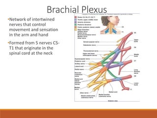
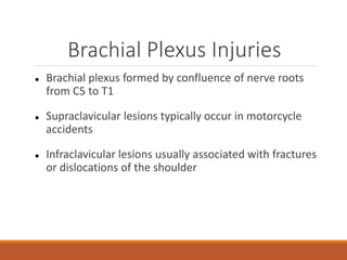
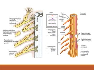
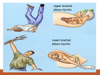
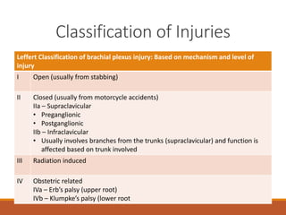
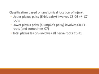
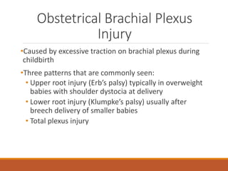
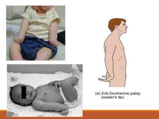
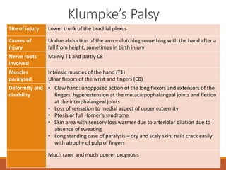
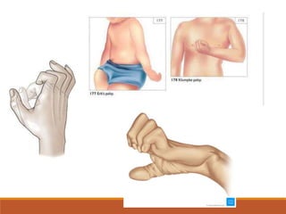
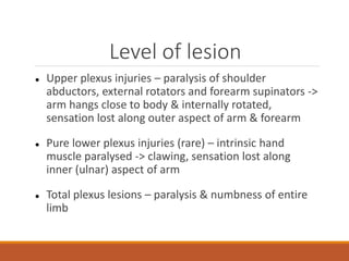
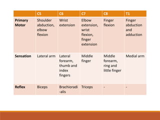
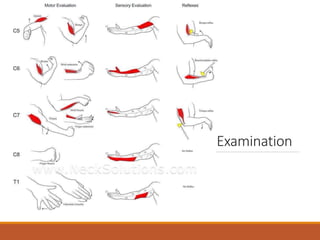
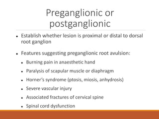
Brachial Plexus Injury
UZAIRIE BIN ANWAR
•Network of intertwined
nerves that control
movement and sensation
in the arm and hand
•Formed from 5 nerves C5-
T1 that originate in the
spinal cord at the neck
Brachial Plexus
Brachial Plexus Injuries
 Brachial plexus formed by confluence of nerve roots
from C5 to T1
 Supraclavicular lesions typically occur in motorcycle
accidents
 Infraclavicular lesions usually associated with fractures
or dislocations of the shoulder
Etiology
•Penetrating wounds
•Injuries related to birth
•Traction/stretch applied to the plexus during falls
•Motor vehicles accidents
•Sport activities
•Radiation
•Motorcycle accidents are the most common cause
Common associated injuries
◦ Fractures to the proximal humerus
◦ Scapula fractures
◦ Clavicle fractures
◦ Fracture of the transverse process of the cervical
vertebrae
◦ Dislocations of the shoulder, acromioclavicular and
sternoclavicular joints
Injury Mechanisms
•Traction/stretch of the
brachial plexus
•Direct blow
•Compression or
impingement of the
brachial plexus
Classification of Injuries
Leffert Classification of brachial plexus injury: Based on mechanism and level of
injury
I Open (usually from stabbing)
II Closed (usually from motorcycle accidents)
IIa – Supraclavicular
• Preganglionic
• Postganglionic
IIb – Infraclavicular
• Usually involves branches from the trunks (supraclavicular) and function is
affected based on trunk involved
III Radiation induced
IV Obstetric related
IVa – Erb’s palsy (upper root)
IVb – Klumpke’s palsy (lower root
Classification based on anatomical location of injury:
◦ Upper plexus palsy (Erb’s palsy) involves C5-C6 +/- C7
roots
◦ Lower plexus palsy (Klumpke’s palsy) involves C8-T1
roots (and sometimes C7)
◦ Total plexus lesions involves all nerve roots C5-T1
Obstetrical Brachial Plexus
Injury
•Caused by excessive traction on brachial plexus during
childbirth
•Three patterns that are commonly seen:
• Upper root injury (Erb’s palsy) typically in overweight
babies with shoulder dystocia at delivery
• Lower root injury (Klumpke’s palsy) usually after
breech delivery of smaller babies
• Total plexus injury
Clinical features
•After a difficult delivery, baby has floppy or flail arm. Further
examination a day or two will define the type of brachial plexus
injury
•Erb’s palsy – arm is held to the side, internally rotated and
pronated
•Klumpke’s palsy – arm supinated, elbow flexed, loss of intrinsic
muscle power of the hand, reflex absent and may be unilateral
Horner’s syndrome
•Total plexus injury – arm is flail and pale, all fingers muscle
paralysed, may be vasomotor impairment and unilateral Horner’s
syndrome
•X-ray should be taken to exclude fracture of shoulder or clavicle
Management
Over the next few weeks, one of the following may happen
◦ Paralysis may recover completely
◦ Paralysis may improve and then remain static
◦ Paralysis may remained unaltered
While waiting for recovery, physiotherapy is applied to keep
joints mobile
If no biceps recovery by 3 months, operative intervention
should be considered
• Roots not avulsed – excise the scar and bridge gap
with free sural nerve grafts
• Roots avulsed – nerve transfer
• Fixed internal rotation and adduction deformity –
subscapularis release and sometimes supplemented
with tendon transfer
• Older children – rotation osteotomy of the humerus
Erb’s Palsy
Site of injury Region of the upper trunk of the brachial plexus that is called Erb’s Point.
Causes of
injury
Undue separation of the head from the shoulder that is commonly
encountered in birth injury, fall on the shoulder
Nerve roots
involved
Mainly C5 and partly C6
Muscles
paralysed
Mainly : biceps, deltoid, brachialis and brachioradialis
Partly : supraspinatus, infraspinatus and supinator
Deformity and
disability
• Arm hangs by the side, adducted and medially rotated
• Forearm extended and pronated
• No abduction - paralysis of deltoid and supraspinatus
• No external rotation - paralysis of infraspinatus and teres minor
• No active flexion - paralysis of biceps, brachialis, brachioradialis
• Pronation of forearm – paralysis of supinator
• Loss of sensation to lateral aspect of upper extremity
More common and better prognosis
Klumpke’s Palsy
Site of injury Lower trunk of the brachial plexus
Causes of
injury
Undue abduction of the arm – clutching something with the hand after a
fall from height, sometimes in birth injury
Nerve roots
involved
Mainly T1 and partly C8
Muscles
paralysed
Intrinsic muscles of the hand (T1)
Ulnar flexors of the wrist and fingers (C8)
Deformity and
disability
• Claw hand: unopposed action of the long flexors and extensors of the
fingers, hyperextension at the metacarpophalangeal joints and flexion
at the interphalangeal joints
• Loss of sensation to medial aspect of upper extremity
• Ptosis or full Horner’s syndrome
• Skin area with sensory loss warmer due to arteriolar dilation due to
absence of sweating
• Long standing case of paralysis – dry and scaly skin, nails crack easily
with atrophy of pulp of fingers
Much rarer and much poorer prognosis
Clinical Features
 Symptoms vary depending type and location of the
injury
 Most common symptoms : weakness, numbness, loss
of sensation, paralysis, pain
 Clinical examination:
 Level of the lesion
 Preganglionic or postganglionic
 Type of damage
Level of lesion
 Upper plexus injuries – paralysis of shoulder
abductors, external rotators and forearm supinators ->
arm hangs close to body & internally rotated,
sensation lost along outer aspect of arm & forearm
 Pure lower plexus injuries (rare) – intrinsic hand
muscle paralysed -> clawing, sensation lost along
inner (ulnar) aspect of arm
 Total plexus lesions – paralysis & numbness of entire
limb
C5 C6 C7 C8 T1
Primary
Motor
Shoulder
abduction,
elbow
flexion
Wrist
extension
Elbow
extension,
wrist
flexion,
finger
extension
Finger
flexion
Finger
abduction
and
adduction
Sensation Lateral arm Lateral
forearm,
thumb and
index
fingers
Middle
finger
Middle
forearm,
ring and
little finger
Medial arm
Reflex Biceps Brachioradi
-alis
Triceps - -
Examination
Preganglionic or
postganglionic
 Establish whether lesion is proximal or distal to dorsal
root ganglion
 Features suggesting preganglionic root avulsion:
 Burning pain in anaesthetic hand
 Paralysis of scapular muscle or diaphragm
 Horner’s syndrome (ptosis, miosis, anhydrosis)
 Severe vascular injury
 Associated fractures of cervical spine
 Spinal cord dysfunction
 Histamine test – intradermal injection of histamine that
normally causes a reflex triple response in surrounding skin
 Central capillary dilatation
 Wheal
 Surrounding flare
 Flare reaction persists in an anaesthetic area of skin ->
preganglionic lesion
 Negative flare reaction -> postganglionic lesion
Type of damage
 With low velocity injuries, a period of observation is
justified
 Neurapraxia & axonotmesis should show signs of
recovery by 6 or 8 weeks
 If neurotmesis seems likely, early operative
exploration is called for
Investigations
•Imaging studies – X-ray
•Electromyogram (EMG)
◦ Serial evaluation of injury to search for signs of reinnervation
•Nerve conduction velocity
•Myelography
• Look forn torn dural sacs around damaged nerve roots -
pseudomeningocoeles
•CT scans
• Establish extent of neck and chest injuries
•MRI
• Assess cervical cord injury and presence of
pseudomeningocoeles
Management
 Emergency surgery required for brachial plexus lesions
associated with penetrating wounds, vascular injury or
severe (high energy) soft tissue damage, whether
open or closed – clean cut nerves should be repaired
or grafted
 Other closed injuries are left until detailed
examination & special investigation completed
 Patients with root avulsion or severe mutilating
injuries – unsuitable for nerve surgery at least until
prognosis of limb function is clear
•Progress of neurological features to be monitored
•If recovery falters or investigations suggests neurotmesis
– surgical exploration and nerve repair, grafting or a
nerve transfer procedure
Goals of Treatment
•For shoulder stability
•For elbow flexion
•For wrist flexion and extension
•For fingers flexion (minimal to grasp cup)
Differential Diagnosis
•CERVICAL INJURY
•CERVICAL SPINE INJURY
•DISLOCATION/FRACTURE OF UPPER EXTREMITIES
•INTRAUTERINE MALADAPTATION PALSY
Prognosis
•Pure upper plexus lesions – best prognosis, hand
function spared and often recover after plexus repair or
nerve transfer
•Lower plexus lesions – poorer prognosis – if shoulder
and elbow movement is restored, loss of hand function
causes severe disability
ASSESS LEVEL OF LESION
DETERMINE PRE OR POST
GANGLIONIC
TYPE OF DAMAGE
IMPORTANT
POINTS
Thank you
References
Solomon, L., Warwick, D. and Nayagam, S., 2014. Apley And Solomon's Concise
System Of Orthopaedics And Trauma, Fourth Edition. Hoboken: CRC Press,
pp.141-143.
S, M., S, T. and D, P., 2007. Instructional Course Lecture: Assessment and Early
Management of Traumatic Brachial Plexus Injury. The Internet Journal of Hand
Surgery, 1(1).
Thatte, M. and Mehta, R., 2011. Obstetric brachial plexus injury. Indian Journal
of Plastic Surgery, 44(3), p.380.
Thatte, M., Babhulkar, S. and Hiremath, A., 2013. Brachial plexus injury in
adults: Diagnosis and surgical treatment strategies. Annals of Indian Academy
of Neurology, 16(1), p.26.
Sakellariou, V., Badilas, N., Mazis, G., Stavropoulos, N., Kotoulas, H.,
Kyriakopoulos, S., Tagkalegkas, I. and Sofianos, I., 2014. Brachial Plexus Injuries
in Adults: Evaluation and Diagnostic Approach. ISRN Orthopedics, 2014, pp.1-9.

BRACHIAL PLEXUS INJURY EDITED.pptx

  • 1.
  • 2.
    •Network of intertwined nervesthat control movement and sensation in the arm and hand •Formed from 5 nerves C5- T1 that originate in the spinal cord at the neck Brachial Plexus
  • 3.
    Brachial Plexus Injuries Brachial plexus formed by confluence of nerve roots from C5 to T1  Supraclavicular lesions typically occur in motorcycle accidents  Infraclavicular lesions usually associated with fractures or dislocations of the shoulder
  • 6.
    Etiology •Penetrating wounds •Injuries relatedto birth •Traction/stretch applied to the plexus during falls •Motor vehicles accidents •Sport activities •Radiation •Motorcycle accidents are the most common cause
  • 7.
    Common associated injuries ◦Fractures to the proximal humerus ◦ Scapula fractures ◦ Clavicle fractures ◦ Fracture of the transverse process of the cervical vertebrae ◦ Dislocations of the shoulder, acromioclavicular and sternoclavicular joints
  • 8.
    Injury Mechanisms •Traction/stretch ofthe brachial plexus •Direct blow •Compression or impingement of the brachial plexus
  • 10.
    Classification of Injuries LeffertClassification of brachial plexus injury: Based on mechanism and level of injury I Open (usually from stabbing) II Closed (usually from motorcycle accidents) IIa – Supraclavicular • Preganglionic • Postganglionic IIb – Infraclavicular • Usually involves branches from the trunks (supraclavicular) and function is affected based on trunk involved III Radiation induced IV Obstetric related IVa – Erb’s palsy (upper root) IVb – Klumpke’s palsy (lower root
  • 11.
    Classification based onanatomical location of injury: ◦ Upper plexus palsy (Erb’s palsy) involves C5-C6 +/- C7 roots ◦ Lower plexus palsy (Klumpke’s palsy) involves C8-T1 roots (and sometimes C7) ◦ Total plexus lesions involves all nerve roots C5-T1
  • 12.
    Obstetrical Brachial Plexus Injury •Causedby excessive traction on brachial plexus during childbirth •Three patterns that are commonly seen: • Upper root injury (Erb’s palsy) typically in overweight babies with shoulder dystocia at delivery • Lower root injury (Klumpke’s palsy) usually after breech delivery of smaller babies • Total plexus injury
  • 13.
    Clinical features •After adifficult delivery, baby has floppy or flail arm. Further examination a day or two will define the type of brachial plexus injury •Erb’s palsy – arm is held to the side, internally rotated and pronated •Klumpke’s palsy – arm supinated, elbow flexed, loss of intrinsic muscle power of the hand, reflex absent and may be unilateral Horner’s syndrome •Total plexus injury – arm is flail and pale, all fingers muscle paralysed, may be vasomotor impairment and unilateral Horner’s syndrome •X-ray should be taken to exclude fracture of shoulder or clavicle
  • 14.
    Management Over the nextfew weeks, one of the following may happen ◦ Paralysis may recover completely ◦ Paralysis may improve and then remain static ◦ Paralysis may remained unaltered While waiting for recovery, physiotherapy is applied to keep joints mobile
  • 15.
    If no bicepsrecovery by 3 months, operative intervention should be considered • Roots not avulsed – excise the scar and bridge gap with free sural nerve grafts • Roots avulsed – nerve transfer • Fixed internal rotation and adduction deformity – subscapularis release and sometimes supplemented with tendon transfer • Older children – rotation osteotomy of the humerus
  • 16.
    Erb’s Palsy Site ofinjury Region of the upper trunk of the brachial plexus that is called Erb’s Point. Causes of injury Undue separation of the head from the shoulder that is commonly encountered in birth injury, fall on the shoulder Nerve roots involved Mainly C5 and partly C6 Muscles paralysed Mainly : biceps, deltoid, brachialis and brachioradialis Partly : supraspinatus, infraspinatus and supinator Deformity and disability • Arm hangs by the side, adducted and medially rotated • Forearm extended and pronated • No abduction - paralysis of deltoid and supraspinatus • No external rotation - paralysis of infraspinatus and teres minor • No active flexion - paralysis of biceps, brachialis, brachioradialis • Pronation of forearm – paralysis of supinator • Loss of sensation to lateral aspect of upper extremity More common and better prognosis
  • 18.
    Klumpke’s Palsy Site ofinjury Lower trunk of the brachial plexus Causes of injury Undue abduction of the arm – clutching something with the hand after a fall from height, sometimes in birth injury Nerve roots involved Mainly T1 and partly C8 Muscles paralysed Intrinsic muscles of the hand (T1) Ulnar flexors of the wrist and fingers (C8) Deformity and disability • Claw hand: unopposed action of the long flexors and extensors of the fingers, hyperextension at the metacarpophalangeal joints and flexion at the interphalangeal joints • Loss of sensation to medial aspect of upper extremity • Ptosis or full Horner’s syndrome • Skin area with sensory loss warmer due to arteriolar dilation due to absence of sweating • Long standing case of paralysis – dry and scaly skin, nails crack easily with atrophy of pulp of fingers Much rarer and much poorer prognosis
  • 20.
    Clinical Features  Symptomsvary depending type and location of the injury  Most common symptoms : weakness, numbness, loss of sensation, paralysis, pain  Clinical examination:  Level of the lesion  Preganglionic or postganglionic  Type of damage
  • 21.
    Level of lesion Upper plexus injuries – paralysis of shoulder abductors, external rotators and forearm supinators -> arm hangs close to body & internally rotated, sensation lost along outer aspect of arm & forearm  Pure lower plexus injuries (rare) – intrinsic hand muscle paralysed -> clawing, sensation lost along inner (ulnar) aspect of arm  Total plexus lesions – paralysis & numbness of entire limb
  • 22.
    C5 C6 C7C8 T1 Primary Motor Shoulder abduction, elbow flexion Wrist extension Elbow extension, wrist flexion, finger extension Finger flexion Finger abduction and adduction Sensation Lateral arm Lateral forearm, thumb and index fingers Middle finger Middle forearm, ring and little finger Medial arm Reflex Biceps Brachioradi -alis Triceps - -
  • 23.
  • 24.
    Preganglionic or postganglionic  Establishwhether lesion is proximal or distal to dorsal root ganglion  Features suggesting preganglionic root avulsion:  Burning pain in anaesthetic hand  Paralysis of scapular muscle or diaphragm  Horner’s syndrome (ptosis, miosis, anhydrosis)  Severe vascular injury  Associated fractures of cervical spine  Spinal cord dysfunction
  • 25.
     Histamine test– intradermal injection of histamine that normally causes a reflex triple response in surrounding skin  Central capillary dilatation  Wheal  Surrounding flare  Flare reaction persists in an anaesthetic area of skin -> preganglionic lesion  Negative flare reaction -> postganglionic lesion
  • 26.
    Type of damage With low velocity injuries, a period of observation is justified  Neurapraxia & axonotmesis should show signs of recovery by 6 or 8 weeks  If neurotmesis seems likely, early operative exploration is called for
  • 27.
    Investigations •Imaging studies –X-ray •Electromyogram (EMG) ◦ Serial evaluation of injury to search for signs of reinnervation •Nerve conduction velocity •Myelography • Look forn torn dural sacs around damaged nerve roots - pseudomeningocoeles •CT scans • Establish extent of neck and chest injuries •MRI • Assess cervical cord injury and presence of pseudomeningocoeles
  • 28.
    Management  Emergency surgeryrequired for brachial plexus lesions associated with penetrating wounds, vascular injury or severe (high energy) soft tissue damage, whether open or closed – clean cut nerves should be repaired or grafted  Other closed injuries are left until detailed examination & special investigation completed  Patients with root avulsion or severe mutilating injuries – unsuitable for nerve surgery at least until prognosis of limb function is clear
  • 29.
    •Progress of neurologicalfeatures to be monitored •If recovery falters or investigations suggests neurotmesis – surgical exploration and nerve repair, grafting or a nerve transfer procedure
  • 30.
    Goals of Treatment •Forshoulder stability •For elbow flexion •For wrist flexion and extension •For fingers flexion (minimal to grasp cup)
  • 31.
    Differential Diagnosis •CERVICAL INJURY •CERVICALSPINE INJURY •DISLOCATION/FRACTURE OF UPPER EXTREMITIES •INTRAUTERINE MALADAPTATION PALSY
  • 32.
    Prognosis •Pure upper plexuslesions – best prognosis, hand function spared and often recover after plexus repair or nerve transfer •Lower plexus lesions – poorer prognosis – if shoulder and elbow movement is restored, loss of hand function causes severe disability
  • 33.
    ASSESS LEVEL OFLESION DETERMINE PRE OR POST GANGLIONIC TYPE OF DAMAGE IMPORTANT POINTS
  • 34.
  • 35.
    References Solomon, L., Warwick,D. and Nayagam, S., 2014. Apley And Solomon's Concise System Of Orthopaedics And Trauma, Fourth Edition. Hoboken: CRC Press, pp.141-143. S, M., S, T. and D, P., 2007. Instructional Course Lecture: Assessment and Early Management of Traumatic Brachial Plexus Injury. The Internet Journal of Hand Surgery, 1(1). Thatte, M. and Mehta, R., 2011. Obstetric brachial plexus injury. Indian Journal of Plastic Surgery, 44(3), p.380. Thatte, M., Babhulkar, S. and Hiremath, A., 2013. Brachial plexus injury in adults: Diagnosis and surgical treatment strategies. Annals of Indian Academy of Neurology, 16(1), p.26. Sakellariou, V., Badilas, N., Mazis, G., Stavropoulos, N., Kotoulas, H., Kyriakopoulos, S., Tagkalegkas, I. and Sofianos, I., 2014. Brachial Plexus Injuries in Adults: Evaluation and Diagnostic Approach. ISRN Orthopedics, 2014, pp.1-9.