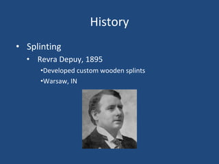
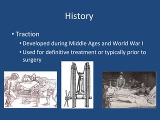
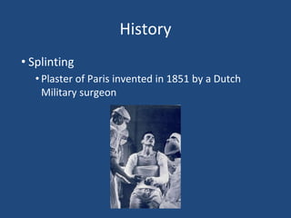
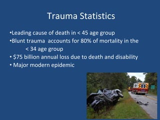
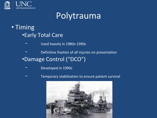
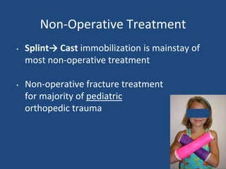
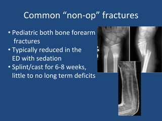
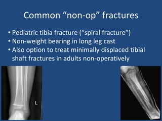
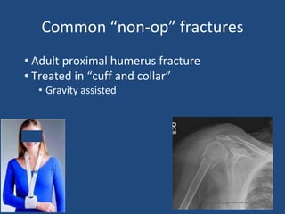
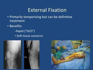
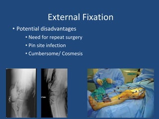
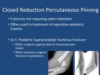
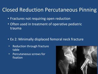
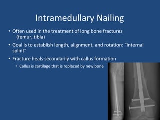
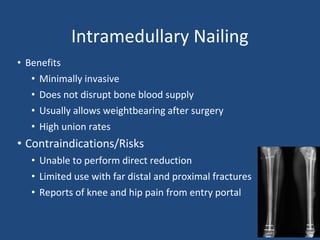
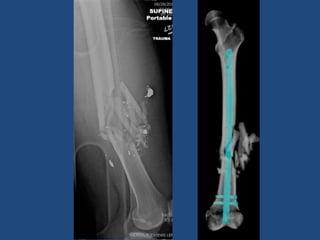
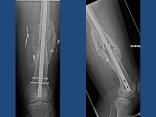
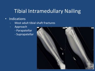
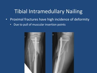
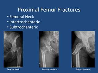
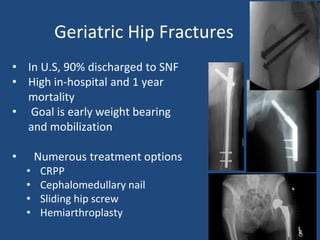
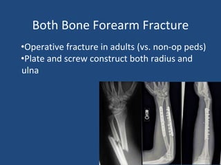
8. Trauma Statistics
•Leading cause of death in < 45 age group
•Blunt trauma accounts for 80% of mortality in the
< 34 age group
• $75 billion annual loss due to death and disability
• Major modern epidemic
9. Level 1 Trauma Activation Criteria
• Traumatic cardiac arrest during transport
• Systolic blood pressure less than 90
• Respiratory compromise/intubation
• Glasgow Coma Scale less than 8
• Traumatic limb paralysis
• Amputation proximal to wrist or ankle
• Vascular compromise of extremity
• Burns with traumatic component
• Penetrating injuries to head, neck chest, abdomen, extremities proximal to the
elbow or knee
PLEASE NOTE: Criteria may vary at centers
depending on State vs. ACS accreditation and
institution policy
10. Level 2 Trauma Activation Criteria
• Glasgow Coma Scale <14, >8
• Vital Sign instability (do not meet RED criteria)
• Flail chest or multiple rib fractures
• Pneumothorax/hemothorax
• Open/depressed skull fractures
• 2 or more long bone fractures
• Crush injury to chest or pelvis
• High level of suspicion related to mechanism
• Burns
11. Mechanism of Injury
•45% Falls
•32% Motor vehicle collision
•8% Assault
•9% Motorcycle collision
•6% Gunshot wounds
12. Polytrauma
•Definition
“blunt trauma patients whose injuries involve multiple body regions or
cavities, compromise patient’s physiology, and potentially cause
dysfunction of uninjured organs”
- Injury
13. Polytrauma
• Timing
•Early Total Care
- Used heavily in 1980s-1990s
- Definitive fixation of all injuries on presentation
•Damage Control (“DCO”)
- Developed in 1990s
- Temporary stabilization to ensure patient survival
15. Non-Operative Treatment
• Splint→ Cast immobilization is mainstay of
most non-operative treatment
• Non-operative fracture treatment
for majority of pediatric
orthopedic trauma
16. Common “non-op” fractures
• Pediatric both bone forearm
fractures
• Typically reduced in the
ED with sedation
• Splint/cast for 6-8 weeks,
little to no long term deficits
17. Common “non-op” fractures
• Pediatric tibia fracture (“spiral fracture”)
• Non-weight bearing in long leg cast
• Also option to treat minimally displaced tibial
shaft fractures in adults non-operatively
18. Common “non-op” fractures
• Adult proximal humerus fracture
• Treated in “cuff and collar”
• Gravity assisted
21. Open Fractures
• Variability in severity
• Surgical “urgency”
• Time to antibiotics and
debridement is crucial
• Antibiotic choice?
• Cefazolin: 1st line
• Clindamycin in PCN allergic
• Aminoglycosides if very
contaminated or Type III
22. Compartment Syndrome
• Surgical emergency
• Typically the result of traumatic injury to soft
tissue +/- fracture
• Excessive pressure within closed space
decreases blood flow to the tissues
• Commonly lower leg but can be thigh, buttock,
hand, forearm, arm, foot
28. Internal Fixation
• Closed reduction percutaneous pinning
• Intramedullary nails
• Plate and screw constructs
29. Closed Reduction Percutaneous Pinning
• Fractures not requiring open reduction
• Often used in treatment of operative pediatric
trauma
• Ex 1: Pediatric Supracondylar Humerus Fracture
• Often surgical urgency due to neurovascular
issues
• Most common surgical
fracture in pediatrics
30. Closed Reduction Percutaneous Pinning
• Fractures not requiring open reduction
• Often used in treatment of operative pediatric
trauma
• Ex 2: Minimally displaced femoral neck fracture
• Reduction through fracture
table
• Percutaneous screws for
fixation
31. Intramedullary Nailing
• Often used in the treatment of long bone fractures
(femur, tibia)
• Goal is to establish length, alignment, and rotation: “internal
splint”
• Fracture heals secondarily with callus formation
• Callus is cartilage that is replaced by new bone
32. Intramedullary Nailing
• Benefits
• Minimally invasive
• Does not disrupt bone blood supply
• Usually allows weightbearing after surgery
• High union rates
• Contraindications/Risks
• Unable to perform direct reduction
• Limited use with far distal and proximal fractures
• Reports of knee and hip pain from entry portal
33.
34.
35. Femoral shaft and tibia fractures
• High energy mechanism
• Commonly treated with intramedullary
nailing
• Typically patients may “WBAT” following
surgery
36. Femoral Intramedullary Nailing
• Antegrade (Through hip)
• Common, and can be used for all shaft fractures
• Starting point options
• Retrograde (Through knee)
• Do not need fracture table
• Bilateral femur fractures
• Floating knee
• Distal fracture
42. Plate and screw constructs
• Plate fixation
• Types: compression, locking, recon, anatomical
• Functions: neutralization, compression, bridging, buttress
• Usually require open reduction
• Multiple plate and screw combinations available
43. Common traumatic injuries and
associated treatments
• Pelvic ring injuries
• Acetabular fractures
• Femur and tibia fractures
• Hip fractures
• Both bone forearm fracture
45. Pelvic Ring Injuries
• Open Book: Initial Management
• Pelvic binder or sheet to close potential
space for hemorrhage
46. Pelvic Ring Injuries
• Typical definitive management
• Sacroiliac screws (posterior injury)
• ORIF of symphysis (anterior injury)
47. Acetabular Fractures
• High energy in young patients
• Low energy in elderly patients
• High incidence of associated injuries
• Extremity 36%
• Head injury 19%
• Chest injury 18%
48. Acetabular Fractures
• Usually treated with ORIF
- Multiple approaches
- Non-weightbearing for 6-12 weeks
• Acute Total Hip Arthroplasty
• Complications
- Post-traumatic arthritis
- Osteonecrosis
- Nerve Palsy
- Heterotopic Ossification
50. Geriatric Hip Fractures
• In U.S, 90% discharged to SNF
• High in-hospital and 1 year
mortality
• Goal is early weight bearing
and mobilization
• Numerous treatment options
• CRPP
• Cephalomedullary nail
• Sliding hip screw
• Hemiarthroplasty
51. Both Bone Forearm Fracture
•Operative fracture in adults (vs. non-op peds)
•Plate and screw construct both radius and
ulna
52. A brief discussion about OR tables
• Regular table
• Fracture table
• OSI Flat top table
53. Regular OR Table
• Not radiolucent
• Useful for procedures that do not require fluoroscopy above
the knee
• Useful for ankle fractures, arm/forearm fractures
54. Fracture Table
• Holds the lower extremity in traction
• Hip fractures requiring traction
• Antegrade Femoral nailing
• Also can be used for total hip arthroplasty
55. OSI Flat Top
• Radiolucent
• Useful for procedures that DO require fluoroscopy
56. Summary
• Orthopaedic traumatic injuries are common and often occur in
setting of high energy trauma with multi-organ involvement
• Mainstay of treatment involves stabilization/fixation
• Goal is early mobilization and function
• Generally acceptable outcomes but occasional severe long
term sequelae
58. References
•Neria Butcher, Zsolt J Balogh. The definition of polytrauma:the need for international consensus. Injury, Int. J. Care Injured (2009)
40S4, S12–S22
•Holstein JH1, Culemann U, Pohlemann T; Working Group Mortality in Pelvic Fracture Patients. What are predictors of mortality in
patients with pelvic fractures? Clin Orthop Relat Res. 2012 Aug;470(8):2090-7. doi: 0.1007/s11999-012-2276-9.
•Taeger G, Ruchholtz S, Waydhas C, Lewan U, Schmidt B, Nast-Kolb D. Damage control orthopedics in patients with multiple injuries is
effective, time saving, and safe. J Trauma. 2005 Aug;59(2):409-16; discussion 417.
• Goodman DC, Fisher ES, Chang CH. After hospitalization: a Dartmouth Atlas report on post-acute care for Medicare beneficiaries. A
report of the Dartmouth Atlas project. 2011.
• Nikkel LE, Kates SL, Schreck M, Maceroli M, Mahmood B, Elfar JC. Length of hospital stay after hip fracture and risk of early mortality
after discharge in New York state: retrospective cohort study. BMJ 2015;351:h6246
•Scott Schnell, MD, Susan M. Friedman, MD, MPH, Daniel A. Mendelson, MS, MD, Karilee W. Bingham, MS, RN, FNP, Stephen L. Kates,
MD. The 1-Year Mortality of Patients Treated in a Hip Fracture Program for Elders. Geriatr Orthop Surg Rehabil. 2010 Sep; 1(1): 6–14.
•Koval KJ1, Friend KD, Aharonoff GB, Zukerman JD. Weight bearing after hip fracture: a prospective series of 596 geriatric hip fracture
patients. J Orthop Trauma. 1996;10(8):526-30.
•Haidukewych GJ. Acetabular fractures: the role of arthroplasty. Orthopedics. United States; 2010;33(9):645.
•Daurka JS, Pastides PS, Lewis A, Rickman M, and Bircher MD. Acetabular fractures in patients aged > 55 years: a systematic review of
the literature. Bone Joint J. England; 2014;96- B(2):157-63.
• Gary JL, Lefaivre KA, Gerold F, Hay MT, Reinert CM, and Starr AJ. Survivorship of the native hip joint after percutaneous repair of
acetabular fractures in the elderly. Injury. Netherlands; 2011;42(10):1144-51. 6.
•Tannast M, Najibi S, and Matta JM. Two to twenty-year survivorship of the hip in 810 patients with operatively treated acetabular
fractures. J Bone Joint Surg Am. United States; 2012;94(17):1559-67.
•O'Toole RV, Hui E, Chandra A, and Nascone JW. How often does open reduction and internal fixation of geriatric acetabular fractures
lead to hip arthroplasty? J Orthop Trauma. United States; 2014;28(3):148-53