Brachial plexus injuries
•Download as PPTX, PDF•
7 likes•362 views

This document discusses brachial plexus injuries, including how to clinically diagnose and classify them as pre- or post-ganglionic and upper or lower plexus injuries. Physical examination findings that help differentiate these types are described. Investigations like X-ray, MRI, EMG and nerve conduction studies can provide further information to accurately diagnose the level and severity of injury. Both non-operative and surgical management options are covered, with the goals of surgery being restoration of elbow flexion, shoulder abduction and sensation in the forearm and hand. Secondary operations like tendon transfers may also be considered if needed.

Recommended
More Related Content
What's hot
What's hot (20)
Similar to Brachial plexus injuries
Similar to Brachial plexus injuries (20)
More from Arvinthran Suguna Seelan
More from Arvinthran Suguna Seelan (13)
Recently uploaded
Recently uploaded (20)
Brachial plexus injuries
- 2. AIM • How to diagnose brachial plexus injuries clinically • How to differentiate pre/post ganglionic injuries • How to classify upper/lower/complete brachial plexus injuries
- 6. History
- 9. Physical Examination • Preganglionic/Postganglionic • Upper / Lower plexus injury
- 10. Preganglionic Postganglionic Horners syndrome No horners Sensory intact Sensory impaired Tinel sign absent Tinel sign present Winged scapula Rhomboid paralysis Supra/infraspinatus paralysis Lattismus dorsi paralysis
- 11. Upper Lesion ( C5,C6), Erbs Palsy Lower Lesion ( C8,T1), Klumpke Palsy Most common obstetric palsy Rare in obstetric palsy Results from excessive displacement of head to opposite side and depression of shoulder on same side Usually avulsion injury caused by excessive abduction Arm adducted , internally rotated, forearm pronated and extended ( waiters tip) Clinically presents as claw hands C5- axillary, suprascapular, musculocutaneous nerve C6 - Radial C8, T1, Ulnar and median nerve Deltoid, teres minor, supraspinatus, infraspinatus, biceps, brachioradialis, supinator Intrinsic muscles of thehand Best prognosis Poor Prognosis
- 12. Physical examination • All nerve groups controlled by the brachial plexus • Horners syndrome • Tinels sign • Serratus anterior and rhomboids Nerve Sensation Motor Musculocutaneous Lateral forearm Elbow flexion Axillary Regimental badge Shoulder abduction Median Pad of index finger Thumb abduction Ulnar Radial little finger Finger abduction Radial Dorsal 1st web Wrist extension
- 13. Investigations • Radiographic evaluation - X ray - Myelography - MRI • Electrophysiology studies - EMG - Nerve conduction velocity - Intraoperative nerve action potential
- 14. X-Ray
- 15. EMG
- 16. CT Myelography
- 17. MRI
- 18. Nerve conduction velocity • Performed along with EMG • Measures sensory nerve action potentials • Distinguishes preganglionic from postganglionic • SNAPs are preserved in lesions proximal to dorsal root ganglia
- 19. Nerve action potentials • Often intraoperative • Tests nerve across a lesion • IF NAP positive across a lesion - Preserved axons or significant regeneration • Can detect reinnervation months before EMG - NAP negative – neuropraxic lesion - NAP positive – axonotmetic lesion
- 22. Follow up at 6/52 • Review symptoms - Functional recovery of muscle - Tinel sign - Pain control • Further imaging - MRI/CT myelography • Operative intervention
- 23. SURGICALGOALS 1)Restoration of elbow flexion 2)Restoration of shoulderabduction 3)Restoration of sensation of medial borderof forearm & hand.
- 24. • Primary neurorrhapy • Nerve grafting • Neurotization Surgical Options
- 26. Secondary Operations 1. Arthrodesis 2. Tendontransfer and 3. Functional free muscle transplantation are favoured treatment options.
- 27. Take home message • BPI’s causes catastrophic injuries and affect the quality of life • Early recognition of the mechanism and level of injury is important to establish early treatment • Thorough clinical examination is essential to obtain an accurate diagnosis • Recent advances in surgical techniques have led to improved outcomes of patients
- 28. References • Sakellariou, V. I., Badilas, N. K., Mazis, G. A., Stavropoulos, N. A., Kotoulas, H. K., Kyriakopoulos, S., … Sofianos, I. P. (2014). Brachial Plexus Injuries in Adults: Evaluation and Diagnostic Approach. ISRN Orthopedics, 2014, 1–9. doi:10.1155/2014/726103 • Ferraresi, S., Garozzo, D., Griffini, C., Resmini, B., Manara, O., Foresti, C., … Ghislandi, I. (1994). Brachial plexus injuries Guidelines for management: Our experience. The Italian Journal of Neurological Sciences, 15(6), 273–284. doi:10.1007/bf02339237 • BMJ Best Practice, Brachial Plexus injuries • Nagano, A. (1998). Treatment of brachial plexus injury*. Journal of Orthopaedic Science, 3(1), 71–80. doi:10.1007/s007760050024 • Sakellariou, V. I., Badilas, N. K., Stavropoulos, N. A., Mazis, G., Kotoulas, H. K., Kyriakopoulos, S., … Sofianos, I. P. (2014). Treatment Options for Brachial Plexus Injuries. ISRN Orthopedics, 2014, 1–10. doi:10.1155/2014/314137 • Park, H. R., Lee, G. S., Kim, I. S., & Chang, J.-C. (2017). Brachial Plexus Injury in Adults. The Nerve, 3(1), 1–11. doi:10.21129/nerve.2017.3.1.1
Editor's Notes
- Severe peripheral nerve injury affecting the upper extremeties, causing functional damage and physical disabilityThe most common cause of adult BPI is a traffic accident, such as motorcycle accidents. Most patients are young men between 15 and 25 years of age. Other traumatic causes include sports injuries, incised wounds, gunshot wounds, carrying a heavy backpack, and inappropriate operative positioning. Non-traumatic causes consist of tumors, irradiation, and congenital abnormalities such as cervical ribs
- it is a network of nerves passing through the cervico-axillary canal to reach axilla and innervates brachium (upper arm), antebrachium (forearm) and hand. The brachial plexus runs within the interscalene triangle bounded by the anterior scalene muscle anteriorly, the middle scalene muscle posteriorly and the superior border of the first rib inferiorly. It traverses the posterior triangle of the neck, formed by the clavicle inferiorly, the trapezius muscle laterally and the posterior border of sternocleidomastoid muscle medially68). Then it passes laterally over the first rib and enters the axilla to supply the upper limb
- Brachial plexus is a somatic nerve plexus formed by the union of anterior rami of C5,C6,C7,C8 and T1. The formation of brachial plexus begins just distal to the scalenus muscles. Function: The brachial plexus is responsible for cutaneous and muscular innervation of the entire upper limb, with two exceptions: the trapezius muscle innervated by the spinal accessory nerve (CN XI) and an area of skin near the axilla innervated by the intercostobrachial nerve. The brachial plexus is composed by 5 anatomical components: 5 roots, 3 trunks, 6 divisions, 3 cords, and 5 terminal branches (Fig. 1). Five roots include the anterior branches of the 4 lowest cervical spinal nerve roots (C5-C8) and the first thoracic nerve root (T1. Rarely the brachial plexus may be pre/post fixed resulting in a slight variation of origin between c4 to t2, Two pairs of nerve roots extend from each segment of the spinal cord; ventral (anterior) and dorsal (posterior) roots. The ventral roots contain motor fibers exiting the spinal cord. The dorsal roots convey sensory fibers that came from the dorsal root ganglion and enter the spinal cord. The ventral and dorsal roots get together beyond the ganglion, and become the spinal nerve. Dorsal scapular C5- rhomboid , stabilisation of scapula Long thoracic C5 – Serratus anterior abduction of scapula Suprascapular – infraspinatus and supraspinatus , abduction of shoulder, expternal rotation of shoulder Medial C8 and Lateral pectoral c7 – pec major and pec minor, adducts shoulder and stabilizes the scapula Subscapular c5 – subscapularis and teres major, internal rotation of shoulder Thoracodorsal c7 – lattismus dorsi , adduction of sjhoulder Musculocutaneous c5 – biceps brachii and brtachialis – flexion of elbow Ulnar c8 t1 – fcu, intrinsic muscleds of hanf – flexors of wrist and fingers, abduction of fingers Median c8 to t1 – pronators of forearm, flexors of wrist and fingers – Radial c 6 to c 8 – supinator , triceps brachii, extensors of wrist and fgingers, Axillary c 5 – deltoid and teres minor – abduction of shoulders
- Seddon classification[4] Based on clinical findings with increasing force of trauma. There are 3 degrees of injury from simple stretch/ compression to complete transaction: • Neurapraxia (first-degree Sunderland) - axon remains intact; includes conduction blocks • Axonotmesis (second-degree Sunderland)- axon is severed, but endoneurium, perineurium, and epineurium are intact • Neurotmesis (fifth-degree Sunderland).- complete injury. Sunderland classification[3] Based on anatomical boundaries within the nerves that are transgressed by increasing force of trauma. There are 5 degrees of injury from simple stretch/compression to complete transaction: • First-degree injury: axon remains intact; includes conduction blocks • Second-degree injury: axon is severed, but endoneurium, perineurium, and epineurium are intact • Third-degree injury: axon and endoneurium damaged leaving fascicular pattern intact • Fourth-degree injury: axon, endoneurium, • Fifth-degree injury: complete injury.
- Closed trauma is the most common cause of adult prachial plexus injury.Most common mechanism is compression or traction. In traction, nerve may be injured, avulsed or significantly stretched from the roots. Traction related injuries can be a result of violent widening of the scapulohumeral anhle , accompanied by the shoulder dislocations and fractures of the humerus, which causes pressure on the inraclavicular neurovascular bundle above the humeral head and injury of the infraclavicular plexus. Because the axillary artery is located close to the medial m lateral and posterior cord, accompanying rupture of the axillary artery has been reported by up to 50% of infraclavicula rplexus injury Mechanism of injury Brachial plexus injury at the level of spinal cord usually produces greater pain than injuries more distant Injuries nearer to the spinal cord may cause a burning numbness known as paraesthesia or dysaethesia
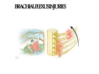
- The patients position and location of the upper limb when the injury occurs is the most important mechanism to understand the mechanism of BPI, Different positions and location of the upper limb will lead to a diffeerent injury mechanism because tension can be applied to various roots. . In this image , if the upper limb is stretched in an elevated position, the greatest tension force will affect the lower roots.
- Leffert is a classification based on the mechanism of injury while Milessi classification is based on the level of injury. However, a much simpler method would be to classifiy the injury based on wheter its pre/ post ganglionic and the anatomical location of injury
- In preganglionic injuries, there is an avulsion proximal to the dorsal root ganglion, involves CNS which does not regenerate, poor prognosis lesions suggesting preganglionic injury: Horner’s syndrome disruption of sympathetic chain winged scapula medially loss of serratus anterior (long thoracic nerve) rhomboids (dorsal scapular nerve) leads to medial winging (inferior border goes medial) presents with motor deficits (flail arm) sensory intact absence of a Tinel sign or tenderness to percussion in the neck normal histamine test (C8-T1 sympathetic ganglion) intact triple response (redness, wheal, flare) elevated hemidiaphragm (phrenic nerve rhomboid paralysis (dorsal scapular nerve) supraspinatus/infraspinatus (suprascapular nerve) latissimus dorsi (thoracodorsal) evaluation EMG may show loss of innervation to cervical paraspinals Horner syndrome is a relatively rare disorder characterized by a constricted pupil (miosis), drooping of the upper eyelid (ptosis), absence of sweating of the face (anhidrosis), and sinking of the eyeball into the bony cavity that protects the eye (enophthalmos). These are the four classic signs of the disorder.
- Deformity Arm: Hangs by the side, it is adducted and medially rotated Forearm: Extended and pronated Abduction impossible because of paralysis of deltoid & supraspinatus m/s. ER impossible because of paralysis of infraspinatus & teres minor m/s. Active flexion impossible because of paralysis biceps, brachialis & brachioradialis. Paralysis of supinator m/s causes pronation deformity of forearm. The deformity is known as "Policeman's tip hand" or "Porter's tip hand". Muscles paralysed: Intrinsic muscles of the hand (T1) Ulnar flexors of the wrist and fingers (C8). Deformity: (position of the hand): claw hand due to the unopposed action of the long flexors and extensors of the fingers. in a claw hand there is hyperextension at the metacarpophalangeal joints and flexion at the interphalangeal joints. Disability: Claw hand Cutaneous anaesthesia and analgesia in a narrow zone along the ulnar border of the forearm and hand. Horner's syndrome: ptosis, miosis, anhydrosis, enophthalmos and loss of ciliospinal reflex- may be associated. This is because of injury to sympathetic fibres to the head and neck that leave the spinal cord through nerve T1.
- Mechanism of injury , high or low energy, -Swelling over the shoulder Palpation -Tinelsign + test: Marked,painfulparaesthesia in the corresponding dermatomes -Palpate for any tenderness over the Clavicle A shooting nerve-like pain on taping along the affected nerves (Tinel sign) suggests an injury farther from the spinal cord. Over time, if the location of the Tinel sign moves down the arm toward the hand, it is a sign that the injury is repairing itself. During the physical examination, assess the arm and shoulder for stability and range of motion Musculocutaneous c5,c6,c7 Axillary c5 c 6 Median c5c7 lateral c8t1 medial Ulna c8 t1
- Imaging studies : X-ray of cervical spine : Fracture of lateral masses of cervical vertebrae are strongly associated with pre-ganglionic injuries. Chest x-ray : May show 1st and 2nd rib fracture or an elevated hemidiaphragm, which denotes phrenic nerve paralysis and proximal injury to upper plexus. Fractures of scapula and clavicle and Humerus may indicate infraclavicular plexus injuries. chest radiograph recommended views PA and lateral fractures to the first or second ribs suggest damage to the overlying brachial plexus evidence of old rib fractures can be important in case intercostal nerve is needed for nerve transfer inspiration and expiration can demonstrate a paralyzed diaphragm (indicates upper nerve root injury) cervical spine series recommended views AP and lateral transverse process fracture likely indicates a root avulsion scapular and shoulder series recommended views at least AP and axillary (or equivalent) scapulothoracic dissociation is associated with root avulsion and major vascular injury clavicle recommended views orthogonal views fracture may indicate brachial plexus injury
- Tests muscle at rest and during activity Fibrillation potential ( denervation changes) Can help distinguish preganglionic from post ganglionic lesions Electromyography (EMG)tests muscles at rest and during activity fibrillation potentials (denervation changes) as early as 10-14 days following injury in proximal muscles as late as 3-6 weeks in distal muscles can help distinguish preganglionic from postganglionic examine proximally innervated muscles that are innervated by root level motor branches rhomboids serratus anterior cervical paraspinals Most important use of EMG studies is for serial evaluation of injury to search for signs of re innervation. A decreased in number of fibrillation potentials and positive sharp potentials typically seen in dennervated muscles regenerating axons have reached the motor end plates. The appearance of prolonged, polyphasic and low-amp indicated re-innervation. Seen several weeks before the onset of voluntary muscle contraction and signify that a further period of observation is in order
- Gold standard for defining nerve root injury Avulsion of nerve roots cause dural sheath to heal with meningocoele Scan should be done 3- 4 weeks after injury - Allows blood clot in the injured area to dissipate and meningocoele to form CT Myelography : If plexus injury is strongly suspected a myelogram and subsequent CT scan should be obtained 2-3 months after injury. It may be inaccurate early after the injury because clotted blood may occlude the opening into the pseudomeningocele. A delay of 6-12 weeks is recommended before myelogram is advised. Advantages: -detect partial root avulsion -excellent visualization of bony structures -no CSF flow artifacts and -multiplanar reconstruction. Disadvantages: - high radiation dose -poor visualization of lower brachial plexus due to bony artifacts.
- Indication Suspect injury distal to nerve roots Can visualise much of the brachial plexus Findings Traumatic neuromas and edema Mass lesions Pseudomeningocoele Empty nerve root sleeves Cord shift away from midline
- Nerve conduction velocity (NCV)performed along with EMG measures sensory nerve action potentials (SNAPs) distinguishes preganglionic from postganglionic SNAPs preserved in lesions proximal to dorsal root ganglia cell body found in dorsal root ganglia if SNAP normal and patient insensate in ulnar nerve distribution preganglionic injury to C8 and T1 if SNAP normal and patient insensate in median nerve distribution preganglionic injury to C5 and C6
- Intra operative nerve action potential (NAP) : This study is performed during surgical exploration of the plexus, which is usually done 3-4 months after injury. If a nerve action potential can be recorded. Substantial number of regenerating axons have traversed the lesion site. Conversely if an action potential cannot be elicited the abnormal segment is resected because spontaneous recovery is likely to be poor. NAP is best for evaluating a neuroma in continuity. If an NAP can be transmitted across the area of injury, the patient has 93% chance of useful motor function will develop in the muscles supplied by that nerve
- In the case of closed BPI wounds and when there are no other emergent injuries, surgical exploration and recovery may not take place immediately. Recommendations include managing pain, and starting rehabilitation. AIMS: to maintain the range of motion of the extremity to strengthen the remaining functional muscles to protect the denervated dermatomes, and to manage pain. Pain management: Significant pain is observed in complete palsy especially in root avulsions. Pain Is excruciating and exhausting for the patient but it can affect the rehabilitation procedure . NSAIDs and opioid drugs useful during the first stages but do not appear to help with neuropathic pain, which requires careful use of antiepileptic drugs (gabapentin and carbamazepine) or antidepressants such as amitriptyline. About 30% of patients report significant pain relief with this type of treatment.
- In the case of closed BPI wounds and when there are no other emergent injuries, surgical exploration and recovery may not take place immediately. Recommendations include managing pain, and starting rehabilitation. AIMS: to maintain the range of motion of the extremity to strengthen the remaining functional muscles to protect the denervated dermatomes, and to manage pain. Pain management: Significant pain is observed in complete palsy especially in root avulsions. Pain Is excruciating and exhausting for the patient but it can affect the rehabilitation procedure . NSAIDs and opioid drugs useful during the first stages but do not appear to help with neuropathic pain, which requires careful use of antiepileptic drugs (gabapentin and carbamazepine) or antidepressants such as amitriptyline. About 30% of patients report significant pain relief with this type of treatment.
- Nerve grafting is the predominant technique for clear cut injuries with a healthy proximal stump and with no axial damage. The outcome is influenced by the length of the nerve graft the presence of scar tissue at the wound site the number of grafts used the presence of a healthy proximal stump available for grafting and the nerve gap to be covered. The sural nerve, the sensory branch of ulnar nerve, and the medial cutaneous nerve of the forearm are the usual donor nerves. Generally, use of nerve grafts shorter than 10 cm results in better functional and clinical outcomes compared with longer grafts This type of procedure is used for preganglionic root injury The nerve transfer may be extraplexus or intraplexus. Intraplexus transfer options include intact nerve roots. Other choices include the use of the medial thoracic nerve and inferior medial cord/ulnar nerve. Oberlin et al. described nerve transfer to the biceps muscle using part of the ulnar nerve for C5-C6 avulsion of the brachial plexus Extraplexus transfer options include the use of intercostal and spinal accessory nerves. The phrenic nerve and deep motor branches of the cervical plexus (C3-C4) may be used as donor nerves. In ROOT avulsion of upper plexus in which no proximal neural stump is available for nerve grafting, neurotization between intercostal nerves or FCU motor fascicles of ulnar nerve & musculocutaneous nerve to restore the ELBOW FLEXION may be considered. NEUROTIZATION of the suprascapular nerve using the spinal accessory nerve and neurotization of the axillary nerve with fascicles of radial nerve innervating the lateral, medial, or long head of triceps can be used to restore SHOULDER ABDUCTION AND EXTERNAL ROTATION After Brachial plexus repair & regeneration 12 to 18 mths required to determine extent of neural regeneration. If recovery inadequate Peripheral reconstruction considered
- In the absence of spontaneous recovery or when the first surgical procedure does not provide satisfactory outcomes then a second operation may be required. In such cases there should be specific signs of neurological denervation or no possibility of neurological recovery, or sufficient time should have passed with no functional improvement. Secondary options include NOTE: The shoulder should be fused with only 20 degrees of abduction, 30 degrees flexion, and 30 degrees of internal rotation to allow the patient to be independent in his daily life with a mean range of 60 degrees abduction and flexion through the scapulothoracic joint Tendon transfers are useful in restoring upper extremity function after BPI. An absolute indication for tendon transfer is upper or lower brachial plexus traumatic injury with only partial paralysis. Many tendon transfer techniques have been described for treating partial shoulder paralysis. the most common procedures are the following: 1.Trapezius to deltoid transfer as described by Elhassan et al. in 2000 to restore abduction of the shoulder 2.Latissimus dorsi transfer as described by L’ Episcopo, to improve external rotation. 3. Anterior transfer of the posterior branch of the deltoid muscle to restore nonfunctional anterior segment. The surgical goal is to restore good muscle strength through a range of elbow motion (30 to 130 degrees). The most commonly used procedures are as follows: i)transfer of the common origin of the flexor forearm muscles to a proximal section as described by Steindler (1918) . The Steindler technique may lead to disappointing outcomes such as elbow stiffness or over pronation; (ii)transfer of latissimus dorsi muscle to the tendon of the biceps brachialis provides great muscle strength, but this muscle is often denervated (iii)transfer of pectoralis major brachial branch tendon to brachial biceps (Clark technique). A fused shoulder is required for the best postoperative result; (iv)transfer of triceps tendon to biceps provides good results