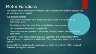
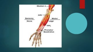
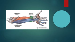
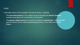
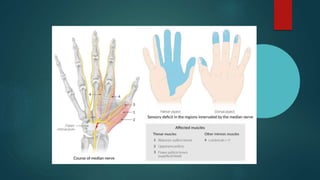
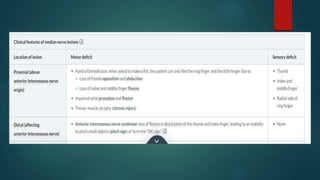
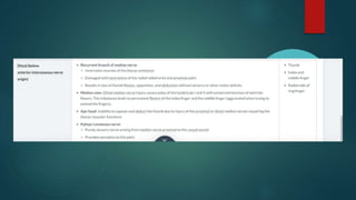
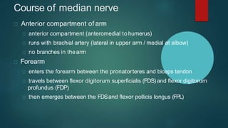
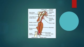
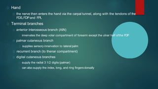
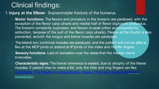
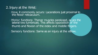
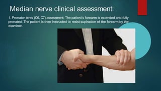
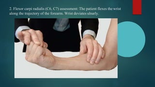
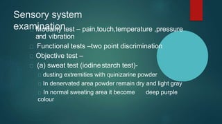
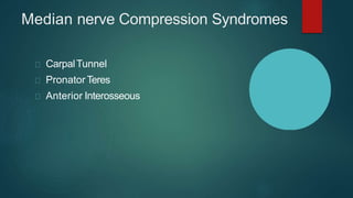
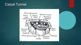
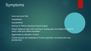
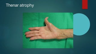
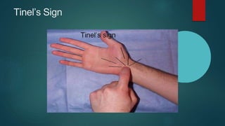
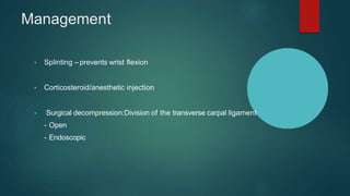
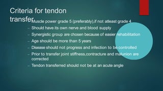
SlideShare a Scribd company logo
MEDIAN NERVE INJURIES
Dr SAURABH AGRAWAL
Assiatant professor
Deptt of orthopedics, AIIMS bedwas
Median nerve introduction
The median nerve is derived from both the lateral and
medial cords of the brachial plexus, with the lateral cord
providing mostly sensory axons from C6 and C7, and the
medial cord providing motor axons from C8 and T1.
it is the only nerve that passes through the carpal
tunnel. Carpal tunnel syndrome is the disability that
results from the median nerve being pressed in the
carpal tunnel.
Also called labourer’s nerve.
Motor Functions
The median nerve innervates the majority of the muscles in the anterior forearm,and
some intrinsic hand muscles.
The Anterior Forearm
Inthe forearm, the median nerve directly innervates muscles in the superficial and
intermediate layers:
Superficial layer: Pronator teres, flexor carpi radialis and palmarislongus.
Intermediate layer:Flexor digitormsuperficialis.
The median nerve also gives rise to the anterior interosseous nerve, which suppliesthe
deep flexors:
Deep layer: Flexor pollicis longus, pronator quadratus, and the lateral half of the
flexor digitorum profundus (the medial half of the muscle is innervated by the ulnar
nerve).
Ingeneral these muscles perform pronation of the forearm, flexion of the wrist and
flexion of the digits of the hand.
HAND
innervates some of the muscles in the hand via two branches.
The recurrent branch of the median nerve innervates the thenar muscles –
muscles associated with movements of the thumb.
The palmar digital branch innervates the lateral twolumbricals – these muscles
perform flexion at the metacarpophalangeal joints of the index and middle
fingers
Sensory innervation:
The median nerve is responsible for the cutaneous
innervation of part of the hand. This is achieved via two
branches:
Palmar cutaneous branch –Arises in the forearm and
travels into the hand. It innervates the lateral aspect of
the palm. This nerve does not pass through the carpal
tunnel, and is spared in carpal tunnel syndrome.
Palmar digital cutaneous branch –Arises in the
hand. Innervates the palmar surface and fingertips of the
lateral three and half digits.
Course of median nerve
Anterior compartment of arm
anterior compartment (anteromedial to humerus)
runs with brachial artery (lateral in upper arm / medial at elbow)
no branches in thearm
Forearm
enters the forearm between the pronatorteres and biceps tendon
travels between flexor digitorum superficialis (FDS)and flexor digitorum
profundus (FDP)
then emerges between the FDSand flexor pollicis longus (FPL)
Hand
the nerve then enters the hand via the carpal tunnel, along with the tendons of the
FDS,FDPand FPL
Terminal branches
anterior interosseous branch (AIN)
innervates the deep volar compartment of forearm except the ulnar half ofthe FDP
palmar cutaneous branch
supplies sensory innervation to lateral palm
recurrent branch (to thenar compartment)
digital cutaneous branches
supply the radial 3 1/2 digits (palmar)
can also supply the index, long, and ring fingers dorsally
Clinical findings:
1. Injury at the Elbow: Supracondylar fracture of the humerus.
Motor functions: The flexors and pronators in the forearm are paralysed, with the
exception of the flexor carpi ulnaris and medial half of flexor digitorum profundus.
The forearm constantly supinated, and flexion is weak (often accompanied by
adduction, because of the pull of the flexor carpi ulnaris). Flexion at the thumb is also
prevented, as both the longus and brevis muscles are paralysed.
The lateral two lumbrical muscles are paralysed, and the patient will not be able to
flex at the MCP joints or extend at IPjoints of the index and middle fingers.
Sensory functions: Lack of sensation over the areas that the median nerve
innervates.
Characteristic signs: The thenar eminence is wasted, due to atrophy of the thenar
muscles. If patient tries to make a fist, only the little and ring fingers can flex
completely. This results in a characteristic shape of the hand, known as hand of
benediction.
2.Injury at the Wrist:
How it commonly occurs: Lacerations just proximal to
the flexor reticaculum.
Motor functions: Thenar muscles paralysed, as are the
lateral two lumbricals. This affects opposition of the
thumb and flexion of the index and middle fingers.
Sensory functions: Same as an injury at the elbow.
Median nerve clinical assessment:
1. Pronator teres (C6, C7) assessment: The patient’s forearm is extended and fully
pronated. The patient is then instructed to resist supination of the forearm by the
examiner.
2. Flexor carpi radialis (C6, C7) assessment: The patient flexes the wrist
along the trajectory of the forearm. Wrist deviates ulnarly.
3. Flexor digitorum superficialis (C8,
T1) assessment: To test proximal
interphalangeal joint flexion, the
supinated forearm and hand are
placed straight. This maneuver
places the finger to be tested
in mild flexion at the metacarpal–
phalangeal (knuckle) joint, and
stabilizes the remaining fingers in
extension, a position that allows
isolation of the flexor digitorum
superficialis. Ask the patient to flex
PIPjt againstresistance.
4. Flexor digitorum profundus (C8,T1) assessment: To assessthe median
innervation of the flexor digitorum profundus one should concentrate on the
index finger. To do so, hold the metacarpal-phalangeal and proximal
interphalangeal joints immobile, and have the patient flex the distal phalanx
against your resistance.
The Thenar Group
5. Flexor pollicis longus (C8, T1)assessment:Immobilize the thumb, except the
interphalangeal joint, and then ask the patient to flex the distal phalanx against
resistance.
6. Okay” or “circle” sign with anterior interosseous
nerve weakness.
A quick way to assessthe flexor digitorum
profundus and flexor pollicis longus innervation
from the anterior interosseous nerve is to ask the
patient to make an okay sign by touching the tips
of the thumb and index finger together.
With weakness in these muscles, the distal
phalanges cannot flex, and instead of the
fingertips touching, the volar surfaces of each
distal phalanx make contact.
8.Abductor pollicis brevis (C8,T1) assessment: Resist movement of the thumb
away from the plane of the palm (palmar abduction), while stabilizing the
metacarpals of the
remaining fingers.
9. Flexor pollicis brevis (C8, T1)
assessment
The patient flexes the thumb at the metacarpal-
phalangeal joint against resistance placed over
both the proximal and distal phalanges.
Make certain the distal interphalangeal joint does
not flex because in allowing this, substitution by
the flexor pollicis longus occurs.
Use your other hand to immobilize the first
metacarpal to reduce substitution by the
opponens pollicis.
Because of its dual innervation, even with
complete thenar motor branch palsies some
thumb flexion still occurs.
10: Opponens pollicis (C8, T1)
assessment:
Have the patient forcibly maintain
contact between the volar pads of the
distal thumb and fifth digit, while you try
to pull the distal first metacarpal away
from the fifth digit. Although thumb
opposition is only innervated by the
median nerve, a combination of thumb
adduction (adductor pollicis, ulnar nerve)
and thumb flexion (flexor pollicis brevis,
deep head, ulnar nerve) may mimic
thumb opposition even when there is
complete median nerve palsy present.
11. Lumbrical of second digit (C8,
T1) assessment:
Stabilize the patient’s index finger
in a hyper-extended position at
the metacarpal-phalangeal joint
and then provide resistance as
the patient extends the finger at
the MCP joint.
Sensory system
examination
Modality test – pain,touch,temperature ,pressure
and vibration
Functional tests –two point discrimination
Objective test –
(a) sweat test (iodinestarch test)-
dusting extremities with quinizarine powder
In denervated area powder remain dry and light gray
In normal sweating area it become deep purple
colour
(b)Skin resistance test
Richter dermometer is used
Absence of sweating demonstrate
increased resistance to passage of
electric current
(c)wrinkle test
denervated skin do not wrinkle on
exposure to water for prolonged period (
4*C for 30 min.)
• (d) Tinel’s sign-
– tested by gentle percussion along course of nerve
from distal to proximal direction.
– Tingling sensation felt by patient in distribution of
nerve.
– Tingling should persist for several seconds
• Importance of Tinel’s sign
– Whether Nerve interrupted
– Whether in Process of regeneration
– Rate of regeneration
– Success of nerve repair
Electrophysiological
study
Electromyography
Around 3 wks after denervation, the
muscle fibres will twitch rhythmically and
involuntarily
These fibrillations cannot be seen clinically
but can be measured by EMG
Fibrillations are called denervation
fibrillation
• Nerve conduction test
– First calculate threshold by stimulating on sound side
– If twice the threshold fails to produce muscle contraction
nerve conduction is absent
– Slow rate of conduction suggest damage to nerve
• Compound muscle action potential can be recorded from both
proximal and distal Forearm extensor muscles
CLASSIFICATIONOFNERVEINJURIES
SEDDON’S CLASSIFICATION
Neuropraxia – temporary paralysis of a nerve
caused by lack of blood flow or by pressure
on the affected nerve with no loss of structural
continuity.
Axonotmesis – neural tube is intact but axons
are disrupted. Nerves are likely to recover.
Neurotmesis – neural tube is severed. Injuries
are likely to be permanent without repair.
SUNDERLAND’S CLASSIFICATION
Median nerve Compression Syndromes
CarpalTunnel
Pronator Teres
Anterior Interosseous
Carpal Tunnel Syndrome
Compressive neuropathy as the nerve passes through the Carpal Tunnel
Causes:
- Idiopathic :Most common
- Inflammatory :Rheumatoid Arthritis
:Wrist osteoarthritis
- Post traumatic :Bone thickening
: Myxoedema
:Acromegaly
- Endocrine
- Pregnancy
- Gout
- Repetitive wrist movts: Typists & Computer users
Carpal Tunnel
Symptoms
Hand and wrist Pain
Paraesthesia
Hypoaesthsia
Sparing of Palmar cutaneous branch supply
Patient wakes at night with burning or aching pain and shakes the hand to
obtain relief and restore sensation
Aggravated by elevation of hand
Thenar atrophy and weakness of thumb opposition and abduction may
develop late
Diagnosis
History
Clinical examination:
- Thenar wasting
- Phalen’s sign
- Tinel’s sign
- Carpal compression test
Electro Diagnostic Studies:
- Very reliable for evaluation
- Atypical casesmay be present
Thenar atrophy
Tinel’s Sign
Carpal Compression test/ Durkan’s test
Management
• Splinting – prevents wrist flexion
• Corticosteroid/anesthetic injection
• Surgical decompression:Division of the transverse carpal ligament
- Open
- Endoscopic
Complications
Injury to palmar cutaneous/recurrent motor branch of the mediannerve
Hypertrophic scarring
Hematoma/Arterial injury
Pronator teres syndrome
• High Compression neuropathy
• It is rare compared to CTSandAIS
• Misnomer  Proximal forearm median nerve compression
Symptoms & signs
Symptoms are similar to those of carpal tunnel syndrome
Sensory disturbances
- Thumb & Index > Middle finger
Night pain is unusual and forearm pain is more common
Hand numbness on gripping
Phalen’s test negative
Symptoms provoked by resisted elbow flexion with forearm
supinated ( tightening of bicipital aponeurosis )
By resisted forearm pronation with the elbow extended
( pronator tension )
Management
No relief with steroids
Surgical decompression
Anterior Interosseous Syndrome
Damage to the Anterior Interosseous Nerve
Pain in the forearm
Weakness of the gripping movement of the thumb and indexfinger(
unable to make ok sign )
Causes:
- Injury to elbow
- Injury during open/closed reduction
Management
Corticosteroids
Surgery:
- Resection/detachment of deep head of PT
Tendon transfer in median nerve injuries
Median nerve palsy is perhaps the most devastating single nerve injury of the
upper extremity. Not only is there a loss of fine motor control and opposition,
but sensibility is lost over the area of the hand used for precision movements
and prehensile functioning.
Tendon transfer procedures to restore movement may be ineffective if
sensibility cannot be restored.
High median nerve palsy is defined as an injury proximal to the innervation of
the forearm muscles. Although PTand FCRfunctions are lost, forearm pronation
and wrist flexion are compensated for by other muscles, and do not need to be
restored.
Although the FDSto all four fingers is lost, flexion is maintained in the ring
and small fingers by the functioning ulnar-innervated FDPmuscle bellies.
However, even though ring and small finger flexion is preserved, grip strength
is diminished.
More importantly, there is a loss of thumb IPJflexion and index and middle
finger DIPJflexion due to loss of the AIN innervated muscles. This results in a
lack of fine motor control of the hand, which is normally provided by precise
movements of the IPJof the thumb and the IPJ’sof the index and middle
fingers.
In addition to these deficits, crucial thumb opposition is lost.
Low median nerve palsy, on the other hand, results in loss of thumb
opposition and sensory loss only. The fact that some degree of sensory
reinnervation is likely when a low median nerve injury has been repaired
makes this a potentially less devastating injury than high median nerve palsy.
General indications of
surgery
In sharp injury exploration for diagnostic as
well as theurapeutic purpose .Nerorrhaphy
can be done at time of exploration or
delayed
In avulsion or blast injury –to identify and
suture of nerve ends for delayed repair
No improvement since last 12 weeks
following close injury
Time of
surgery
Primary repair within 6-8 hours gives
best results
Delayed primary repair – between 7- 18
days
Secondary repair - 3 to 6 weeks later
.preferable in crushed
,avulsed,contaminated wounds where
patients life is seriously endangerd
Surgical
techniques Coaptation
Approximatingthe cut ends of nerve
in such a way that motor fasiculi
meets another motor fasiculi and
sensory to sensory
Conventionally done by 8-0 to 10-0
nylon suture
Sutureless methods includes fibrin
clots, adhesive tapes ,collagen
Neurolysi
s Neurolysis is dissection to free nerve
fibers
Exo Neurolysis-
removal of neuroma or binding
cicatrix
Endo Neurolysis-
seperation of of each of fasciculi
from surrounding non fascicular
tissue
Neurorrhaph
yNeurorrhaphy is end to end suturing of
nerve
Types
Partial Neurorrhaphy
Epineural Neurorrhaphy
Perineural (fascicular) Neurorrhaphy
Epiperineural Neurorrhaphy
Interfascicular nerve grafting
Epineural
neurorrhaphy
Perineural
neurorrhaphy
Epiperineural
neurorrhaphy
Inter fascicular nerve
grafting
Nerve
grafting
A gap between cut ends more than 2.5-4
cm is indication of nerve graft
Types of grafts
Trunk graft
Cable graft
Pedicle nerve graft
Inter fascicular nerve graft
Pre vascularised nerve graft
Critical Limit of Delay of Suture
Return of motor function should not be
expected when suture has been delayed for
more than 15 months.
Reconstructive
procedure
1 Tendon transfer
2 Arthrodesis
When neighboring tendons are intact and if all
criteria for tendon transfer met ,then tendon transfer
is treatment of choice
Tendon transfer should be delayed for 6 months
Criteria for tendon
transfer
– Muscle power grade 5 (preferably),if not atleast grade 4
– Should have its own nerve and blood supply
– Synergistic group are chosen because of easier rehabilitation
– Age should be more than 5 years
– Disease should not progress and infection to be controlled
– Prior to transfer joint stiffness,contracture and malunion are
corrected
– Tendon transferred should not be at an acute angle
COMPLICATIONS:
Tendon adhesions,
transfer rupture,
Transfer weakness
Infection
Neuro vascular injuries
Thank
you

More Related Content

What's hot

Degloving wound.pptx
Degloving wound.pptxDegloving wound.pptx
Degloving wound.pptx
PhuNguyen759685
 
Deformities in leprosy Dr Manasa Shettisara Janney
Deformities in leprosy Dr Manasa Shettisara JanneyDeformities in leprosy Dr Manasa Shettisara Janney
Deformities in leprosy Dr Manasa Shettisara Janney
Manasa Janney
 
Anatomy and biomechanics of hand
Anatomy and biomechanics of handAnatomy and biomechanics of hand
Anatomy and biomechanics of hand
Dr.Amit kumar choudhary
 
Anatomy of radial nerve and wrist drop
Anatomy of radial nerve and wrist dropAnatomy of radial nerve and wrist drop
Anatomy of radial nerve and wrist drop
BipulBorthakur
 
13.2 Different types of massage techniques.pptx
13.2 Different types of massage techniques.pptx13.2 Different types of massage techniques.pptx
13.2 Different types of massage techniques.pptx
alpadhanani
 
Ultraviolet Radiation (UVR)
Ultraviolet Radiation (UVR)Ultraviolet Radiation (UVR)
Ultraviolet Radiation (UVR)
Ibrahim Farag
 
Ultrasound Therapy.pdf
Ultrasound Therapy.pdfUltrasound Therapy.pdf
Ultrasound Therapy.pdf
Trevor Perkes
 
Supracondylar fractures in children
Supracondylar fractures in childrenSupracondylar fractures in children
Supracondylar fractures in childrenorthoprince
 
Hand anatomy 1
Hand anatomy 1 Hand anatomy 1
Hand anatomy 1
Mahmoud Gouda
 
Flexortendoninjuries1 140504003542-phpapp01
Flexortendoninjuries1 140504003542-phpapp01Flexortendoninjuries1 140504003542-phpapp01
Flexortendoninjuries1 140504003542-phpapp01
ssanjibanisudha
 
Flexor tendon injuries
Flexor tendon injuriesFlexor tendon injuries
Flexor tendon injuriesorthoprince
 
The foot
The footThe foot
The foot
Idris Siddiqui
 
Ulnar nerve
Ulnar nerveUlnar nerve
Ulnar nerve
NeurologyKota
 
Anatomy of elbow joint
Anatomy of elbow jointAnatomy of elbow joint
Anatomy of elbow joint
BipulBorthakur
 
Atlantoaxial and occipital joint
Atlantoaxial and occipital jointAtlantoaxial and occipital joint
Atlantoaxial and occipital joint
ntkhab24
 
Odontoid and hangman fracture
Odontoid and hangman fractureOdontoid and hangman fracture
Odontoid and hangman fracture
Dikpal Singh
 
Nerve injury
Nerve injuryNerve injury
Nerve injury
Madhukar Reddy
 
Hand spaces anatomy & infections
Hand spaces anatomy & infectionsHand spaces anatomy & infections
Hand spaces anatomy & infections
NRI MEDICAL COLLEGE
 
Fracture of distal radius
Fracture of distal radiusFracture of distal radius
Fracture of distal radius
hanisahwarrior
 
Median nerve
Median nerveMedian nerve
Median nerve
Harsha Nandini
 

What's hot (20)

Degloving wound.pptx
Degloving wound.pptxDegloving wound.pptx
Degloving wound.pptx
 
Deformities in leprosy Dr Manasa Shettisara Janney
Deformities in leprosy Dr Manasa Shettisara JanneyDeformities in leprosy Dr Manasa Shettisara Janney
Deformities in leprosy Dr Manasa Shettisara Janney
 
Anatomy and biomechanics of hand
Anatomy and biomechanics of handAnatomy and biomechanics of hand
Anatomy and biomechanics of hand
 
Anatomy of radial nerve and wrist drop
Anatomy of radial nerve and wrist dropAnatomy of radial nerve and wrist drop
Anatomy of radial nerve and wrist drop
 
13.2 Different types of massage techniques.pptx
13.2 Different types of massage techniques.pptx13.2 Different types of massage techniques.pptx
13.2 Different types of massage techniques.pptx
 
Ultraviolet Radiation (UVR)
Ultraviolet Radiation (UVR)Ultraviolet Radiation (UVR)
Ultraviolet Radiation (UVR)
 
Ultrasound Therapy.pdf
Ultrasound Therapy.pdfUltrasound Therapy.pdf
Ultrasound Therapy.pdf
 
Supracondylar fractures in children
Supracondylar fractures in childrenSupracondylar fractures in children
Supracondylar fractures in children
 
Hand anatomy 1
Hand anatomy 1 Hand anatomy 1
Hand anatomy 1
 
Flexortendoninjuries1 140504003542-phpapp01
Flexortendoninjuries1 140504003542-phpapp01Flexortendoninjuries1 140504003542-phpapp01
Flexortendoninjuries1 140504003542-phpapp01
 
Flexor tendon injuries
Flexor tendon injuriesFlexor tendon injuries
Flexor tendon injuries
 
The foot
The footThe foot
The foot
 
Ulnar nerve
Ulnar nerveUlnar nerve
Ulnar nerve
 
Anatomy of elbow joint
Anatomy of elbow jointAnatomy of elbow joint
Anatomy of elbow joint
 
Atlantoaxial and occipital joint
Atlantoaxial and occipital jointAtlantoaxial and occipital joint
Atlantoaxial and occipital joint
 
Odontoid and hangman fracture
Odontoid and hangman fractureOdontoid and hangman fracture
Odontoid and hangman fracture
 
Nerve injury
Nerve injuryNerve injury
Nerve injury
 
Hand spaces anatomy & infections
Hand spaces anatomy & infectionsHand spaces anatomy & infections
Hand spaces anatomy & infections
 
Fracture of distal radius
Fracture of distal radiusFracture of distal radius
Fracture of distal radius
 
Median nerve
Median nerveMedian nerve
Median nerve
 

Similar to median nerve injuries.pptx

Median nerve injuries
Median nerve injuriesMedian nerve injuries
Median nerve injuries
Mahak Jain
 
Median Nerve .pptx
Median Nerve .pptxMedian Nerve .pptx
Median Nerve .pptx
Rupesh Prasad
 
MEDIAN NERVE PALSY AND TENDON TRANSFERS
MEDIAN NERVE PALSY AND TENDON TRANSFERSMEDIAN NERVE PALSY AND TENDON TRANSFERS
MEDIAN NERVE PALSY AND TENDON TRANSFERS
Benthungo Tungoe
 
peripheral nerve injuries
peripheral nerve injuriesperipheral nerve injuries
peripheral nerve injuries
Vivek Mathew Philip
 
ULNAR AND MEDIAN NERVE INJURY.pptx
ULNAR AND MEDIAN NERVE INJURY.pptxULNAR AND MEDIAN NERVE INJURY.pptx
ULNAR AND MEDIAN NERVE INJURY.pptx
RasigaRaj
 
Ulnarnerveseminar BY KARNA VENKATESWARA REDDY
Ulnarnerveseminar BY KARNA VENKATESWARA REDDYUlnarnerveseminar BY KARNA VENKATESWARA REDDY
Ulnarnerveseminar BY KARNA VENKATESWARA REDDY
KARNA VENKATESWARA REDDY
 
807_Ulnar-nerve-and-its-lesions.pptx
807_Ulnar-nerve-and-its-lesions.pptx807_Ulnar-nerve-and-its-lesions.pptx
807_Ulnar-nerve-and-its-lesions.pptx
GUNASEKARANM20
 
ULNAR NERVE PALSY AND TENDON TRANSFERS
ULNAR NERVE PALSY AND TENDON TRANSFERSULNAR NERVE PALSY AND TENDON TRANSFERS
ULNAR NERVE PALSY AND TENDON TRANSFERS
Benthungo Tungoe
 
Median nerve palsy final
Median nerve palsy finalMedian nerve palsy final
Median nerve palsy final
animesh kunwar
 
Entrapment syndrome
Entrapment syndromeEntrapment syndrome
Entrapment syndrome
Hazel Panabe
 
Median nerve palsy
Median nerve palsy Median nerve palsy
Median nerve palsy
Bijay Mehta
 
Peripheral nerve injuries Dr Aditya shrimal
Peripheral nerve injuries Dr Aditya shrimalPeripheral nerve injuries Dr Aditya shrimal
Peripheral nerve injuries Dr Aditya shrimal
Saurabh Chahar
 
RADIAL NERVE PALSY AND TENDON TRANSFER
RADIAL NERVE PALSY AND TENDON TRANSFERRADIAL NERVE PALSY AND TENDON TRANSFER
RADIAL NERVE PALSY AND TENDON TRANSFER
Benthungo Tungoe
 
Ulnar nerve seminar
Ulnar nerve seminarUlnar nerve seminar
Ulnar nerve seminar
Subhakanta Mohapatra
 
Median nerve
Median nerveMedian nerve
Median nerve
Idris Siddiqui
 
Carpal Tunnel Syndrome
Carpal Tunnel SyndromeCarpal Tunnel Syndrome
Carpal Tunnel Syndrome
Hemant Aggarwal
 
mediannerve-.ppt
mediannerve-.pptmediannerve-.ppt
mediannerve-.ppt
drahmedpt1
 
Nil
NilNil
Nil
DRJS74
 
Median nerve injuries
Median nerve injuries Median nerve injuries
Median nerve injuries
Dr. Hardik Dodia
 
Ulnar_nerve_palsy_and_Tendon_transfer.pptx
Ulnar_nerve_palsy_and_Tendon_transfer.pptxUlnar_nerve_palsy_and_Tendon_transfer.pptx
Ulnar_nerve_palsy_and_Tendon_transfer.pptx
MuhammadAnwarKhilji
 

Similar to median nerve injuries.pptx (20)

Median nerve injuries
Median nerve injuriesMedian nerve injuries
Median nerve injuries
 
Median Nerve .pptx
Median Nerve .pptxMedian Nerve .pptx
Median Nerve .pptx
 
MEDIAN NERVE PALSY AND TENDON TRANSFERS
MEDIAN NERVE PALSY AND TENDON TRANSFERSMEDIAN NERVE PALSY AND TENDON TRANSFERS
MEDIAN NERVE PALSY AND TENDON TRANSFERS
 
peripheral nerve injuries
peripheral nerve injuriesperipheral nerve injuries
peripheral nerve injuries
 
ULNAR AND MEDIAN NERVE INJURY.pptx
ULNAR AND MEDIAN NERVE INJURY.pptxULNAR AND MEDIAN NERVE INJURY.pptx
ULNAR AND MEDIAN NERVE INJURY.pptx
 
Ulnarnerveseminar BY KARNA VENKATESWARA REDDY
Ulnarnerveseminar BY KARNA VENKATESWARA REDDYUlnarnerveseminar BY KARNA VENKATESWARA REDDY
Ulnarnerveseminar BY KARNA VENKATESWARA REDDY
 
807_Ulnar-nerve-and-its-lesions.pptx
807_Ulnar-nerve-and-its-lesions.pptx807_Ulnar-nerve-and-its-lesions.pptx
807_Ulnar-nerve-and-its-lesions.pptx
 
ULNAR NERVE PALSY AND TENDON TRANSFERS
ULNAR NERVE PALSY AND TENDON TRANSFERSULNAR NERVE PALSY AND TENDON TRANSFERS
ULNAR NERVE PALSY AND TENDON TRANSFERS
 
Median nerve palsy final
Median nerve palsy finalMedian nerve palsy final
Median nerve palsy final
 
Entrapment syndrome
Entrapment syndromeEntrapment syndrome
Entrapment syndrome
 
Median nerve palsy
Median nerve palsy Median nerve palsy
Median nerve palsy
 
Peripheral nerve injuries Dr Aditya shrimal
Peripheral nerve injuries Dr Aditya shrimalPeripheral nerve injuries Dr Aditya shrimal
Peripheral nerve injuries Dr Aditya shrimal
 
RADIAL NERVE PALSY AND TENDON TRANSFER
RADIAL NERVE PALSY AND TENDON TRANSFERRADIAL NERVE PALSY AND TENDON TRANSFER
RADIAL NERVE PALSY AND TENDON TRANSFER
 
Ulnar nerve seminar
Ulnar nerve seminarUlnar nerve seminar
Ulnar nerve seminar
 
Median nerve
Median nerveMedian nerve
Median nerve
 
Carpal Tunnel Syndrome
Carpal Tunnel SyndromeCarpal Tunnel Syndrome
Carpal Tunnel Syndrome
 
mediannerve-.ppt
mediannerve-.pptmediannerve-.ppt
mediannerve-.ppt
 
Nil
NilNil
Nil
 
Median nerve injuries
Median nerve injuries Median nerve injuries
Median nerve injuries
 
Ulnar_nerve_palsy_and_Tendon_transfer.pptx
Ulnar_nerve_palsy_and_Tendon_transfer.pptxUlnar_nerve_palsy_and_Tendon_transfer.pptx
Ulnar_nerve_palsy_and_Tendon_transfer.pptx
 

More from Saurabh Agrawal

Complications of orthopedic fracture.pptx
Complications of orthopedic fracture.pptxComplications of orthopedic fracture.pptx
Complications of orthopedic fracture.pptx
Saurabh Agrawal
 
ilizarov technique.pptx
ilizarov technique.pptxilizarov technique.pptx
ilizarov technique.pptx
Saurabh Agrawal
 
torticollis- cervical rib.pptx
torticollis- cervical rib.pptxtorticollis- cervical rib.pptx
torticollis- cervical rib.pptx
Saurabh Agrawal
 
fracture shaft of femur.pptx
fracture shaft of femur.pptxfracture shaft of femur.pptx
fracture shaft of femur.pptx
Saurabh Agrawal
 
DEFORMITIES OF SPINE.pptx
DEFORMITIES OF SPINE.pptxDEFORMITIES OF SPINE.pptx
DEFORMITIES OF SPINE.pptx
Saurabh Agrawal
 
fractureshaftoffemurfinal-180203132536 (2).pptx
fractureshaftoffemurfinal-180203132536 (2).pptxfractureshaftoffemurfinal-180203132536 (2).pptx
fractureshaftoffemurfinal-180203132536 (2).pptx
Saurabh Agrawal
 
Tuberculosis of Hip joint and Management-30-06-2021.pptx
Tuberculosis of Hip joint and Management-30-06-2021.pptxTuberculosis of Hip joint and Management-30-06-2021.pptx
Tuberculosis of Hip joint and Management-30-06-2021.pptx
Saurabh Agrawal
 
knee dislocation.pptx
knee dislocation.pptxknee dislocation.pptx
knee dislocation.pptx
Saurabh Agrawal
 
OSTEOARTHRITIS introduction.pptx
OSTEOARTHRITIS introduction.pptxOSTEOARTHRITIS introduction.pptx
OSTEOARTHRITIS introduction.pptx
Saurabh Agrawal
 
fractures of proximal tibia.pptx
fractures of proximal tibia.pptxfractures of proximal tibia.pptx
fractures of proximal tibia.pptx
Saurabh Agrawal
 

More from Saurabh Agrawal (10)

Complications of orthopedic fracture.pptx
Complications of orthopedic fracture.pptxComplications of orthopedic fracture.pptx
Complications of orthopedic fracture.pptx
 
ilizarov technique.pptx
ilizarov technique.pptxilizarov technique.pptx
ilizarov technique.pptx
 
torticollis- cervical rib.pptx
torticollis- cervical rib.pptxtorticollis- cervical rib.pptx
torticollis- cervical rib.pptx
 
fracture shaft of femur.pptx
fracture shaft of femur.pptxfracture shaft of femur.pptx
fracture shaft of femur.pptx
 
DEFORMITIES OF SPINE.pptx
DEFORMITIES OF SPINE.pptxDEFORMITIES OF SPINE.pptx
DEFORMITIES OF SPINE.pptx
 
fractureshaftoffemurfinal-180203132536 (2).pptx
fractureshaftoffemurfinal-180203132536 (2).pptxfractureshaftoffemurfinal-180203132536 (2).pptx
fractureshaftoffemurfinal-180203132536 (2).pptx
 
Tuberculosis of Hip joint and Management-30-06-2021.pptx
Tuberculosis of Hip joint and Management-30-06-2021.pptxTuberculosis of Hip joint and Management-30-06-2021.pptx
Tuberculosis of Hip joint and Management-30-06-2021.pptx
 
knee dislocation.pptx
knee dislocation.pptxknee dislocation.pptx
knee dislocation.pptx
 
OSTEOARTHRITIS introduction.pptx
OSTEOARTHRITIS introduction.pptxOSTEOARTHRITIS introduction.pptx
OSTEOARTHRITIS introduction.pptx
 
fractures of proximal tibia.pptx
fractures of proximal tibia.pptxfractures of proximal tibia.pptx
fractures of proximal tibia.pptx
 

Recently uploaded

Surat @ℂall @Girls ꧁❤8527049040❤꧂@ℂall @Girls Service Vip Top Model Safe
Surat @ℂall @Girls ꧁❤8527049040❤꧂@ℂall @Girls Service Vip Top Model SafeSurat @ℂall @Girls ꧁❤8527049040❤꧂@ℂall @Girls Service Vip Top Model Safe
Surat @ℂall @Girls ꧁❤8527049040❤꧂@ℂall @Girls Service Vip Top Model Safe
Savita Shen $i11
 
Triangles of Neck and Clinical Correlation by Dr. RIG.pptx
Triangles of Neck and Clinical Correlation by Dr. RIG.pptxTriangles of Neck and Clinical Correlation by Dr. RIG.pptx
Triangles of Neck and Clinical Correlation by Dr. RIG.pptx
Dr. Rabia Inam Gandapore
 
New Drug Discovery and Development .....
New Drug Discovery and Development .....New Drug Discovery and Development .....
New Drug Discovery and Development .....
NEHA GUPTA
 
Report Back from SGO 2024: What’s the Latest in Cervical Cancer?
Report Back from SGO 2024: What’s the Latest in Cervical Cancer?Report Back from SGO 2024: What’s the Latest in Cervical Cancer?
Report Back from SGO 2024: What’s the Latest in Cervical Cancer?
bkling
 
For Better Surat #ℂall #Girl Service ❤85270-49040❤ Surat #ℂall #Girls
For Better Surat #ℂall #Girl Service ❤85270-49040❤ Surat #ℂall #GirlsFor Better Surat #ℂall #Girl Service ❤85270-49040❤ Surat #ℂall #Girls
For Better Surat #ℂall #Girl Service ❤85270-49040❤ Surat #ℂall #Girls
Savita Shen $i11
 
Ozempic: Preoperative Management of Patients on GLP-1 Receptor Agonists
Ozempic: Preoperative Management of Patients on GLP-1 Receptor Agonists  Ozempic: Preoperative Management of Patients on GLP-1 Receptor Agonists
Ozempic: Preoperative Management of Patients on GLP-1 Receptor Agonists
Saeid Safari
 
The Normal Electrocardiogram - Part I of II
The Normal Electrocardiogram - Part I of IIThe Normal Electrocardiogram - Part I of II
The Normal Electrocardiogram - Part I of II
MedicoseAcademics
 
Physiology of Special Chemical Sensation of Taste
Physiology of Special Chemical Sensation of TastePhysiology of Special Chemical Sensation of Taste
Physiology of Special Chemical Sensation of Taste
MedicoseAcademics
 
24 Upakrama.pptx class ppt useful in all
24 Upakrama.pptx class ppt useful in all24 Upakrama.pptx class ppt useful in all
24 Upakrama.pptx class ppt useful in all
DrSathishMS1
 
Charaka Samhita Sutra sthana Chapter 15 Upakalpaniyaadhyaya
Charaka Samhita Sutra sthana Chapter 15 UpakalpaniyaadhyayaCharaka Samhita Sutra sthana Chapter 15 Upakalpaniyaadhyaya
Charaka Samhita Sutra sthana Chapter 15 Upakalpaniyaadhyaya
Dr KHALID B.M
 
Lung Cancer: Artificial Intelligence, Synergetics, Complex System Analysis, S...
Lung Cancer: Artificial Intelligence, Synergetics, Complex System Analysis, S...Lung Cancer: Artificial Intelligence, Synergetics, Complex System Analysis, S...
Lung Cancer: Artificial Intelligence, Synergetics, Complex System Analysis, S...
Oleg Kshivets
 
basicmodesofventilation2022-220313203758.pdf
basicmodesofventilation2022-220313203758.pdfbasicmodesofventilation2022-220313203758.pdf
basicmodesofventilation2022-220313203758.pdf
aljamhori teaching hospital
 
Alcohol_Dr. Jeenal Mistry MD Pharmacology.pdf
Alcohol_Dr. Jeenal Mistry MD Pharmacology.pdfAlcohol_Dr. Jeenal Mistry MD Pharmacology.pdf
Alcohol_Dr. Jeenal Mistry MD Pharmacology.pdf
Dr Jeenal Mistry
 
Couples presenting to the infertility clinic- Do they really have infertility...
Couples presenting to the infertility clinic- Do they really have infertility...Couples presenting to the infertility clinic- Do they really have infertility...
Couples presenting to the infertility clinic- Do they really have infertility...
Sujoy Dasgupta
 
KDIGO 2024 guidelines for diabetologists
KDIGO 2024 guidelines for diabetologistsKDIGO 2024 guidelines for diabetologists
KDIGO 2024 guidelines for diabetologists
د.محمود نجيب
 
TEST BANK for Operations Management, 14th Edition by William J. Stevenson, Ve...
TEST BANK for Operations Management, 14th Edition by William J. Stevenson, Ve...TEST BANK for Operations Management, 14th Edition by William J. Stevenson, Ve...
TEST BANK for Operations Management, 14th Edition by William J. Stevenson, Ve...
kevinkariuki227
 
ARTIFICIAL INTELLIGENCE IN HEALTHCARE.pdf
ARTIFICIAL INTELLIGENCE IN  HEALTHCARE.pdfARTIFICIAL INTELLIGENCE IN  HEALTHCARE.pdf
ARTIFICIAL INTELLIGENCE IN HEALTHCARE.pdf
Anujkumaranit
 
Pharynx and Clinical Correlations BY Dr.Rabia Inam Gandapore.pptx
Pharynx and Clinical Correlations BY Dr.Rabia Inam Gandapore.pptxPharynx and Clinical Correlations BY Dr.Rabia Inam Gandapore.pptx
Pharynx and Clinical Correlations BY Dr.Rabia Inam Gandapore.pptx
Dr. Rabia Inam Gandapore
 
How to Give Better Lectures: Some Tips for Doctors
How to Give Better Lectures: Some Tips for DoctorsHow to Give Better Lectures: Some Tips for Doctors
How to Give Better Lectures: Some Tips for Doctors
LanceCatedral
 
ARTHROLOGY PPT NCISM SYLLABUS AYURVEDA STUDENTS
ARTHROLOGY PPT NCISM SYLLABUS AYURVEDA STUDENTSARTHROLOGY PPT NCISM SYLLABUS AYURVEDA STUDENTS
ARTHROLOGY PPT NCISM SYLLABUS AYURVEDA STUDENTS
Dr. Vinay Pareek
 

Recently uploaded (20)

Surat @ℂall @Girls ꧁❤8527049040❤꧂@ℂall @Girls Service Vip Top Model Safe
Surat @ℂall @Girls ꧁❤8527049040❤꧂@ℂall @Girls Service Vip Top Model SafeSurat @ℂall @Girls ꧁❤8527049040❤꧂@ℂall @Girls Service Vip Top Model Safe
Surat @ℂall @Girls ꧁❤8527049040❤꧂@ℂall @Girls Service Vip Top Model Safe
 
Triangles of Neck and Clinical Correlation by Dr. RIG.pptx
Triangles of Neck and Clinical Correlation by Dr. RIG.pptxTriangles of Neck and Clinical Correlation by Dr. RIG.pptx
Triangles of Neck and Clinical Correlation by Dr. RIG.pptx
 
New Drug Discovery and Development .....
New Drug Discovery and Development .....New Drug Discovery and Development .....
New Drug Discovery and Development .....
 
Report Back from SGO 2024: What’s the Latest in Cervical Cancer?
Report Back from SGO 2024: What’s the Latest in Cervical Cancer?Report Back from SGO 2024: What’s the Latest in Cervical Cancer?
Report Back from SGO 2024: What’s the Latest in Cervical Cancer?
 
For Better Surat #ℂall #Girl Service ❤85270-49040❤ Surat #ℂall #Girls
For Better Surat #ℂall #Girl Service ❤85270-49040❤ Surat #ℂall #GirlsFor Better Surat #ℂall #Girl Service ❤85270-49040❤ Surat #ℂall #Girls
For Better Surat #ℂall #Girl Service ❤85270-49040❤ Surat #ℂall #Girls
 
Ozempic: Preoperative Management of Patients on GLP-1 Receptor Agonists
Ozempic: Preoperative Management of Patients on GLP-1 Receptor Agonists  Ozempic: Preoperative Management of Patients on GLP-1 Receptor Agonists
Ozempic: Preoperative Management of Patients on GLP-1 Receptor Agonists
 
The Normal Electrocardiogram - Part I of II
The Normal Electrocardiogram - Part I of IIThe Normal Electrocardiogram - Part I of II
The Normal Electrocardiogram - Part I of II
 
Physiology of Special Chemical Sensation of Taste
Physiology of Special Chemical Sensation of TastePhysiology of Special Chemical Sensation of Taste
Physiology of Special Chemical Sensation of Taste
 
24 Upakrama.pptx class ppt useful in all
24 Upakrama.pptx class ppt useful in all24 Upakrama.pptx class ppt useful in all
24 Upakrama.pptx class ppt useful in all
 
Charaka Samhita Sutra sthana Chapter 15 Upakalpaniyaadhyaya
Charaka Samhita Sutra sthana Chapter 15 UpakalpaniyaadhyayaCharaka Samhita Sutra sthana Chapter 15 Upakalpaniyaadhyaya
Charaka Samhita Sutra sthana Chapter 15 Upakalpaniyaadhyaya
 
Lung Cancer: Artificial Intelligence, Synergetics, Complex System Analysis, S...
Lung Cancer: Artificial Intelligence, Synergetics, Complex System Analysis, S...Lung Cancer: Artificial Intelligence, Synergetics, Complex System Analysis, S...
Lung Cancer: Artificial Intelligence, Synergetics, Complex System Analysis, S...
 
basicmodesofventilation2022-220313203758.pdf
basicmodesofventilation2022-220313203758.pdfbasicmodesofventilation2022-220313203758.pdf
basicmodesofventilation2022-220313203758.pdf
 
Alcohol_Dr. Jeenal Mistry MD Pharmacology.pdf
Alcohol_Dr. Jeenal Mistry MD Pharmacology.pdfAlcohol_Dr. Jeenal Mistry MD Pharmacology.pdf
Alcohol_Dr. Jeenal Mistry MD Pharmacology.pdf
 
Couples presenting to the infertility clinic- Do they really have infertility...
Couples presenting to the infertility clinic- Do they really have infertility...Couples presenting to the infertility clinic- Do they really have infertility...
Couples presenting to the infertility clinic- Do they really have infertility...
 
KDIGO 2024 guidelines for diabetologists
KDIGO 2024 guidelines for diabetologistsKDIGO 2024 guidelines for diabetologists
KDIGO 2024 guidelines for diabetologists
 
TEST BANK for Operations Management, 14th Edition by William J. Stevenson, Ve...
TEST BANK for Operations Management, 14th Edition by William J. Stevenson, Ve...TEST BANK for Operations Management, 14th Edition by William J. Stevenson, Ve...
TEST BANK for Operations Management, 14th Edition by William J. Stevenson, Ve...
 
ARTIFICIAL INTELLIGENCE IN HEALTHCARE.pdf
ARTIFICIAL INTELLIGENCE IN  HEALTHCARE.pdfARTIFICIAL INTELLIGENCE IN  HEALTHCARE.pdf
ARTIFICIAL INTELLIGENCE IN HEALTHCARE.pdf
 
Pharynx and Clinical Correlations BY Dr.Rabia Inam Gandapore.pptx
Pharynx and Clinical Correlations BY Dr.Rabia Inam Gandapore.pptxPharynx and Clinical Correlations BY Dr.Rabia Inam Gandapore.pptx
Pharynx and Clinical Correlations BY Dr.Rabia Inam Gandapore.pptx
 
How to Give Better Lectures: Some Tips for Doctors
How to Give Better Lectures: Some Tips for DoctorsHow to Give Better Lectures: Some Tips for Doctors
How to Give Better Lectures: Some Tips for Doctors
 
ARTHROLOGY PPT NCISM SYLLABUS AYURVEDA STUDENTS
ARTHROLOGY PPT NCISM SYLLABUS AYURVEDA STUDENTSARTHROLOGY PPT NCISM SYLLABUS AYURVEDA STUDENTS
ARTHROLOGY PPT NCISM SYLLABUS AYURVEDA STUDENTS
 

median nerve injuries.pptx

  • 1. MEDIAN NERVE INJURIES Dr SAURABH AGRAWAL Assiatant professor Deptt of orthopedics, AIIMS bedwas
  • 2. Median nerve introduction The median nerve is derived from both the lateral and medial cords of the brachial plexus, with the lateral cord providing mostly sensory axons from C6 and C7, and the medial cord providing motor axons from C8 and T1. it is the only nerve that passes through the carpal tunnel. Carpal tunnel syndrome is the disability that results from the median nerve being pressed in the carpal tunnel. Also called labourer’s nerve.
  • 3.
  • 4. Motor Functions The median nerve innervates the majority of the muscles in the anterior forearm,and some intrinsic hand muscles. The Anterior Forearm Inthe forearm, the median nerve directly innervates muscles in the superficial and intermediate layers: Superficial layer: Pronator teres, flexor carpi radialis and palmarislongus. Intermediate layer:Flexor digitormsuperficialis. The median nerve also gives rise to the anterior interosseous nerve, which suppliesthe deep flexors: Deep layer: Flexor pollicis longus, pronator quadratus, and the lateral half of the flexor digitorum profundus (the medial half of the muscle is innervated by the ulnar nerve). Ingeneral these muscles perform pronation of the forearm, flexion of the wrist and flexion of the digits of the hand.
  • 5.
  • 6.
  • 7. HAND innervates some of the muscles in the hand via two branches. The recurrent branch of the median nerve innervates the thenar muscles – muscles associated with movements of the thumb. The palmar digital branch innervates the lateral twolumbricals – these muscles perform flexion at the metacarpophalangeal joints of the index and middle fingers
  • 8.
  • 9.
  • 10.
  • 11. Sensory innervation: The median nerve is responsible for the cutaneous innervation of part of the hand. This is achieved via two branches: Palmar cutaneous branch –Arises in the forearm and travels into the hand. It innervates the lateral aspect of the palm. This nerve does not pass through the carpal tunnel, and is spared in carpal tunnel syndrome. Palmar digital cutaneous branch –Arises in the hand. Innervates the palmar surface and fingertips of the lateral three and half digits.
  • 12. Course of median nerve Anterior compartment of arm anterior compartment (anteromedial to humerus) runs with brachial artery (lateral in upper arm / medial at elbow) no branches in thearm Forearm enters the forearm between the pronatorteres and biceps tendon travels between flexor digitorum superficialis (FDS)and flexor digitorum profundus (FDP) then emerges between the FDSand flexor pollicis longus (FPL)
  • 13.
  • 14. Hand the nerve then enters the hand via the carpal tunnel, along with the tendons of the FDS,FDPand FPL Terminal branches anterior interosseous branch (AIN) innervates the deep volar compartment of forearm except the ulnar half ofthe FDP palmar cutaneous branch supplies sensory innervation to lateral palm recurrent branch (to thenar compartment) digital cutaneous branches supply the radial 3 1/2 digits (palmar) can also supply the index, long, and ring fingers dorsally
  • 15. Clinical findings: 1. Injury at the Elbow: Supracondylar fracture of the humerus. Motor functions: The flexors and pronators in the forearm are paralysed, with the exception of the flexor carpi ulnaris and medial half of flexor digitorum profundus. The forearm constantly supinated, and flexion is weak (often accompanied by adduction, because of the pull of the flexor carpi ulnaris). Flexion at the thumb is also prevented, as both the longus and brevis muscles are paralysed. The lateral two lumbrical muscles are paralysed, and the patient will not be able to flex at the MCP joints or extend at IPjoints of the index and middle fingers. Sensory functions: Lack of sensation over the areas that the median nerve innervates. Characteristic signs: The thenar eminence is wasted, due to atrophy of the thenar muscles. If patient tries to make a fist, only the little and ring fingers can flex completely. This results in a characteristic shape of the hand, known as hand of benediction.
  • 16.
  • 17. 2.Injury at the Wrist: How it commonly occurs: Lacerations just proximal to the flexor reticaculum. Motor functions: Thenar muscles paralysed, as are the lateral two lumbricals. This affects opposition of the thumb and flexion of the index and middle fingers. Sensory functions: Same as an injury at the elbow.
  • 18. Median nerve clinical assessment: 1. Pronator teres (C6, C7) assessment: The patient’s forearm is extended and fully pronated. The patient is then instructed to resist supination of the forearm by the examiner.
  • 19. 2. Flexor carpi radialis (C6, C7) assessment: The patient flexes the wrist along the trajectory of the forearm. Wrist deviates ulnarly.
  • 20. 3. Flexor digitorum superficialis (C8, T1) assessment: To test proximal interphalangeal joint flexion, the supinated forearm and hand are placed straight. This maneuver places the finger to be tested in mild flexion at the metacarpal– phalangeal (knuckle) joint, and stabilizes the remaining fingers in extension, a position that allows isolation of the flexor digitorum superficialis. Ask the patient to flex PIPjt againstresistance.
  • 21. 4. Flexor digitorum profundus (C8,T1) assessment: To assessthe median innervation of the flexor digitorum profundus one should concentrate on the index finger. To do so, hold the metacarpal-phalangeal and proximal interphalangeal joints immobile, and have the patient flex the distal phalanx against your resistance.
  • 22. The Thenar Group 5. Flexor pollicis longus (C8, T1)assessment:Immobilize the thumb, except the interphalangeal joint, and then ask the patient to flex the distal phalanx against resistance.
  • 23. 6. Okay” or “circle” sign with anterior interosseous nerve weakness. A quick way to assessthe flexor digitorum profundus and flexor pollicis longus innervation from the anterior interosseous nerve is to ask the patient to make an okay sign by touching the tips of the thumb and index finger together. With weakness in these muscles, the distal phalanges cannot flex, and instead of the fingertips touching, the volar surfaces of each distal phalanx make contact.
  • 24. 8.Abductor pollicis brevis (C8,T1) assessment: Resist movement of the thumb away from the plane of the palm (palmar abduction), while stabilizing the metacarpals of the remaining fingers.
  • 25. 9. Flexor pollicis brevis (C8, T1) assessment The patient flexes the thumb at the metacarpal- phalangeal joint against resistance placed over both the proximal and distal phalanges. Make certain the distal interphalangeal joint does not flex because in allowing this, substitution by the flexor pollicis longus occurs. Use your other hand to immobilize the first metacarpal to reduce substitution by the opponens pollicis. Because of its dual innervation, even with complete thenar motor branch palsies some thumb flexion still occurs.
  • 26. 10: Opponens pollicis (C8, T1) assessment: Have the patient forcibly maintain contact between the volar pads of the distal thumb and fifth digit, while you try to pull the distal first metacarpal away from the fifth digit. Although thumb opposition is only innervated by the median nerve, a combination of thumb adduction (adductor pollicis, ulnar nerve) and thumb flexion (flexor pollicis brevis, deep head, ulnar nerve) may mimic thumb opposition even when there is complete median nerve palsy present.
  • 27. 11. Lumbrical of second digit (C8, T1) assessment: Stabilize the patient’s index finger in a hyper-extended position at the metacarpal-phalangeal joint and then provide resistance as the patient extends the finger at the MCP joint.
  • 28. Sensory system examination Modality test – pain,touch,temperature ,pressure and vibration Functional tests –two point discrimination Objective test – (a) sweat test (iodinestarch test)- dusting extremities with quinizarine powder In denervated area powder remain dry and light gray In normal sweating area it become deep purple colour
  • 29. (b)Skin resistance test Richter dermometer is used Absence of sweating demonstrate increased resistance to passage of electric current (c)wrinkle test denervated skin do not wrinkle on exposure to water for prolonged period ( 4*C for 30 min.)
  • 30. • (d) Tinel’s sign- – tested by gentle percussion along course of nerve from distal to proximal direction. – Tingling sensation felt by patient in distribution of nerve. – Tingling should persist for several seconds • Importance of Tinel’s sign – Whether Nerve interrupted – Whether in Process of regeneration – Rate of regeneration – Success of nerve repair
  • 31. Electrophysiological study Electromyography Around 3 wks after denervation, the muscle fibres will twitch rhythmically and involuntarily These fibrillations cannot be seen clinically but can be measured by EMG Fibrillations are called denervation fibrillation
  • 32. • Nerve conduction test – First calculate threshold by stimulating on sound side – If twice the threshold fails to produce muscle contraction nerve conduction is absent – Slow rate of conduction suggest damage to nerve • Compound muscle action potential can be recorded from both proximal and distal Forearm extensor muscles
  • 33. CLASSIFICATIONOFNERVEINJURIES SEDDON’S CLASSIFICATION Neuropraxia – temporary paralysis of a nerve caused by lack of blood flow or by pressure on the affected nerve with no loss of structural continuity. Axonotmesis – neural tube is intact but axons are disrupted. Nerves are likely to recover. Neurotmesis – neural tube is severed. Injuries are likely to be permanent without repair.
  • 35. Median nerve Compression Syndromes CarpalTunnel Pronator Teres Anterior Interosseous
  • 36. Carpal Tunnel Syndrome Compressive neuropathy as the nerve passes through the Carpal Tunnel Causes: - Idiopathic :Most common - Inflammatory :Rheumatoid Arthritis :Wrist osteoarthritis - Post traumatic :Bone thickening : Myxoedema :Acromegaly - Endocrine - Pregnancy - Gout - Repetitive wrist movts: Typists & Computer users
  • 38. Symptoms Hand and wrist Pain Paraesthesia Hypoaesthsia Sparing of Palmar cutaneous branch supply Patient wakes at night with burning or aching pain and shakes the hand to obtain relief and restore sensation Aggravated by elevation of hand Thenar atrophy and weakness of thumb opposition and abduction may develop late
  • 39. Diagnosis History Clinical examination: - Thenar wasting - Phalen’s sign - Tinel’s sign - Carpal compression test Electro Diagnostic Studies: - Very reliable for evaluation - Atypical casesmay be present
  • 41.
  • 43. Carpal Compression test/ Durkan’s test
  • 44. Management • Splinting – prevents wrist flexion • Corticosteroid/anesthetic injection • Surgical decompression:Division of the transverse carpal ligament - Open - Endoscopic
  • 45.
  • 46.
  • 47.
  • 48. Complications Injury to palmar cutaneous/recurrent motor branch of the mediannerve Hypertrophic scarring Hematoma/Arterial injury
  • 49. Pronator teres syndrome • High Compression neuropathy • It is rare compared to CTSandAIS • Misnomer  Proximal forearm median nerve compression
  • 50. Symptoms & signs Symptoms are similar to those of carpal tunnel syndrome Sensory disturbances - Thumb & Index > Middle finger Night pain is unusual and forearm pain is more common Hand numbness on gripping Phalen’s test negative Symptoms provoked by resisted elbow flexion with forearm supinated ( tightening of bicipital aponeurosis ) By resisted forearm pronation with the elbow extended ( pronator tension )
  • 51. Management No relief with steroids Surgical decompression
  • 52. Anterior Interosseous Syndrome Damage to the Anterior Interosseous Nerve Pain in the forearm Weakness of the gripping movement of the thumb and indexfinger( unable to make ok sign ) Causes: - Injury to elbow - Injury during open/closed reduction
  • 54. Tendon transfer in median nerve injuries Median nerve palsy is perhaps the most devastating single nerve injury of the upper extremity. Not only is there a loss of fine motor control and opposition, but sensibility is lost over the area of the hand used for precision movements and prehensile functioning. Tendon transfer procedures to restore movement may be ineffective if sensibility cannot be restored. High median nerve palsy is defined as an injury proximal to the innervation of the forearm muscles. Although PTand FCRfunctions are lost, forearm pronation and wrist flexion are compensated for by other muscles, and do not need to be restored.
  • 55. Although the FDSto all four fingers is lost, flexion is maintained in the ring and small fingers by the functioning ulnar-innervated FDPmuscle bellies. However, even though ring and small finger flexion is preserved, grip strength is diminished. More importantly, there is a loss of thumb IPJflexion and index and middle finger DIPJflexion due to loss of the AIN innervated muscles. This results in a lack of fine motor control of the hand, which is normally provided by precise movements of the IPJof the thumb and the IPJ’sof the index and middle fingers. In addition to these deficits, crucial thumb opposition is lost. Low median nerve palsy, on the other hand, results in loss of thumb opposition and sensory loss only. The fact that some degree of sensory reinnervation is likely when a low median nerve injury has been repaired makes this a potentially less devastating injury than high median nerve palsy.
  • 56. General indications of surgery In sharp injury exploration for diagnostic as well as theurapeutic purpose .Nerorrhaphy can be done at time of exploration or delayed In avulsion or blast injury –to identify and suture of nerve ends for delayed repair No improvement since last 12 weeks following close injury
  • 57. Time of surgery Primary repair within 6-8 hours gives best results Delayed primary repair – between 7- 18 days Secondary repair - 3 to 6 weeks later .preferable in crushed ,avulsed,contaminated wounds where patients life is seriously endangerd
  • 58. Surgical techniques Coaptation Approximatingthe cut ends of nerve in such a way that motor fasiculi meets another motor fasiculi and sensory to sensory Conventionally done by 8-0 to 10-0 nylon suture Sutureless methods includes fibrin clots, adhesive tapes ,collagen
  • 59. Neurolysi s Neurolysis is dissection to free nerve fibers Exo Neurolysis- removal of neuroma or binding cicatrix Endo Neurolysis- seperation of of each of fasciculi from surrounding non fascicular tissue
  • 60. Neurorrhaph yNeurorrhaphy is end to end suturing of nerve Types Partial Neurorrhaphy Epineural Neurorrhaphy Perineural (fascicular) Neurorrhaphy Epiperineural Neurorrhaphy Interfascicular nerve grafting
  • 65. Nerve grafting A gap between cut ends more than 2.5-4 cm is indication of nerve graft Types of grafts Trunk graft Cable graft Pedicle nerve graft Inter fascicular nerve graft Pre vascularised nerve graft
  • 66. Critical Limit of Delay of Suture Return of motor function should not be expected when suture has been delayed for more than 15 months.
  • 67. Reconstructive procedure 1 Tendon transfer 2 Arthrodesis When neighboring tendons are intact and if all criteria for tendon transfer met ,then tendon transfer is treatment of choice Tendon transfer should be delayed for 6 months
  • 68. Criteria for tendon transfer – Muscle power grade 5 (preferably),if not atleast grade 4 – Should have its own nerve and blood supply – Synergistic group are chosen because of easier rehabilitation – Age should be more than 5 years – Disease should not progress and infection to be controlled – Prior to transfer joint stiffness,contracture and malunion are corrected – Tendon transferred should not be at an acute angle
  • 69. COMPLICATIONS: Tendon adhesions, transfer rupture, Transfer weakness Infection Neuro vascular injuries