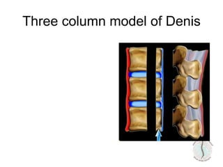
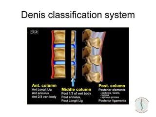
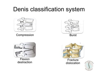
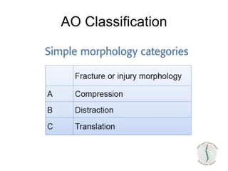
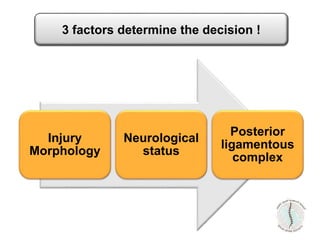
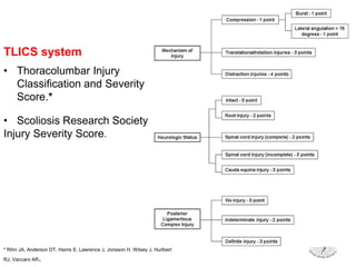
Thoraco-lumbar Fractures
Sami Al Eissa, MD
Consultant Orthopedic & Spine Surgery
Epidemiology
• United State
– 150000 to 160000 vertebral column fracture/ year.
• 10000 -12000 spine cord injury
– 15000 major thoraco-lumbar fracture/ year
• 4700 – 5000 significant neurological deficit
• Saudi Arabia
– No national statistic exist
– In one trauma center in Riyadh;
• Over 100 patients admitted yearly with major T/L fracture
from MVA only
Biomechanics
T2 – T9
• Shielded by;
– Paraspinal musculature
– Sternum & Rigid thoracic rib stiffness
– Coronal alignment of facet joint
» Resist flexion / Extension
» Minimal resistance to torsion
– Physiological kyphosis
compression/ flexion injuries
Biomechanics
• T11 – L1; Transition zone between
Kyphotic immobile segment &
Lordotic mobile segment
–Predispose to injury by rotational and shearing
forces
• Rib are not present
• Facet have not re-oriented completely
60% of TL fractures occur at this junction
Biomechanics
• Upper thoracic spine:
Center of gravity is anterior to the
spine. Axial loading will result
in compressive forces
anteriorly, tensile forces
posteriorly. This will result in
flexion-type of injuries.
• lumbar spine:
Center of gravity is posteriorly.
Flexion type of injuries will
straigthen the lumbar spine
and result in axial loading. In
this area we will see many
burst fractures.
Biomechanics
Three column model of Denis
Thoracolumbar fractures
• 75% to 90% of spinal fractures occur in the thoracic and
lumbar spine
• Most of these occurring at thoracolumbar junction (T10-
L2).
• Little consensus regarding injury classification and
management.
• Treatment varies widely, from bracing to
circumferential fusion, based on geographical,
institutional, and surgeon preferences rather than on
scientific evidence.
General guidelines
• Stability
• Neurological compromise
• Deformity
How can we decide?
How
can not
decide?
Surgery
Brace or
no brace
Anterior approach
Posterior approach
Conservative
Bed rest
How many levels?
General guidelines
Spine structure Neurologic Treatment
stable Normal Non surgical
stable Complete Non surgical
stable Incomplete Decompression &
stabilization
Unstable complete stabilization
Unstable incomplete Decompression &
stabilization
Adopted by Capen DA, Spine, 2003
Classification
• Many systems are convoluted, with an
impractical number of variables. Others
are too simple, lacking sufficient detail to
provide clinically relevant information.
lack of a widely accepted classification
system
Data Supporting the Common Classification Schemes
• Several classification systems
• Most commonly used are
– Denis classification system
– Load sharing classification described by
McComack
– AO classification system
Classification
Denis classification system
Compression
Fracture
dislocation
Burst
Flexion
destraction
• Not sufficiently detailed to account for all
fracture types
• Does not provide prognostic information
for the neurological status of
does not adequately aid surgical decision
making.
Denis classification system
AO classification
• Simple Morphology
• Neurological Injury
• Modefiers
AO Classification
AO Classification
AO Classification
AO Classification
AO Classification
AO classification
Algorithm for AO fracture type
classification
Thoracolumbar Injury Classification and
Severity Score
• Introduced by the Spine Trauma Study Group in 2005
• The TLICS is the first system to incorporate the
neurologic status of the patient
Injury
Morphology
Neurological
status
Posterior
ligamentous
complex
3 factors determine the decision !
TLICS system
• Thoracolumbar Injury
Classification and Severity
Score.*
• Scoliosis Research Society
Injury Severity Score.
* Rihn JA, Anderson DT, Harris E, Lawrence J, Jonsson H, Wilsey J, Hurlbert
RJ, Vaccaro AR.
Injury Morphology
• Compression injuries:
Loss of height of the vertebral
body or disruption through the
vertebral end plate. This
includes;
– Traditional compression (ie,
anterior column)
– Burst (ie, anterior column,
middle column)
Injury Morphology
• Rotation/translation injury
horizontal displacement of one
thoracolumbar vertebral body
with respect to another.
– Unilateral/ bilateral dislocations,
facet fracture-dislocations, as
well as bilateral pedicle or pars
fractures with vertebral
subluxation.
• Distraction injury; anatomic
dissociation in the vertical axis,
such as a hyperextension injury
– Disruption of the anterior longitudinal
ligament, with subsequent widening of
the anterior disk space.
– Fractures of the posterior elements (ie,
facet, lamina, spinous process) may
also be present in distraction injury.
– Severe kyphotic deformities caused by
tensile failure of the posterior
ligamentous structures,
Injury Morphology
Mechanism-Point
System
Compression
1 point
Distraction
4 points
Translation
Rotation
3 points
Injury Morphology
Neurologic status
• Described in increasing order of urgency:
neurologically
– Intact
– Nerve root injury
– Complete (motor and sensory) spinal cord or cauda equina
injury
– Incomplete (motor or sensory) spinal cord or cauda equina
injury.
Neurology point
system
cord
complete
2 points
incomplete
3 points
Cauda
equina
3 points
Nerve root
1 point
intact
0 point
Neurological status
Posterior Ligamentous
Complex Integrity
• Anatomic structures of the PLC include the supraspinous
ligament, interspinous ligament, ligamentum flavum, and
facet joint capsules.
• plays a critical role in protecting the spine and spinal
cord against excessive flexion, rotation, translation, and
distraction.
Once disrupted, the ligamentous structures
demonstrate poor healing ability
• Categorized
– Intact, Indeterminate, and Disrupted.
• Assessment based on
– Clinical exam
– Plain radiographs
– CT scans
– magnetic resonance
Widening of the interspinous space or of the facet
joints, empty facet joints, facet perch or
subluxation, Dislocation of the spine
Posterior Ligamentous
Complex Integrity
Posterior
longitudinal
ligament
Not Intact
3 points
intact
0 point
Stability-Soft Tissue Point System
Next Step - Direct TX
Assign Points
Conservative Surgery
• Fractures with 4 points or less = non
operative.
• Fractures with 5 points or more =
surgery
Treatment
Compression ( mechanism) - 1
Intact (neurology) - 0
PLC (ligament) no injury - 0
Anterior Compression Fx
Total 1 points Non Op
Example
Compression (mechanism) : 1+ 1
Intact ( neurology) - 0
PLC (ligament) no injury :0
Stable Burst Fracture
Total 2 points Non Op
Example
Compression + burst (mechanism): 1 + 1
Complete (neurology) : 2
PLC (ligament) injury : 3
Unstable Burst-Complete Neuro
Injury
Total 7 points Surgery
Example
Translation/rotation - distraction
(mechanism): 3
Complete (neurology): 2
PLC (ligament) injury: 3
Fracture Dislocation
Total 8 points Surgery
Example
18 yr-old
MVA
Normal
neurological exam
63 yr-old
Fall from hight
Normal
neurological exam
Limitation of TLICS system
• Not for pediatric population
• For acute injuries
• cannot be applied to;
– Symptomatic epidural hematoma
– Spinal cord injury without radiographic abnormalities
– posttraumatic deformity
– Iatrogenic spinal instability
– Pathologic fractures associated with tumor or infection.
• TLICS is a reliable system for assessing fractures of the
thoracic and lumbar spine when used by experts.
• the posterior ligamentous complex subcomponent score
was the least reliable component.
Timing of surgery
• Remain unclear.
• Lack of class one evidence, no standard guidelines.
Timing of surgery
• Preclinical studies suggest that early surgical
decompression of the spinal cord is important in
mitigating secondary injury.
• The completeness of SCI injury seems to be the key
prognostic factor
• To date …. there is no robust evidence to suggest that
early surgical intervention in tSCI is superior.
• Surgical decompression performed before 24 h post
injury has the potential to result in superior motor
recovery in comparison with late surgery performed at or
after 24 h post injury
General guidelines
Spine structure Neurologic Treatment
stable Normal Non surgical
stable Complete Non surgical
stable Incomplete Decompression &
stabilization
Unstable complete stabilization
Unstable incomplete Decompression &
stabilization
Adopted by Capen DA, Spine, 2003
Conclusion
•Have a clear understanding to nature of the fracture
and it’s consequences.
Stability, deformity, and neurological picture will
remain the main factors determining the surgical
decision

Thoracolumbar-spine-fracture-.ppt

  • 1.
    Thoraco-lumbar Fractures Sami AlEissa, MD Consultant Orthopedic & Spine Surgery
  • 2.
    Epidemiology • United State –150000 to 160000 vertebral column fracture/ year. • 10000 -12000 spine cord injury – 15000 major thoraco-lumbar fracture/ year • 4700 – 5000 significant neurological deficit • Saudi Arabia – No national statistic exist – In one trauma center in Riyadh; • Over 100 patients admitted yearly with major T/L fracture from MVA only
  • 3.
  • 4.
    T2 – T9 •Shielded by; – Paraspinal musculature – Sternum & Rigid thoracic rib stiffness – Coronal alignment of facet joint » Resist flexion / Extension » Minimal resistance to torsion – Physiological kyphosis compression/ flexion injuries Biomechanics
  • 5.
    • T11 –L1; Transition zone between Kyphotic immobile segment & Lordotic mobile segment –Predispose to injury by rotational and shearing forces • Rib are not present • Facet have not re-oriented completely 60% of TL fractures occur at this junction Biomechanics
  • 6.
    • Upper thoracicspine: Center of gravity is anterior to the spine. Axial loading will result in compressive forces anteriorly, tensile forces posteriorly. This will result in flexion-type of injuries. • lumbar spine: Center of gravity is posteriorly. Flexion type of injuries will straigthen the lumbar spine and result in axial loading. In this area we will see many burst fractures. Biomechanics
  • 7.
  • 8.
  • 9.
    Thoracolumbar fractures • 75%to 90% of spinal fractures occur in the thoracic and lumbar spine • Most of these occurring at thoracolumbar junction (T10- L2). • Little consensus regarding injury classification and management. • Treatment varies widely, from bracing to circumferential fusion, based on geographical, institutional, and surgeon preferences rather than on scientific evidence.
  • 10.
    General guidelines • Stability •Neurological compromise • Deformity
  • 11.
    How can wedecide?
  • 12.
    How can not decide? Surgery Brace or nobrace Anterior approach Posterior approach Conservative Bed rest How many levels?
  • 13.
    General guidelines Spine structureNeurologic Treatment stable Normal Non surgical stable Complete Non surgical stable Incomplete Decompression & stabilization Unstable complete stabilization Unstable incomplete Decompression & stabilization Adopted by Capen DA, Spine, 2003
  • 14.
    Classification • Many systemsare convoluted, with an impractical number of variables. Others are too simple, lacking sufficient detail to provide clinically relevant information. lack of a widely accepted classification system
  • 15.
    Data Supporting theCommon Classification Schemes
  • 16.
    • Several classificationsystems • Most commonly used are – Denis classification system – Load sharing classification described by McComack – AO classification system Classification
  • 17.
  • 18.
  • 19.
    • Not sufficientlydetailed to account for all fracture types • Does not provide prognostic information for the neurological status of does not adequately aid surgical decision making. Denis classification system
  • 20.
    AO classification • SimpleMorphology • Neurological Injury • Modefiers
  • 21.
  • 22.
  • 23.
  • 24.
  • 25.
  • 26.
  • 27.
    Algorithm for AOfracture type classification
  • 28.
    Thoracolumbar Injury Classificationand Severity Score • Introduced by the Spine Trauma Study Group in 2005 • The TLICS is the first system to incorporate the neurologic status of the patient
  • 29.
  • 30.
    TLICS system • ThoracolumbarInjury Classification and Severity Score.* • Scoliosis Research Society Injury Severity Score. * Rihn JA, Anderson DT, Harris E, Lawrence J, Jonsson H, Wilsey J, Hurlbert RJ, Vaccaro AR.
  • 31.
    Injury Morphology • Compressioninjuries: Loss of height of the vertebral body or disruption through the vertebral end plate. This includes; – Traditional compression (ie, anterior column) – Burst (ie, anterior column, middle column)
  • 32.
    Injury Morphology • Rotation/translationinjury horizontal displacement of one thoracolumbar vertebral body with respect to another. – Unilateral/ bilateral dislocations, facet fracture-dislocations, as well as bilateral pedicle or pars fractures with vertebral subluxation.
  • 33.
    • Distraction injury;anatomic dissociation in the vertical axis, such as a hyperextension injury – Disruption of the anterior longitudinal ligament, with subsequent widening of the anterior disk space. – Fractures of the posterior elements (ie, facet, lamina, spinous process) may also be present in distraction injury. – Severe kyphotic deformities caused by tensile failure of the posterior ligamentous structures, Injury Morphology
  • 34.
  • 35.
    Neurologic status • Describedin increasing order of urgency: neurologically – Intact – Nerve root injury – Complete (motor and sensory) spinal cord or cauda equina injury – Incomplete (motor or sensory) spinal cord or cauda equina injury.
  • 36.
    Neurology point system cord complete 2 points incomplete 3points Cauda equina 3 points Nerve root 1 point intact 0 point Neurological status
  • 37.
    Posterior Ligamentous Complex Integrity •Anatomic structures of the PLC include the supraspinous ligament, interspinous ligament, ligamentum flavum, and facet joint capsules. • plays a critical role in protecting the spine and spinal cord against excessive flexion, rotation, translation, and distraction. Once disrupted, the ligamentous structures demonstrate poor healing ability
  • 38.
    • Categorized – Intact,Indeterminate, and Disrupted. • Assessment based on – Clinical exam – Plain radiographs – CT scans – magnetic resonance Widening of the interspinous space or of the facet joints, empty facet joints, facet perch or subluxation, Dislocation of the spine Posterior Ligamentous Complex Integrity
  • 39.
    Posterior longitudinal ligament Not Intact 3 points intact 0point Stability-Soft Tissue Point System
  • 40.
    Next Step -Direct TX Assign Points Conservative Surgery
  • 42.
    • Fractures with4 points or less = non operative. • Fractures with 5 points or more = surgery Treatment
  • 43.
    Compression ( mechanism)- 1 Intact (neurology) - 0 PLC (ligament) no injury - 0 Anterior Compression Fx Total 1 points Non Op Example
  • 44.
    Compression (mechanism) :1+ 1 Intact ( neurology) - 0 PLC (ligament) no injury :0 Stable Burst Fracture Total 2 points Non Op Example
  • 45.
    Compression + burst(mechanism): 1 + 1 Complete (neurology) : 2 PLC (ligament) injury : 3 Unstable Burst-Complete Neuro Injury Total 7 points Surgery Example
  • 46.
    Translation/rotation - distraction (mechanism):3 Complete (neurology): 2 PLC (ligament) injury: 3 Fracture Dislocation Total 8 points Surgery Example
  • 47.
    18 yr-old MVA Normal neurological exam 63yr-old Fall from hight Normal neurological exam
  • 49.
  • 50.
    • Not forpediatric population • For acute injuries • cannot be applied to; – Symptomatic epidural hematoma – Spinal cord injury without radiographic abnormalities – posttraumatic deformity – Iatrogenic spinal instability – Pathologic fractures associated with tumor or infection.
  • 51.
    • TLICS isa reliable system for assessing fractures of the thoracic and lumbar spine when used by experts. • the posterior ligamentous complex subcomponent score was the least reliable component.
  • 52.
    Timing of surgery •Remain unclear. • Lack of class one evidence, no standard guidelines.
  • 53.
    Timing of surgery •Preclinical studies suggest that early surgical decompression of the spinal cord is important in mitigating secondary injury. • The completeness of SCI injury seems to be the key prognostic factor • To date …. there is no robust evidence to suggest that early surgical intervention in tSCI is superior. • Surgical decompression performed before 24 h post injury has the potential to result in superior motor recovery in comparison with late surgery performed at or after 24 h post injury
  • 54.
    General guidelines Spine structureNeurologic Treatment stable Normal Non surgical stable Complete Non surgical stable Incomplete Decompression & stabilization Unstable complete stabilization Unstable incomplete Decompression & stabilization Adopted by Capen DA, Spine, 2003
  • 55.
    Conclusion •Have a clearunderstanding to nature of the fracture and it’s consequences. Stability, deformity, and neurological picture will remain the main factors determining the surgical decision