SlideShare a Scribd company logo
Trauma Radiography
Trauma CentersTrauma Centers
• Many types of facilities provide emergency
medical care, ranging from major
metropolitan medical center to small
outpatient clinics in rural areas.
• The term “Trauma Center” signifies a
specific level of emergency medical care
as defined by the American College of
Surgeons Commission on Trauma.
Trauma LevelsTrauma Levels
Level I = is the most comprehensive,
usually a university-based center,
research facility, or large medical center,
complete imaging capabilities 24 hours a
day, specialty physicians are available on
site 24 hours a day
Trauma LevelsTrauma Levels
Level II = same as level one, but not a
research facility, may not have as many
specialists
Level III = no specialists, can stabilize
patient for transport to a higher level
center, may not have 24 hour imaging
Level IV = clinics, attend minor injuries,
some stabilization before transfer
IntroductionIntroduction
Trauma is defined as a sudden,Trauma is defined as a sudden,
unexpected, dramatic, forceful, ordramatic, forceful, or violentviolentunexpected,
event
Blunt, penetrating,
event
Blunt, explosive, and thermaland thermalpenetrating, explosive,
forces are common causes of traumatictraumaticforces are common causes of
injuriesinjuries
IntroductionIntroduction
Trauma affects persons in all age rangesin all age rangesTrauma affects persons
Radiographers in the emergencyRadiographers in the emergency
department (ED) mustmust be prepared forbe prepared for aa
variety of procedures on patients in all age
department (ED)
variety of procedures on patients in all age
groupsgroups
Preliminary ConsiderationsPreliminary Considerations
Specialized trauma imaging systemsSpecialized trauma imaging systems
reduce the amount ofof time required totime required to
obtain diagnostic images
reduce the amount
obtain diagnostic images
– One type provides greaterOne type provides greater flexibility in IR/CRflexibility in IR/CR
maneuverabilitymaneuverability
– AnotherAnother type scans the entire body in a fewtype scans the entire body in a few
secondsseconds
S
T
A
T
S
C
A
N
Mobile radiography is often used for EDis often used for EDMobile radiography
proceduresprocedures
Preliminary ConsiderationsPreliminary Considerations
Mobile fluoroscopy units, orunits, or C-arms,C-arms, maymay
be used in fracture reduction or foreign
Mobile fluoroscopy
be used in fracture reduction or foreign
body localizations
Immobilization devices are a necessity
localizations
Immobilization devices are a necessity in
body
in
trauma imagingtrauma imaging
– Trauma patients often cannotTrauma patients often cannot hold thehold the
required positionrequired position
Radiographer’s Role in TraumaRadiographer’s Role in Trauma
Depends upon departmentupon department protocol andprotocol andDepends
staffing
Primary responsibilities
staffing
Primary responsibilities
– Perform qualityPerform quality diagnostic imagingdiagnostic imaging
procedures
Practice ethical radiation protection
procedures
– Practice ethical radiation protection
– Provide patient careProvide patient care
Radiographer’s Role in TraumaRadiographer’s Role in Trauma
Patient level of consciousnessPatient level of consciousness changeschanges
are common in traumaare common in trauma
“Best Practices” in Trauma“Best Practices” in Trauma
RadiographyRadiography
SpeedSpeed
– Efficiency in producing qualityEfficiency in producing quality images in theimages in the
shortest possible timeshortest possible time
AccuracyAccuracy
– Optimum image quality, minimum repeatsOptimum image quality, minimum repeats
QualityQuality
– Quality cannot be sacrificed for speedQuality cannot be sacrificed for speed
– Do not use patient condition as an excuse forDo not use patient condition as an excuse for
poor quality imagespoor quality images
“Best Practices” in Trauma“Best Practices” in Trauma
RadiographyRadiography
PositioningPositioning
– ImportantImportant notnot to aggravate patient’s conditionto aggravate patient’s condition
when obtaining imageswhen obtaining images
– Move tube and IR, instead of patient,Move tube and IR, instead of patient,
whenever possiblewhenever possible
Practice standard precautionsPractice standard precautions
– Expect to be exposed to bodyExpect to be exposed to body fluidsfluids in EDin ED
– Do not touch a patient without gloves!Do not touch a patient without gloves!
Disclaimer:
The following three slides are
Disclaimer:
The following three slides are
gruesome.gruesome.
“Best Practices” in Trauma“Best Practices” in Trauma
RadiographyRadiography
AnticipationAnticipation
– Some injuriesSome injuries require follow-up procedures;require follow-up procedures;
knowing what to do increases appreciation forappreciation forknowing what to do increases
radiographer’s role in EDrole in EDradiographer’s
“Best Practices” in Trauma“Best Practices” in Trauma
RadiographyRadiography
Attention to detailAttention to detail
– Pay carefulPay careful attention to patient’s condition,attention to patient’s condition,
which could change at anyany timetimewhich could change at
Attention to ED protocol and scope ofand scope ofAttention to ED protocol
practicepractice
– Know the protocolKnow the protocol and scope ofand scope of practice inpractice in
your facilityfacilityyour
ProfessionalismProfessionalism
– Adhere to Code ofAdhere to Code of EthicsEthics
Radiographic Procedures inRadiographic Procedures in
TraumaTrauma
Slide 28
General ProceduralGeneral Procedural
GuidelinesGuidelines
Slide 29
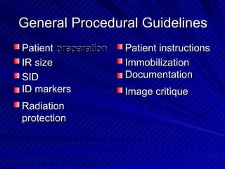
General Procedural GuidelinesGeneral Procedural Guidelines
Patient preparationpreparationPatient
IR sizeIR size
SID
ID markers
SID
ID markers
Radiation
protection
Radiation
protection
Patient instructionsPatient instructions
Immobilization
Documentation
Immobilization
Documentation
Image critiqueImage critique
Patient PreparationPatient Preparation
Use good communication skills with
appropriate touch and eye contact
Use good communication skills with
appropriate touch and eye contact
– Trauma often causesTrauma often causes anxietyanxiety
Check patient forfor potential artifactspotential artifactsCheck patient
– Explain whatExplain what you are removing and whyyou are removing and why
– Secure all personalSecure all personal effectseffects using properusing proper
procedure for your facilityprocedure for your facility
IR SizeIR Size
IR size for trauma procedures are thetrauma procedures are theIR size for
same as for routine proceduresroutine proceduressame as for
Use smallest IR thatIR that will demonstratewill demonstrateUse smallest
anatomy
Collimate field size to anatomy of
anatomy
Collimate field size to anatomy interestof interest
SIDSID
SID is standardized as a parta part ofofSID is standardized as
procedural protocolprocedural protocol
– When SID is notWhen SID is not specified underspecified under a projection,a projection,
40 to 48′′40 to 48
– 60 to 72′′
60 to 72′′
′′ SID recommended forSID recommended for projectionsprojections
with increased OIDwith increased OID
ID MarkersID Markers
Right or left side markers must beRight or left side markers must be
included on each imageincluded on each image
Other required ID markersrequired ID markers mustmust be in thebe in the
blocker or elsewhere on the final image
Other
blocker or elsewhere on the final image
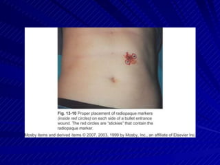
Markers used for penetrating trauma toused for penetrating trauma toMarkers
identify entrance and exit woundswoundsidentify entrance and exit
Just Kidding…..
Radiation ProtectionRadiation Protection
Shield pediatric patients and patientspatients and patients ofofShield pediatric
reproductive agereproductive age
Warn other staffstaff ofof exposure whenexposure when
performing mobile imaging
Warn other
performing mobile imaging
Other radiation protection measuresradiation protection measuresOther
– Close collimationClose collimation
– Optimum technique factorsOptimum technique factors
Patient InstructionsPatient Instructions
Explain and demonstrate positions, when
possible
Explain and demonstrate positions, when
possible
Explain respiration instructionsExplain respiration instructions forfor patientspatients
who can cooperatewho can cooperate
Use short exposure times to eliminateexposure times to eliminateUse short
possibility of imaging motionimaging motionpossibility of
ImmobilizationImmobilization
Many ED patientsED patients arrive in some sortarrive in some sort ofofMany
immobilization device
Immobilization devices are not
immobilization device
Immobilization devices are not to beto be
removed unless ordered by a physicianremoved unless ordered by a physician
Imaging procedures are often performedare often performed
without removal of the immobilization
Imaging procedures
without removal of the immobilization
Images are used to rule out injury andinjury and
show if it is safe to remove immobilization
Images are used to rule out
show if it is safe to remove immobilization
DocumentationDocumentation
Because deviation or adjustmentadjustment ofof
routine procedures is often required to
Because deviation or
routine procedures is often required to
accommodate a patient’s injury,injury,
documentation is important
accommodate a patient’s
documentation is important
Make sure that deviation from routine isdeviation from routine is
still within your scope of practice!
Make sure that
still within your scope of practice!
Document deviation (AP, X-table,deviation (AP, X-table, etc.),etc.),Document
time, portabletime, portable
Image Critique CriteriaImage Critique Criteria
Image evaluation for trauma procedures istrauma procedures is
the same as for routine procedures
Image evaluation for
the same as for routine procedures
Image quality is critical foris critical for an accuratean accurateImage quality
diagnosisdiagnosis
It is pooris poor practice to acceptpractice to accept lowerlower qualityqualityIt
images due to patient condition or difficultydifficultyimages due to patient condition or
of procedureof procedure
Trauma ProjectionsTrauma Projections
Lateral Cervical SpineCervical SpineLateral
Horizontal CR centered to midpoint ofof IRIRHorizontal CR centered to midpoint
Pre-vertebral soft tissue must betissue must bePre-vertebral soft
visualized
Image should demonstrate entire C-
visualized
Image should demonstrate entire C-
spine from sella turcica to top of T1T1spine from sella turcica to top of
– If allall seven cervicalseven cervical vertebrae are notvertebrae are not seen,seen,If
then a swimmer’s view is requiredthen a swimmer’s view is required
Lateral Cervical SpineCervical SpineLateral
Patient and IR centered for trauma lateral of C-spineand IR centered for trauma lateral of C-spinePatient
Lateral Cervical SpineCervical SpineLateral
Lateral projection of C-spine in dorsal decubitus position; dislocation of C3-
C4; C7 not demonstrated, so swimmer’s view is needed
Swimmer’s (cervicothoracic)(cervicothoracic)Swimmer’s
Required if C7 and top of T1 notC7 and top of T1 not
demonstrated on lateral C-spine
Required if
demonstrated on lateral C-spine
Trauma usually requires dorsal decubitusrequires dorsal decubitusTrauma usually
positionposition
Patient supine withoutsupine without rotation
Ask patient to raise arm opposite the x-ray
rotation
Ask patient to raise arm opposite the x-ray
Patient
tube over headheadtube over
– Assist patientAssist patient and provide supportand provide support
Cervicothoracic SpineCervicothoracic Spine
Relax shouldershoulder closercloser to x-rayto x-ray tubetube
Vertical IR centered just above jugular
Relax
Vertical IR centered just above jugular
notchnotch
Horizontal CR centered to C7-T1Horizontal CR centered to C7-T1
interspace and midcoronal planeinterspace and midcoronal plane
Use breathing technique if possiblepossibleUse breathing technique if
– BlurBlur ribs and lung markings to betterribs and lung markings to better
demonstrate spinedemonstrate spine
Cervicothoracic SpineCervicothoracic Spine
Image demonstrates lower cervical andcervical and
upper thoracic vertebrae in profile
Image demonstrates lower
upper thoracic vertebrae in profile
between the shouldersbetween the shoulders
Cervicothoracic SpineCervicothoracic Spine
Patient and IR positioned for trauma lateral projection of
cervicothoracic vertebrae using dorsal decubitus position
Cervicothoracic SpineCervicothoracic Spine
Lateral projection, dorsal decubitus position of cervicothoracic
vertebrae
AP Axial CervicalCervical SpineSpineAP Axial
Patient is supineis supinePatient
– UsuallyUsually immobilized with collarimmobilized with collar and spineand spine
boardboard
Place IR under spine board,spine board, ifif present,present,
centered to C4 (Adam’s apple)
Place IR under
centered to C4 (Adam’s apple)
Head and shoulders without rotationwithout rotationHead and shoulders
– Ask patientAsk patient to lookto look straightstraight aheadahead
AP Axial CervicalCervical SpineSpineAP Axial
CR directed 15 to 20 degrees cephalad tocephalad toCR directed 15 to 20 degrees
enter MSP atMSP at C4
Image demonstrates C3-T1 or T2,
C4
Image demonstrates C3-T1 or T2,
enter
including all soft tissuestissuesincluding all soft
– IfIf backboard is present,backboard is present, unavoidable artifactsunavoidable artifacts
may be seenmay be seen
AP Axial CervicalCervical SpineSpineAP Axial
Patient and IR positioned for trauma AP axial C-spine
AP Axial CervicalCervical SpineSpineAP Axial
Trauma AP axial C-spine; complete dislocation at C2-C3
AP Axial Oblique Cervical SpineOblique Cervical SpineAP Axial
TRAUMA OBLIQUESTRAUMA OBLIQUES
Patient is supineis supinePatient
– UsuallyUsually immobilized with collarimmobilized with collar and spineand spine
boardboard
Place IR under spine board (notspine board (not bucky),bucky), ififPlace IR under
present, centered to C4 and adjacent
mastoid process
present, centered to C4 and adjacent
mastoid process
– AboutAbout 3′′3′′ laterallateral to MSPto MSP
AP Axial Oblique Cervical SpineOblique Cervical SpineAP Axial
Head and shoulders without rotationwithout rotationHead and shoulders
– Ask patientAsk patient to lookto look straightstraight aheadahead
CR has double angleCR has double angle
– 45 degrees45 degrees lateromediallylateromedially
– 15 to 20 degrees15 to 20 degrees cephaliccephalic
CR enters lateral to MSP at level of C4lateral to MSP at level of C4CR enters
AP Axial Oblique Cervical SpineOblique Cervical SpineAP Axial
CR exit should be in centershould be in center of IR
Image demonstrates side opposite CR
of IR
Image demonstrates side opposite CR
CR exit
– C1-T1 orC1-T1 or T2 bodies and disk spacesT2 bodies and disk spaces
– IntervertebralIntervertebral foramina openforamina open
– If backboard is present,If backboard is present, unavoidable artifactsunavoidable artifacts
may be seenmay be seen
AP Axial Oblique Cervical SpineOblique Cervical SpineAP Axial
Patient and IR positioned for trauma AP axial oblique C-spine
AP Axial Oblique Cervical SpineOblique Cervical SpineAP Axial
Trauma AP axial oblique C-spine
Thoracic and Lumbar SpineSpineThoracic and Lumbar
X-table laterals performed firstX-table laterals performed first
Vertical grid and IRVertical grid and IR
– Top ofTop of IR 1.5IR 1.5′′ to 2′′ to 2′′ (3.8 to 5 cm)′′ (3.8 to 5 cm) aboveabove
shoulders forfor thoracicthoracic spinespineshoulders
– Centered to level of iliacCentered to level of iliac crests forcrests for lumbarlumbar
spinespine
Have patient crosscross armsarms on anterioron anterior chestchestHave patient
Thoracic and Lumbar SpineSpineThoracic and Lumbar
CR horizontalCR horizontal
– Centered to spine and IR
Breathing technique improves
Centered to spine and IR
Breathing technique improves
visualization of thoracic vertebrae
Exposure made on suspended respiration
thoracic vertebrae
Exposure made on suspended respiration
visualization of
for lumbarlumbar vertebraevertebraefor
Thoracic and Lumbar SpineSpineThoracic and Lumbar
Thoracic image demonstrates T3 or T4 toT4 toThoracic image demonstrates T3 or
L1
Lumbar image demonstrates T12 to
L1
Lumbar image demonstrates T12 to
sacrum
Vertebral bodies and spinous processes
sacrum
Vertebral bodies and spinous inprocesses in
profileprofile
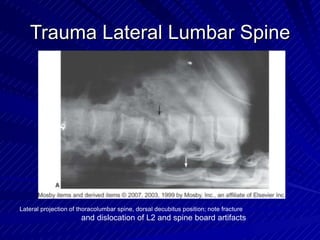
Trauma Lateral Lumbar SpineTrauma Lateral Lumbar Spine
CR and IR positioned for trauma lateral projection of lumbar spine using dorsal
decubitus position
Trauma Lateral Lumbar SpineTrauma Lateral Lumbar Spine
Lateral projection of thoracolumbar spine, dorsal decubitus position; note fracture
and dislocation of L2 and spine board artifacts
ChestChest
Supine position used if generalgeneral surveysurveySupine position used if
image of chestchest desireddesired
Check for need to demonstrate air-fluid
image of
Check for need to demonstrate air-fluid
levelslevels
– IfIf air-fluid levelsair-fluid levels are suspected,are suspected, use X-tableuse X-table
laterallateral
– IfIf patient’spatient’s condition permits, lateralcondition permits, lateral decubitusdecubitus
position with patient lying on affected side willposition with patient lying on affected side will
also show air-fluid levelsalso show air-fluid levels
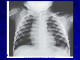
Trauma AP ChestTrauma AP Chest
Obtain help to lift patientpatient forfor IR placementIR placementObtain help to lift
– Top ofTop of IR placed aboutIR placed about 1.51.5′′ to 2′′ to 2′′ above′′ above
shouldersshoulders
Arms abducted
MCP parallel to IR
Use maximum SID to reduce heart
abducted
MCP parallel to IR
Use maximum SID to reduce heart
magnification
Arms
magnification
Trauma AP ChestTrauma AP Chest
Ensure chin extended out ofof anatomyanatomy ofofEnsure chin extended out
interest
CR directed perpendicular to center of
interest
CR directed perpendicular to center IRof IR
– look forlook for lightlight field slightlyfield slightly above shoulders andabove shoulders and
on sides of chest, CW or LWLWon sides of chest, CW or
Exposure made upon second fullExposure made upon second full
inhalation, ifif possiblepossibleinhalation,
Trauma AP ChestTrauma AP Chest
Image demonstrates lung fields in theirin theirImage demonstrates lung fields
entiretyentirety
– MinimalMinimal rotation and distortion presentrotation and distortion present
AbdomenAbdomen
If transfertransfer to x-rayto x-ray table is not possible,table is not possible,
obtain lift help for IR placement
If
obtain lift help for IR placement
IR centered to MSP at level oflevel of iliaciliac crests
Check for possibility of fluid accumulation
crests
Check for possibility of fluid accumulation
in abdominal cavity
IR centered to MSP at
in abdominal cavity
– AffectsAffects exposure factorsexposure factors
– RequiresRequires close monitoring ofclose monitoring of patientpatient forfor statusstatus
change during procedureschange during procedures
AbdomenAbdomen
Mark entrance and exit wounds,entrance and exit wounds, ifif presentpresentMark
Align shoulders and hips in same planeAlign shoulders and hips in same plane
MCP parallel to tableMCP parallel to table
CR perpendicular to centerto center ofof IR
Image demonstrates entire abdomen with
IR
Image demonstrates entire abdomen with
CR perpendicular
pubic symphysis visible at lower borderborderpubic symphysis visible at lower
PelvisPelvis
Pelvic fractures have a high riskfractures have a high risk ofof
hemorrhage – pay close attention to
Pelvic
hemorrhage – pay close attention to
patient for statusstatus changechangepatient for
Obtain lift help for IR placement ifIR placement if transfertransferObtain lift help for
to x-ray table is not possiblepossible
IR centered 2′′ above pubic symphysis or
to x-ray table is not
IR centered 2′′ above pubic symphysis or
2′′ below ASIS2′′ below ASIS
MCP parallel to IRMCP parallel to IR
PelvisPelvis
Lower limbslimbs internally rotated only ifinternally rotated only ifLower
possible
Ensure arms are not in anatomy
possible
Ensure arms of interestinterestare not in anatomy of
CR perpendicular to centerto center ofof IR
Exposure made on suspended respiration
IR
Exposure made on suspended respiration
CR perpendicular
Image demonstrates entire pelvis and
proximal femora
Image demonstrates entire pelvis and
proximal femora
Trauma AP PelvisTrauma AP Pelvis
Trauma AP pelvis; note fracture of left ilium and separation of pubic bones
CraniumCranium
Patients with head trauma are oftenwith head trauma are often
referred to CT first
Patients
referred to CT first
When x-rays are ordered,When x-rays are ordered, a generala general
survey requires AP and lateral projectionssurvey requires AP and lateral projections
Generally, the patientthe patient is supineis supineGenerally,
– LateralLateral projection uses dorsalprojection uses dorsal decubitusdecubitus
positionposition
Trauma Lateral CraniumTrauma Lateral Cranium
Elevate head on radiolucent supportElevate head on radiolucent support
– Ensure C-spine injury hasEnsure C-spine injury has been ruled outbeen ruled out
Trauma Lateral CraniumTrauma Lateral Cranium
Trauma lateral projection of cranium; note multiple fractures in frontal bone
Trauma AP CraniumTrauma AP Cranium
Check with physician to determineCheck with physician to determine
anatomy of interestof interestanatomy
– AP projection demonstratesAP projection demonstrates anterioranterior craniumcranium
– AP axial projection (Towne) demonstratesAP axial projection (Towne) demonstrates
posterior craniumcraniumposterior
Trauma CraniumTrauma Cranium
Patient and IR positioned for
trauma AP cranium
Patient and IR positioned for
trauma AP axial cranium
Trauma AP CraniumTrauma AP Cranium
Trauma AP cranium; note fracture line
Facial BonesBonesFacial
Patients with facialwith facial bone injuriesbone injuries are oftenare oftenPatients
referred to CT first
Anticipate profuse bleeding and use
universal precautions
referred to CT first
Anticipate profuse bleeding and use
universal precautions
Upper and Lowerand Lower LimbsLimbsUpper
Obtain lift help forhelp for IR placementIR placement
Injured limbs should be lifted with support
Obtain lift
Injured limbs should be lifted with support
at both jointsat both joints
– LiftLift only enough to place IRonly enough to place IR
Two projections at 90 degrees from eachTwo projections at 90 degrees from each
other requiredother required
– Do not attempt to rotate severely injured limbsDo not attempt to rotate severely injured limbs
for true positionsfor true positions
Upper and Lowerand Lower LimbsLimbsUpper
Long bones require demonstration ofrequire demonstration of
adjacent joints
Long bones
adjacent joints
– Take separate projections,Take separate projections, ifif necessarynecessary
Maximize patient safetysafety and comfortand comfort byby
moving IR and CR, rather than injured
Maximize patient
moving IR and CR, rather than injured
limblimb
Other Imaging Procedures inOther Imaging Procedures in
TraumaTrauma
Slide 96
Other Imaging in TraumaOther Imaging in Trauma
CT is extensively used in trauma patientsextensively used in trauma patientsCT is
– Often,Often, CT isCT is modalitymodality ofof choicechoice
Angiography may be used for vascularvascularAngiography may be used for
injuries
Contrast
injuries
studies are often ordered forContrast studies are often ordered for
evaluation of urinary systemevaluation of urinary system
– BluntBlunt abdominalabdominal trauma and suspected pelvictrauma and suspected pelvic
fractures often resultoften result in injury to urinaryin injury to urinaryfractures
systemsystem
Time for the “good stuff”!the “good stuff”!Time for
Thankyou
Nitish Virmani

More Related Content

What's hot

soft tissue radiography
soft tissue radiographysoft tissue radiography
soft tissue radiography
Dinesh Darshana
 
Dacrocystography and sialography
Dacrocystography and sialographyDacrocystography and sialography
Dacrocystography and sialography
Yashawant Yadav
 
Digital fluoro
Digital fluoroDigital fluoro
Digital fluoro
Rad Tech
 
PATIENT CARE IN RADIOLOGY DEPARTMENT.pptx
PATIENT CARE IN RADIOLOGY DEPARTMENT.pptxPATIENT CARE IN RADIOLOGY DEPARTMENT.pptx
PATIENT CARE IN RADIOLOGY DEPARTMENT.pptx
mohamedabushanab1
 
Emergency radiography
Emergency radiographyEmergency radiography
Emergency radiography
Vignesh Viki
 
Pediatric radiography
Pediatric radiographyPediatric radiography
Pediatric radiography
Julie Parsons
 
Emergency radiology
Emergency  radiologyEmergency  radiology
Emergency radiology
Anupam Niraula
 
Quality Assurance Programme in Computed Tomography
Quality Assurance Programme in Computed TomographyQuality Assurance Programme in Computed Tomography
Quality Assurance Programme in Computed Tomography
Ramzee Small
 
Reject film Analysis
Reject film AnalysisReject film Analysis
Reject film Analysis
sunil seksana
 
CT History
CT HistoryCT History
CT History
bartonect
 
Ct head protocols
Ct head protocolsCt head protocols
Ct head protocols
Maajid Mohi ud din
 
Patient care [autosaved]
Patient care [autosaved]Patient care [autosaved]
Patient care [autosaved]
Leafeanking
 
Mammography positioning technique for Lateral Views (LM/ML)
Mammography positioning technique for Lateral Views (LM/ML)Mammography positioning technique for Lateral Views (LM/ML)
Mammography positioning technique for Lateral Views (LM/ML)
Selin Prasad
 
Dacrocystography
DacrocystographyDacrocystography
Dacrocystography
Ankit Mishra
 
CR, DR and recent advances
CR, DR and recent advancesCR, DR and recent advances
CR, DR and recent advances
Vishal Sankpal
 
Computed radiography and digital radiography- CR/DR
Computed radiography and digital radiography- CR/DRComputed radiography and digital radiography- CR/DR
Computed radiography and digital radiography- CR/DR
Ashim Budhathoki
 
Emegency drugs in radiology department
Emegency drugs in radiology department Emegency drugs in radiology department
Emegency drugs in radiology department
AbubakarMustaphaAman
 
Artifacts in conventional radiology.pptx
Artifacts in conventional radiology.pptxArtifacts in conventional radiology.pptx
Artifacts in conventional radiology.pptx
VANI PUSHPA MUDAVATH
 
Arthrography of the Shoulder, Ankle and Wrist.pptx
Arthrography of the Shoulder, Ankle and Wrist.pptxArthrography of the Shoulder, Ankle and Wrist.pptx
Arthrography of the Shoulder, Ankle and Wrist.pptx
Deneicer Guy
 
Mammography technique
Mammography techniqueMammography technique
Mammography technique
vishwanath0908
 

What's hot (20)

soft tissue radiography
soft tissue radiographysoft tissue radiography
soft tissue radiography
 
Dacrocystography and sialography
Dacrocystography and sialographyDacrocystography and sialography
Dacrocystography and sialography
 
Digital fluoro
Digital fluoroDigital fluoro
Digital fluoro
 
PATIENT CARE IN RADIOLOGY DEPARTMENT.pptx
PATIENT CARE IN RADIOLOGY DEPARTMENT.pptxPATIENT CARE IN RADIOLOGY DEPARTMENT.pptx
PATIENT CARE IN RADIOLOGY DEPARTMENT.pptx
 
Emergency radiography
Emergency radiographyEmergency radiography
Emergency radiography
 
Pediatric radiography
Pediatric radiographyPediatric radiography
Pediatric radiography
 
Emergency radiology
Emergency  radiologyEmergency  radiology
Emergency radiology
 
Quality Assurance Programme in Computed Tomography
Quality Assurance Programme in Computed TomographyQuality Assurance Programme in Computed Tomography
Quality Assurance Programme in Computed Tomography
 
Reject film Analysis
Reject film AnalysisReject film Analysis
Reject film Analysis
 
CT History
CT HistoryCT History
CT History
 
Ct head protocols
Ct head protocolsCt head protocols
Ct head protocols
 
Patient care [autosaved]
Patient care [autosaved]Patient care [autosaved]
Patient care [autosaved]
 
Mammography positioning technique for Lateral Views (LM/ML)
Mammography positioning technique for Lateral Views (LM/ML)Mammography positioning technique for Lateral Views (LM/ML)
Mammography positioning technique for Lateral Views (LM/ML)
 
Dacrocystography
DacrocystographyDacrocystography
Dacrocystography
 
CR, DR and recent advances
CR, DR and recent advancesCR, DR and recent advances
CR, DR and recent advances
 
Computed radiography and digital radiography- CR/DR
Computed radiography and digital radiography- CR/DRComputed radiography and digital radiography- CR/DR
Computed radiography and digital radiography- CR/DR
 
Emegency drugs in radiology department
Emegency drugs in radiology department Emegency drugs in radiology department
Emegency drugs in radiology department
 
Artifacts in conventional radiology.pptx
Artifacts in conventional radiology.pptxArtifacts in conventional radiology.pptx
Artifacts in conventional radiology.pptx
 
Arthrography of the Shoulder, Ankle and Wrist.pptx
Arthrography of the Shoulder, Ankle and Wrist.pptxArthrography of the Shoulder, Ankle and Wrist.pptx
Arthrography of the Shoulder, Ankle and Wrist.pptx
 
Mammography technique
Mammography techniqueMammography technique
Mammography technique
 

Similar to Trauma radiograph

Emergency Radiography.pptx
Emergency Radiography.pptxEmergency Radiography.pptx
Emergency Radiography.pptx
Latifaomary
 
C.V Hany
C.V HanyC.V Hany
C.V Hany
Hany Kerbalou
 
Setting up a Neurointervention cath lab
Setting up a Neurointervention cath labSetting up a Neurointervention cath lab
Setting up a Neurointervention cath lab
NeurologyKota
 
How are computers used in medicine - Lalitmohan Gurjar
How are computers used in medicine - Lalitmohan GurjarHow are computers used in medicine - Lalitmohan Gurjar
How are computers used in medicine - Lalitmohan Gurjar
lalitmohangurjar
 
Patient safety
Patient safetyPatient safety
Patient safety
hab1978
 
Safe practices in imaging dept.
Safe practices in imaging dept.Safe practices in imaging dept.
Safe practices in imaging dept.
Tapendu Mondal
 
Introduction to operating room
Introduction to operating roomIntroduction to operating room
Introduction to operating room
Syed Ahmed Uddin (BSc, REEGT, CNIM)
 
What is the Process to become an x-ray Technician.pdf
What is the Process to become an x-ray Technician.pdfWhat is the Process to become an x-ray Technician.pdf
What is the Process to become an x-ray Technician.pdf
Mr. Business Magazine
 
AN OVERVIEW ON CYBERKNIFE
AN OVERVIEW ON CYBERKNIFEAN OVERVIEW ON CYBERKNIFE
AN OVERVIEW ON CYBERKNIFE
Subrata Roy
 
RADIATION PROTECTION .pptx
RADIATION PROTECTION .pptxRADIATION PROTECTION .pptx
RADIATION PROTECTION .pptx
DentalYoutube
 
Hospital safety committee ptlls assignment 1
Hospital safety committee ptlls assignment 1Hospital safety committee ptlls assignment 1
Hospital safety committee ptlls assignment 1
Dr. Salma Azeez
 
Role of Radiographers.pptx
Role of Radiographers.pptxRole of Radiographers.pptx
Role of Radiographers.pptx
NangSuKabyar
 
X-RAY BASICS
X-RAY BASICSX-RAY BASICS
X-RAY BASICS
AbhijeetSingh567
 
Occupational radiation safety in Radiological imaging, Dr. Roshan S Livingstone
Occupational radiation safety in Radiological imaging, Dr. Roshan S LivingstoneOccupational radiation safety in Radiological imaging, Dr. Roshan S Livingstone
Occupational radiation safety in Radiological imaging, Dr. Roshan S Livingstone
ohscmcvellore
 
Pre-Clinical-Radiation-Protection_-(002).pptx
Pre-Clinical-Radiation-Protection_-(002).pptxPre-Clinical-Radiation-Protection_-(002).pptx
Pre-Clinical-Radiation-Protection_-(002).pptx
EmmanuelOluseyi1
 
Radiograph guidelines for orthodontists by Almuzian
Radiograph guidelines for orthodontists by AlmuzianRadiograph guidelines for orthodontists by Almuzian
Radiograph guidelines for orthodontists by Almuzian
University of Sydney and Edinbugh
 
Initial assesment atls
Initial assesment  atlsInitial assesment  atls
Initial assesment atls
Saleem Al adhrai
 
Critical_Observations_for_the_Infection_Preventionist_in_the_OR_and_SPD.pptx
Critical_Observations_for_the_Infection_Preventionist_in_the_OR_and_SPD.pptxCritical_Observations_for_the_Infection_Preventionist_in_the_OR_and_SPD.pptx
Critical_Observations_for_the_Infection_Preventionist_in_the_OR_and_SPD.pptx
KhalilSemlali
 
Radiation hazard and safety measure of radiation hazard.pptx
Radiation hazard and safety measure of radiation hazard.pptxRadiation hazard and safety measure of radiation hazard.pptx
Radiation hazard and safety measure of radiation hazard.pptx
Deathhunter009
 
Radiation hazard and safety measure of radiation hazard.pptx
Radiation hazard and safety measure of radiation hazard.pptxRadiation hazard and safety measure of radiation hazard.pptx
Radiation hazard and safety measure of radiation hazard.pptx
Deathhunter009
 

Similar to Trauma radiograph (20)

Emergency Radiography.pptx
Emergency Radiography.pptxEmergency Radiography.pptx
Emergency Radiography.pptx
 
C.V Hany
C.V HanyC.V Hany
C.V Hany
 
Setting up a Neurointervention cath lab
Setting up a Neurointervention cath labSetting up a Neurointervention cath lab
Setting up a Neurointervention cath lab
 
How are computers used in medicine - Lalitmohan Gurjar
How are computers used in medicine - Lalitmohan GurjarHow are computers used in medicine - Lalitmohan Gurjar
How are computers used in medicine - Lalitmohan Gurjar
 
Patient safety
Patient safetyPatient safety
Patient safety
 
Safe practices in imaging dept.
Safe practices in imaging dept.Safe practices in imaging dept.
Safe practices in imaging dept.
 
Introduction to operating room
Introduction to operating roomIntroduction to operating room
Introduction to operating room
 
What is the Process to become an x-ray Technician.pdf
What is the Process to become an x-ray Technician.pdfWhat is the Process to become an x-ray Technician.pdf
What is the Process to become an x-ray Technician.pdf
 
AN OVERVIEW ON CYBERKNIFE
AN OVERVIEW ON CYBERKNIFEAN OVERVIEW ON CYBERKNIFE
AN OVERVIEW ON CYBERKNIFE
 
RADIATION PROTECTION .pptx
RADIATION PROTECTION .pptxRADIATION PROTECTION .pptx
RADIATION PROTECTION .pptx
 
Hospital safety committee ptlls assignment 1
Hospital safety committee ptlls assignment 1Hospital safety committee ptlls assignment 1
Hospital safety committee ptlls assignment 1
 
Role of Radiographers.pptx
Role of Radiographers.pptxRole of Radiographers.pptx
Role of Radiographers.pptx
 
X-RAY BASICS
X-RAY BASICSX-RAY BASICS
X-RAY BASICS
 
Occupational radiation safety in Radiological imaging, Dr. Roshan S Livingstone
Occupational radiation safety in Radiological imaging, Dr. Roshan S LivingstoneOccupational radiation safety in Radiological imaging, Dr. Roshan S Livingstone
Occupational radiation safety in Radiological imaging, Dr. Roshan S Livingstone
 
Pre-Clinical-Radiation-Protection_-(002).pptx
Pre-Clinical-Radiation-Protection_-(002).pptxPre-Clinical-Radiation-Protection_-(002).pptx
Pre-Clinical-Radiation-Protection_-(002).pptx
 
Radiograph guidelines for orthodontists by Almuzian
Radiograph guidelines for orthodontists by AlmuzianRadiograph guidelines for orthodontists by Almuzian
Radiograph guidelines for orthodontists by Almuzian
 
Initial assesment atls
Initial assesment  atlsInitial assesment  atls
Initial assesment atls
 
Critical_Observations_for_the_Infection_Preventionist_in_the_OR_and_SPD.pptx
Critical_Observations_for_the_Infection_Preventionist_in_the_OR_and_SPD.pptxCritical_Observations_for_the_Infection_Preventionist_in_the_OR_and_SPD.pptx
Critical_Observations_for_the_Infection_Preventionist_in_the_OR_and_SPD.pptx
 
Radiation hazard and safety measure of radiation hazard.pptx
Radiation hazard and safety measure of radiation hazard.pptxRadiation hazard and safety measure of radiation hazard.pptx
Radiation hazard and safety measure of radiation hazard.pptx
 
Radiation hazard and safety measure of radiation hazard.pptx
Radiation hazard and safety measure of radiation hazard.pptxRadiation hazard and safety measure of radiation hazard.pptx
Radiation hazard and safety measure of radiation hazard.pptx
 

More from Nitish Virmani

Role of artificial intellegence (a.i) in radiology department nitish virmani
Role of artificial intellegence (a.i) in radiology department nitish virmaniRole of artificial intellegence (a.i) in radiology department nitish virmani
Role of artificial intellegence (a.i) in radiology department nitish virmani
Nitish Virmani
 
Tomosynthesis & Dexa Scan (BMD)- Its Application & Techniques
Tomosynthesis & Dexa Scan (BMD)- Its Application & Techniques  Tomosynthesis & Dexa Scan (BMD)- Its Application & Techniques
Tomosynthesis & Dexa Scan (BMD)- Its Application & Techniques
Nitish Virmani
 
MRI spectroscopy- Its Application, Principle & Techniques
MRI spectroscopy- Its Application, Principle & Techniques MRI spectroscopy- Its Application, Principle & Techniques
MRI spectroscopy- Its Application, Principle & Techniques
Nitish Virmani
 
MRI shoulder and knee- Anatomy, Scan Planning & Its Techniques
MRI shoulder and knee- Anatomy, Scan Planning & Its Techniques  MRI shoulder and knee- Anatomy, Scan Planning & Its Techniques
MRI shoulder and knee- Anatomy, Scan Planning & Its Techniques
Nitish Virmani
 
Magnetic Resonance Cholangiopancreatography- MRCP
Magnetic Resonance Cholangiopancreatography- MRCPMagnetic Resonance Cholangiopancreatography- MRCP
Magnetic Resonance Cholangiopancreatography- MRCP
Nitish Virmani
 
MRI Contrast Agent & Its Principle
MRI Contrast Agent & Its Principle  MRI Contrast Agent & Its Principle
MRI Contrast Agent & Its Principle
Nitish Virmani
 
Radiographic Image contrast & image resolution
Radiographic Image contrast & image resolutionRadiographic Image contrast & image resolution
Radiographic Image contrast & image resolution
Nitish Virmani
 
Radiation protection
Radiation protectionRadiation protection
Radiation protection
Nitish Virmani
 
Radiation detection instrumentation fundamentals
Radiation detection instrumentation fundamentalsRadiation detection instrumentation fundamentals
Radiation detection instrumentation fundamentals
Nitish Virmani
 
Neonatal or and pediatric radiography
Neonatal or and pediatric radiographyNeonatal or and pediatric radiography
Neonatal or and pediatric radiography
Nitish Virmani
 

More from Nitish Virmani (10)

Role of artificial intellegence (a.i) in radiology department nitish virmani
Role of artificial intellegence (a.i) in radiology department nitish virmaniRole of artificial intellegence (a.i) in radiology department nitish virmani
Role of artificial intellegence (a.i) in radiology department nitish virmani
 
Tomosynthesis & Dexa Scan (BMD)- Its Application & Techniques
Tomosynthesis & Dexa Scan (BMD)- Its Application & Techniques  Tomosynthesis & Dexa Scan (BMD)- Its Application & Techniques
Tomosynthesis & Dexa Scan (BMD)- Its Application & Techniques
 
MRI spectroscopy- Its Application, Principle & Techniques
MRI spectroscopy- Its Application, Principle & Techniques MRI spectroscopy- Its Application, Principle & Techniques
MRI spectroscopy- Its Application, Principle & Techniques
 
MRI shoulder and knee- Anatomy, Scan Planning & Its Techniques
MRI shoulder and knee- Anatomy, Scan Planning & Its Techniques  MRI shoulder and knee- Anatomy, Scan Planning & Its Techniques
MRI shoulder and knee- Anatomy, Scan Planning & Its Techniques
 
Magnetic Resonance Cholangiopancreatography- MRCP
Magnetic Resonance Cholangiopancreatography- MRCPMagnetic Resonance Cholangiopancreatography- MRCP
Magnetic Resonance Cholangiopancreatography- MRCP
 
MRI Contrast Agent & Its Principle
MRI Contrast Agent & Its Principle  MRI Contrast Agent & Its Principle
MRI Contrast Agent & Its Principle
 
Radiographic Image contrast & image resolution
Radiographic Image contrast & image resolutionRadiographic Image contrast & image resolution
Radiographic Image contrast & image resolution
 
Radiation protection
Radiation protectionRadiation protection
Radiation protection
 
Radiation detection instrumentation fundamentals
Radiation detection instrumentation fundamentalsRadiation detection instrumentation fundamentals
Radiation detection instrumentation fundamentals
 
Neonatal or and pediatric radiography
Neonatal or and pediatric radiographyNeonatal or and pediatric radiography
Neonatal or and pediatric radiography
 

Recently uploaded

HUMAN BRAIN.pptx.PRIYA BHOJWANI@GAMIL.COM
HUMAN BRAIN.pptx.PRIYA BHOJWANI@GAMIL.COMHUMAN BRAIN.pptx.PRIYA BHOJWANI@GAMIL.COM
HUMAN BRAIN.pptx.PRIYA BHOJWANI@GAMIL.COM
priyabhojwani1200
 
R3 Stem Cell Therapy: A New Hope for Women with Ovarian Failure
R3 Stem Cell Therapy: A New Hope for Women with Ovarian FailureR3 Stem Cell Therapy: A New Hope for Women with Ovarian Failure
R3 Stem Cell Therapy: A New Hope for Women with Ovarian Failure
R3 Stem Cell
 
DELIRIUM BY DR JAGMOHAN PRAJAPATI.......
DELIRIUM BY DR JAGMOHAN PRAJAPATI.......DELIRIUM BY DR JAGMOHAN PRAJAPATI.......
DELIRIUM BY DR JAGMOHAN PRAJAPATI.......
DR Jag Mohan Prajapati
 
Innovative Minds France's Most Impactful Healthcare Leaders.pdf
Innovative Minds France's Most Impactful Healthcare Leaders.pdfInnovative Minds France's Most Impactful Healthcare Leaders.pdf
Innovative Minds France's Most Impactful Healthcare Leaders.pdf
eurohealthleaders
 
PET CT beginners Guide covers some of the underrepresented topics in PET CT
PET CT  beginners Guide  covers some of the underrepresented topics  in PET CTPET CT  beginners Guide  covers some of the underrepresented topics  in PET CT
PET CT beginners Guide covers some of the underrepresented topics in PET CT
MiadAlsulami
 
PrudentRx: A Resource for Patient Education and Engagement
PrudentRx: A Resource for Patient Education and EngagementPrudentRx: A Resource for Patient Education and Engagement
PrudentRx: A Resource for Patient Education and Engagement
PrudentRx Program
 
2024 HIPAA Compliance Training Guide to the Compliance Officers
2024 HIPAA Compliance Training Guide to the Compliance Officers2024 HIPAA Compliance Training Guide to the Compliance Officers
2024 HIPAA Compliance Training Guide to the Compliance Officers
Conference Panel
 
LGBTQ+ Adults: Unique Opportunities and Inclusive Approaches to Care
LGBTQ+ Adults: Unique Opportunities and Inclusive Approaches to CareLGBTQ+ Adults: Unique Opportunities and Inclusive Approaches to Care
LGBTQ+ Adults: Unique Opportunities and Inclusive Approaches to Care
VITASAuthor
 
MBC Support Group for Black Women – Insights in Genetic Testing.pdf
MBC Support Group for Black Women – Insights in Genetic Testing.pdfMBC Support Group for Black Women – Insights in Genetic Testing.pdf
MBC Support Group for Black Women – Insights in Genetic Testing.pdf
bkling
 
Vicarious movements or trick movements_AB.pdf
Vicarious movements or trick movements_AB.pdfVicarious movements or trick movements_AB.pdf
Vicarious movements or trick movements_AB.pdf
Arunima620542
 
NEEDLE STICK INJURY - JOURNAL CLUB PRESENTATION - DR SHAMIN EABENSON
NEEDLE STICK INJURY - JOURNAL CLUB PRESENTATION - DR SHAMIN EABENSONNEEDLE STICK INJURY - JOURNAL CLUB PRESENTATION - DR SHAMIN EABENSON
NEEDLE STICK INJURY - JOURNAL CLUB PRESENTATION - DR SHAMIN EABENSON
SHAMIN EABENSON
 
DR SHAMIN EABENSON - JOURNAL CLUB - NEEDLE STICK INJURY
DR SHAMIN EABENSON - JOURNAL CLUB - NEEDLE STICK INJURYDR SHAMIN EABENSON - JOURNAL CLUB - NEEDLE STICK INJURY
DR SHAMIN EABENSON - JOURNAL CLUB - NEEDLE STICK INJURY
SHAMIN EABENSON
 
CCSN_June_06 2024_jones. Cancer Rehabpptx
CCSN_June_06 2024_jones. Cancer RehabpptxCCSN_June_06 2024_jones. Cancer Rehabpptx
CCSN_June_06 2024_jones. Cancer Rehabpptx
Canadian Cancer Survivor Network
 
Top massage center in ajman chandrima Spa
Top massage center in ajman chandrima  SpaTop massage center in ajman chandrima  Spa
Top massage center in ajman chandrima Spa
Chandrima Spa Ajman
 
Champions of Health Spotlight On Leaders Shaping Germany's Healthcare.pdf
Champions of Health Spotlight On Leaders Shaping Germany's Healthcare.pdfChampions of Health Spotlight On Leaders Shaping Germany's Healthcare.pdf
Champions of Health Spotlight On Leaders Shaping Germany's Healthcare.pdf
eurohealthleaders
 
CANSA support - Caring for Cancer Patients' Caregivers
CANSA support - Caring for Cancer Patients' CaregiversCANSA support - Caring for Cancer Patients' Caregivers
CANSA support - Caring for Cancer Patients' Caregivers
CANSA The Cancer Association of South Africa
 
Exploring the Benefits of Binaural Hearing: Why Two Hearing Aids Are Better T...
Exploring the Benefits of Binaural Hearing: Why Two Hearing Aids Are Better T...Exploring the Benefits of Binaural Hearing: Why Two Hearing Aids Are Better T...
Exploring the Benefits of Binaural Hearing: Why Two Hearing Aids Are Better T...
Ear Solutions (ESPL)
 
DRAFT Ventilator Rapid Reference version 2.4.pdf
DRAFT Ventilator Rapid Reference  version  2.4.pdfDRAFT Ventilator Rapid Reference  version  2.4.pdf
DRAFT Ventilator Rapid Reference version 2.4.pdf
Robert Cole
 
Tips for Pet Care in winters How to take care of pets.
Tips for Pet Care in winters How to take care of pets.Tips for Pet Care in winters How to take care of pets.
Tips for Pet Care in winters How to take care of pets.
Dinesh Chauhan
 
The Power of Superfoods and Exercise.pdf
The Power of Superfoods and Exercise.pdfThe Power of Superfoods and Exercise.pdf
The Power of Superfoods and Exercise.pdf
Dr Rachana Gujar
 

Recently uploaded (20)

HUMAN BRAIN.pptx.PRIYA BHOJWANI@GAMIL.COM
HUMAN BRAIN.pptx.PRIYA BHOJWANI@GAMIL.COMHUMAN BRAIN.pptx.PRIYA BHOJWANI@GAMIL.COM
HUMAN BRAIN.pptx.PRIYA BHOJWANI@GAMIL.COM
 
R3 Stem Cell Therapy: A New Hope for Women with Ovarian Failure
R3 Stem Cell Therapy: A New Hope for Women with Ovarian FailureR3 Stem Cell Therapy: A New Hope for Women with Ovarian Failure
R3 Stem Cell Therapy: A New Hope for Women with Ovarian Failure
 
DELIRIUM BY DR JAGMOHAN PRAJAPATI.......
DELIRIUM BY DR JAGMOHAN PRAJAPATI.......DELIRIUM BY DR JAGMOHAN PRAJAPATI.......
DELIRIUM BY DR JAGMOHAN PRAJAPATI.......
 
Innovative Minds France's Most Impactful Healthcare Leaders.pdf
Innovative Minds France's Most Impactful Healthcare Leaders.pdfInnovative Minds France's Most Impactful Healthcare Leaders.pdf
Innovative Minds France's Most Impactful Healthcare Leaders.pdf
 
PET CT beginners Guide covers some of the underrepresented topics in PET CT
PET CT  beginners Guide  covers some of the underrepresented topics  in PET CTPET CT  beginners Guide  covers some of the underrepresented topics  in PET CT
PET CT beginners Guide covers some of the underrepresented topics in PET CT
 
PrudentRx: A Resource for Patient Education and Engagement
PrudentRx: A Resource for Patient Education and EngagementPrudentRx: A Resource for Patient Education and Engagement
PrudentRx: A Resource for Patient Education and Engagement
 
2024 HIPAA Compliance Training Guide to the Compliance Officers
2024 HIPAA Compliance Training Guide to the Compliance Officers2024 HIPAA Compliance Training Guide to the Compliance Officers
2024 HIPAA Compliance Training Guide to the Compliance Officers
 
LGBTQ+ Adults: Unique Opportunities and Inclusive Approaches to Care
LGBTQ+ Adults: Unique Opportunities and Inclusive Approaches to CareLGBTQ+ Adults: Unique Opportunities and Inclusive Approaches to Care
LGBTQ+ Adults: Unique Opportunities and Inclusive Approaches to Care
 
MBC Support Group for Black Women – Insights in Genetic Testing.pdf
MBC Support Group for Black Women – Insights in Genetic Testing.pdfMBC Support Group for Black Women – Insights in Genetic Testing.pdf
MBC Support Group for Black Women – Insights in Genetic Testing.pdf
 
Vicarious movements or trick movements_AB.pdf
Vicarious movements or trick movements_AB.pdfVicarious movements or trick movements_AB.pdf
Vicarious movements or trick movements_AB.pdf
 
NEEDLE STICK INJURY - JOURNAL CLUB PRESENTATION - DR SHAMIN EABENSON
NEEDLE STICK INJURY - JOURNAL CLUB PRESENTATION - DR SHAMIN EABENSONNEEDLE STICK INJURY - JOURNAL CLUB PRESENTATION - DR SHAMIN EABENSON
NEEDLE STICK INJURY - JOURNAL CLUB PRESENTATION - DR SHAMIN EABENSON
 
DR SHAMIN EABENSON - JOURNAL CLUB - NEEDLE STICK INJURY
DR SHAMIN EABENSON - JOURNAL CLUB - NEEDLE STICK INJURYDR SHAMIN EABENSON - JOURNAL CLUB - NEEDLE STICK INJURY
DR SHAMIN EABENSON - JOURNAL CLUB - NEEDLE STICK INJURY
 
CCSN_June_06 2024_jones. Cancer Rehabpptx
CCSN_June_06 2024_jones. Cancer RehabpptxCCSN_June_06 2024_jones. Cancer Rehabpptx
CCSN_June_06 2024_jones. Cancer Rehabpptx
 
Top massage center in ajman chandrima Spa
Top massage center in ajman chandrima  SpaTop massage center in ajman chandrima  Spa
Top massage center in ajman chandrima Spa
 
Champions of Health Spotlight On Leaders Shaping Germany's Healthcare.pdf
Champions of Health Spotlight On Leaders Shaping Germany's Healthcare.pdfChampions of Health Spotlight On Leaders Shaping Germany's Healthcare.pdf
Champions of Health Spotlight On Leaders Shaping Germany's Healthcare.pdf
 
CANSA support - Caring for Cancer Patients' Caregivers
CANSA support - Caring for Cancer Patients' CaregiversCANSA support - Caring for Cancer Patients' Caregivers
CANSA support - Caring for Cancer Patients' Caregivers
 
Exploring the Benefits of Binaural Hearing: Why Two Hearing Aids Are Better T...
Exploring the Benefits of Binaural Hearing: Why Two Hearing Aids Are Better T...Exploring the Benefits of Binaural Hearing: Why Two Hearing Aids Are Better T...
Exploring the Benefits of Binaural Hearing: Why Two Hearing Aids Are Better T...
 
DRAFT Ventilator Rapid Reference version 2.4.pdf
DRAFT Ventilator Rapid Reference  version  2.4.pdfDRAFT Ventilator Rapid Reference  version  2.4.pdf
DRAFT Ventilator Rapid Reference version 2.4.pdf
 
Tips for Pet Care in winters How to take care of pets.
Tips for Pet Care in winters How to take care of pets.Tips for Pet Care in winters How to take care of pets.
Tips for Pet Care in winters How to take care of pets.
 
The Power of Superfoods and Exercise.pdf
The Power of Superfoods and Exercise.pdfThe Power of Superfoods and Exercise.pdf
The Power of Superfoods and Exercise.pdf
 

Trauma radiograph

  • 2. Trauma CentersTrauma Centers • Many types of facilities provide emergency medical care, ranging from major metropolitan medical center to small outpatient clinics in rural areas. • The term “Trauma Center” signifies a specific level of emergency medical care as defined by the American College of Surgeons Commission on Trauma.
  • 3. Trauma LevelsTrauma Levels Level I = is the most comprehensive, usually a university-based center, research facility, or large medical center, complete imaging capabilities 24 hours a day, specialty physicians are available on site 24 hours a day
  • 4. Trauma LevelsTrauma Levels Level II = same as level one, but not a research facility, may not have as many specialists Level III = no specialists, can stabilize patient for transport to a higher level center, may not have 24 hour imaging Level IV = clinics, attend minor injuries, some stabilization before transfer
  • 5. IntroductionIntroduction Trauma is defined as a sudden,Trauma is defined as a sudden, unexpected, dramatic, forceful, ordramatic, forceful, or violentviolentunexpected, event Blunt, penetrating, event Blunt, explosive, and thermaland thermalpenetrating, explosive, forces are common causes of traumatictraumaticforces are common causes of injuriesinjuries
  • 6. IntroductionIntroduction Trauma affects persons in all age rangesin all age rangesTrauma affects persons Radiographers in the emergencyRadiographers in the emergency department (ED) mustmust be prepared forbe prepared for aa variety of procedures on patients in all age department (ED) variety of procedures on patients in all age groupsgroups
  • 7. Preliminary ConsiderationsPreliminary Considerations Specialized trauma imaging systemsSpecialized trauma imaging systems reduce the amount ofof time required totime required to obtain diagnostic images reduce the amount obtain diagnostic images – One type provides greaterOne type provides greater flexibility in IR/CRflexibility in IR/CR maneuverabilitymaneuverability – AnotherAnother type scans the entire body in a fewtype scans the entire body in a few secondsseconds
  • 8.
  • 10.
  • 11.
  • 12. Mobile radiography is often used for EDis often used for EDMobile radiography proceduresprocedures
  • 13. Preliminary ConsiderationsPreliminary Considerations Mobile fluoroscopy units, orunits, or C-arms,C-arms, maymay be used in fracture reduction or foreign Mobile fluoroscopy be used in fracture reduction or foreign body localizations Immobilization devices are a necessity localizations Immobilization devices are a necessity in body in trauma imagingtrauma imaging – Trauma patients often cannotTrauma patients often cannot hold thehold the required positionrequired position
  • 14. Radiographer’s Role in TraumaRadiographer’s Role in Trauma Depends upon departmentupon department protocol andprotocol andDepends staffing Primary responsibilities staffing Primary responsibilities – Perform qualityPerform quality diagnostic imagingdiagnostic imaging procedures Practice ethical radiation protection procedures – Practice ethical radiation protection – Provide patient careProvide patient care
  • 15. Radiographer’s Role in TraumaRadiographer’s Role in Trauma Patient level of consciousnessPatient level of consciousness changeschanges are common in traumaare common in trauma
  • 16. “Best Practices” in Trauma“Best Practices” in Trauma RadiographyRadiography SpeedSpeed – Efficiency in producing qualityEfficiency in producing quality images in theimages in the shortest possible timeshortest possible time AccuracyAccuracy – Optimum image quality, minimum repeatsOptimum image quality, minimum repeats QualityQuality – Quality cannot be sacrificed for speedQuality cannot be sacrificed for speed – Do not use patient condition as an excuse forDo not use patient condition as an excuse for poor quality imagespoor quality images
  • 17. “Best Practices” in Trauma“Best Practices” in Trauma RadiographyRadiography PositioningPositioning – ImportantImportant notnot to aggravate patient’s conditionto aggravate patient’s condition when obtaining imageswhen obtaining images – Move tube and IR, instead of patient,Move tube and IR, instead of patient, whenever possiblewhenever possible Practice standard precautionsPractice standard precautions – Expect to be exposed to bodyExpect to be exposed to body fluidsfluids in EDin ED – Do not touch a patient without gloves!Do not touch a patient without gloves!
  • 18. Disclaimer: The following three slides are Disclaimer: The following three slides are gruesome.gruesome.
  • 19. “Best Practices” in Trauma“Best Practices” in Trauma RadiographyRadiography AnticipationAnticipation – Some injuriesSome injuries require follow-up procedures;require follow-up procedures; knowing what to do increases appreciation forappreciation forknowing what to do increases radiographer’s role in EDrole in EDradiographer’s
  • 20. “Best Practices” in Trauma“Best Practices” in Trauma RadiographyRadiography Attention to detailAttention to detail – Pay carefulPay careful attention to patient’s condition,attention to patient’s condition, which could change at anyany timetimewhich could change at Attention to ED protocol and scope ofand scope ofAttention to ED protocol practicepractice – Know the protocolKnow the protocol and scope ofand scope of practice inpractice in your facilityfacilityyour ProfessionalismProfessionalism – Adhere to Code ofAdhere to Code of EthicsEthics
  • 21. Radiographic Procedures inRadiographic Procedures in TraumaTrauma Slide 28
  • 23. General Procedural GuidelinesGeneral Procedural Guidelines Patient preparationpreparationPatient IR sizeIR size SID ID markers SID ID markers Radiation protection Radiation protection Patient instructionsPatient instructions Immobilization Documentation Immobilization Documentation Image critiqueImage critique
  • 24. Patient PreparationPatient Preparation Use good communication skills with appropriate touch and eye contact Use good communication skills with appropriate touch and eye contact – Trauma often causesTrauma often causes anxietyanxiety Check patient forfor potential artifactspotential artifactsCheck patient – Explain whatExplain what you are removing and whyyou are removing and why – Secure all personalSecure all personal effectseffects using properusing proper procedure for your facilityprocedure for your facility
  • 25. IR SizeIR Size IR size for trauma procedures are thetrauma procedures are theIR size for same as for routine proceduresroutine proceduressame as for Use smallest IR thatIR that will demonstratewill demonstrateUse smallest anatomy Collimate field size to anatomy of anatomy Collimate field size to anatomy interestof interest
  • 26. SIDSID SID is standardized as a parta part ofofSID is standardized as procedural protocolprocedural protocol – When SID is notWhen SID is not specified underspecified under a projection,a projection, 40 to 48′′40 to 48 – 60 to 72′′ 60 to 72′′ ′′ SID recommended forSID recommended for projectionsprojections with increased OIDwith increased OID
  • 27. ID MarkersID Markers Right or left side markers must beRight or left side markers must be included on each imageincluded on each image Other required ID markersrequired ID markers mustmust be in thebe in the blocker or elsewhere on the final image Other blocker or elsewhere on the final image Markers used for penetrating trauma toused for penetrating trauma toMarkers identify entrance and exit woundswoundsidentify entrance and exit
  • 28.
  • 30. Radiation ProtectionRadiation Protection Shield pediatric patients and patientspatients and patients ofofShield pediatric reproductive agereproductive age Warn other staffstaff ofof exposure whenexposure when performing mobile imaging Warn other performing mobile imaging Other radiation protection measuresradiation protection measuresOther – Close collimationClose collimation – Optimum technique factorsOptimum technique factors
  • 31. Patient InstructionsPatient Instructions Explain and demonstrate positions, when possible Explain and demonstrate positions, when possible Explain respiration instructionsExplain respiration instructions forfor patientspatients who can cooperatewho can cooperate Use short exposure times to eliminateexposure times to eliminateUse short possibility of imaging motionimaging motionpossibility of
  • 32. ImmobilizationImmobilization Many ED patientsED patients arrive in some sortarrive in some sort ofofMany immobilization device Immobilization devices are not immobilization device Immobilization devices are not to beto be removed unless ordered by a physicianremoved unless ordered by a physician Imaging procedures are often performedare often performed without removal of the immobilization Imaging procedures without removal of the immobilization Images are used to rule out injury andinjury and show if it is safe to remove immobilization Images are used to rule out show if it is safe to remove immobilization
  • 33.
  • 34.
  • 35. DocumentationDocumentation Because deviation or adjustmentadjustment ofof routine procedures is often required to Because deviation or routine procedures is often required to accommodate a patient’s injury,injury, documentation is important accommodate a patient’s documentation is important Make sure that deviation from routine isdeviation from routine is still within your scope of practice! Make sure that still within your scope of practice! Document deviation (AP, X-table,deviation (AP, X-table, etc.),etc.),Document time, portabletime, portable
  • 36. Image Critique CriteriaImage Critique Criteria Image evaluation for trauma procedures istrauma procedures is the same as for routine procedures Image evaluation for the same as for routine procedures Image quality is critical foris critical for an accuratean accurateImage quality diagnosisdiagnosis It is pooris poor practice to acceptpractice to accept lowerlower qualityqualityIt images due to patient condition or difficultydifficultyimages due to patient condition or of procedureof procedure
  • 38. Lateral Cervical SpineCervical SpineLateral Horizontal CR centered to midpoint ofof IRIRHorizontal CR centered to midpoint Pre-vertebral soft tissue must betissue must bePre-vertebral soft visualized Image should demonstrate entire C- visualized Image should demonstrate entire C- spine from sella turcica to top of T1T1spine from sella turcica to top of – If allall seven cervicalseven cervical vertebrae are notvertebrae are not seen,seen,If then a swimmer’s view is requiredthen a swimmer’s view is required
  • 39. Lateral Cervical SpineCervical SpineLateral Patient and IR centered for trauma lateral of C-spineand IR centered for trauma lateral of C-spinePatient
  • 40. Lateral Cervical SpineCervical SpineLateral Lateral projection of C-spine in dorsal decubitus position; dislocation of C3- C4; C7 not demonstrated, so swimmer’s view is needed
  • 41. Swimmer’s (cervicothoracic)(cervicothoracic)Swimmer’s Required if C7 and top of T1 notC7 and top of T1 not demonstrated on lateral C-spine Required if demonstrated on lateral C-spine Trauma usually requires dorsal decubitusrequires dorsal decubitusTrauma usually positionposition Patient supine withoutsupine without rotation Ask patient to raise arm opposite the x-ray rotation Ask patient to raise arm opposite the x-ray Patient tube over headheadtube over – Assist patientAssist patient and provide supportand provide support
  • 42. Cervicothoracic SpineCervicothoracic Spine Relax shouldershoulder closercloser to x-rayto x-ray tubetube Vertical IR centered just above jugular Relax Vertical IR centered just above jugular notchnotch Horizontal CR centered to C7-T1Horizontal CR centered to C7-T1 interspace and midcoronal planeinterspace and midcoronal plane Use breathing technique if possiblepossibleUse breathing technique if – BlurBlur ribs and lung markings to betterribs and lung markings to better demonstrate spinedemonstrate spine
  • 43. Cervicothoracic SpineCervicothoracic Spine Image demonstrates lower cervical andcervical and upper thoracic vertebrae in profile Image demonstrates lower upper thoracic vertebrae in profile between the shouldersbetween the shoulders
  • 44. Cervicothoracic SpineCervicothoracic Spine Patient and IR positioned for trauma lateral projection of cervicothoracic vertebrae using dorsal decubitus position
  • 45. Cervicothoracic SpineCervicothoracic Spine Lateral projection, dorsal decubitus position of cervicothoracic vertebrae
  • 46. AP Axial CervicalCervical SpineSpineAP Axial Patient is supineis supinePatient – UsuallyUsually immobilized with collarimmobilized with collar and spineand spine boardboard Place IR under spine board,spine board, ifif present,present, centered to C4 (Adam’s apple) Place IR under centered to C4 (Adam’s apple) Head and shoulders without rotationwithout rotationHead and shoulders – Ask patientAsk patient to lookto look straightstraight aheadahead
  • 47. AP Axial CervicalCervical SpineSpineAP Axial CR directed 15 to 20 degrees cephalad tocephalad toCR directed 15 to 20 degrees enter MSP atMSP at C4 Image demonstrates C3-T1 or T2, C4 Image demonstrates C3-T1 or T2, enter including all soft tissuestissuesincluding all soft – IfIf backboard is present,backboard is present, unavoidable artifactsunavoidable artifacts may be seenmay be seen
  • 48. AP Axial CervicalCervical SpineSpineAP Axial Patient and IR positioned for trauma AP axial C-spine
  • 49. AP Axial CervicalCervical SpineSpineAP Axial Trauma AP axial C-spine; complete dislocation at C2-C3
  • 50. AP Axial Oblique Cervical SpineOblique Cervical SpineAP Axial TRAUMA OBLIQUESTRAUMA OBLIQUES Patient is supineis supinePatient – UsuallyUsually immobilized with collarimmobilized with collar and spineand spine boardboard Place IR under spine board (notspine board (not bucky),bucky), ififPlace IR under present, centered to C4 and adjacent mastoid process present, centered to C4 and adjacent mastoid process – AboutAbout 3′′3′′ laterallateral to MSPto MSP
  • 51. AP Axial Oblique Cervical SpineOblique Cervical SpineAP Axial Head and shoulders without rotationwithout rotationHead and shoulders – Ask patientAsk patient to lookto look straightstraight aheadahead CR has double angleCR has double angle – 45 degrees45 degrees lateromediallylateromedially – 15 to 20 degrees15 to 20 degrees cephaliccephalic CR enters lateral to MSP at level of C4lateral to MSP at level of C4CR enters
  • 52. AP Axial Oblique Cervical SpineOblique Cervical SpineAP Axial CR exit should be in centershould be in center of IR Image demonstrates side opposite CR of IR Image demonstrates side opposite CR CR exit – C1-T1 orC1-T1 or T2 bodies and disk spacesT2 bodies and disk spaces – IntervertebralIntervertebral foramina openforamina open – If backboard is present,If backboard is present, unavoidable artifactsunavoidable artifacts may be seenmay be seen
  • 53. AP Axial Oblique Cervical SpineOblique Cervical SpineAP Axial Patient and IR positioned for trauma AP axial oblique C-spine
  • 54. AP Axial Oblique Cervical SpineOblique Cervical SpineAP Axial Trauma AP axial oblique C-spine
  • 55. Thoracic and Lumbar SpineSpineThoracic and Lumbar X-table laterals performed firstX-table laterals performed first Vertical grid and IRVertical grid and IR – Top ofTop of IR 1.5IR 1.5′′ to 2′′ to 2′′ (3.8 to 5 cm)′′ (3.8 to 5 cm) aboveabove shoulders forfor thoracicthoracic spinespineshoulders – Centered to level of iliacCentered to level of iliac crests forcrests for lumbarlumbar spinespine Have patient crosscross armsarms on anterioron anterior chestchestHave patient
  • 56. Thoracic and Lumbar SpineSpineThoracic and Lumbar CR horizontalCR horizontal – Centered to spine and IR Breathing technique improves Centered to spine and IR Breathing technique improves visualization of thoracic vertebrae Exposure made on suspended respiration thoracic vertebrae Exposure made on suspended respiration visualization of for lumbarlumbar vertebraevertebraefor
  • 57. Thoracic and Lumbar SpineSpineThoracic and Lumbar Thoracic image demonstrates T3 or T4 toT4 toThoracic image demonstrates T3 or L1 Lumbar image demonstrates T12 to L1 Lumbar image demonstrates T12 to sacrum Vertebral bodies and spinous processes sacrum Vertebral bodies and spinous inprocesses in profileprofile
  • 58. Trauma Lateral Lumbar SpineTrauma Lateral Lumbar Spine CR and IR positioned for trauma lateral projection of lumbar spine using dorsal decubitus position
  • 59. Trauma Lateral Lumbar SpineTrauma Lateral Lumbar Spine Lateral projection of thoracolumbar spine, dorsal decubitus position; note fracture and dislocation of L2 and spine board artifacts
  • 60. ChestChest Supine position used if generalgeneral surveysurveySupine position used if image of chestchest desireddesired Check for need to demonstrate air-fluid image of Check for need to demonstrate air-fluid levelslevels – IfIf air-fluid levelsair-fluid levels are suspected,are suspected, use X-tableuse X-table laterallateral – IfIf patient’spatient’s condition permits, lateralcondition permits, lateral decubitusdecubitus position with patient lying on affected side willposition with patient lying on affected side will also show air-fluid levelsalso show air-fluid levels
  • 61. Trauma AP ChestTrauma AP Chest Obtain help to lift patientpatient forfor IR placementIR placementObtain help to lift – Top ofTop of IR placed aboutIR placed about 1.51.5′′ to 2′′ to 2′′ above′′ above shouldersshoulders Arms abducted MCP parallel to IR Use maximum SID to reduce heart abducted MCP parallel to IR Use maximum SID to reduce heart magnification Arms magnification
  • 62. Trauma AP ChestTrauma AP Chest Ensure chin extended out ofof anatomyanatomy ofofEnsure chin extended out interest CR directed perpendicular to center of interest CR directed perpendicular to center IRof IR – look forlook for lightlight field slightlyfield slightly above shoulders andabove shoulders and on sides of chest, CW or LWLWon sides of chest, CW or Exposure made upon second fullExposure made upon second full inhalation, ifif possiblepossibleinhalation,
  • 63.
  • 64. Trauma AP ChestTrauma AP Chest Image demonstrates lung fields in theirin theirImage demonstrates lung fields entiretyentirety – MinimalMinimal rotation and distortion presentrotation and distortion present
  • 65.
  • 66.
  • 67. AbdomenAbdomen If transfertransfer to x-rayto x-ray table is not possible,table is not possible, obtain lift help for IR placement If obtain lift help for IR placement IR centered to MSP at level oflevel of iliaciliac crests Check for possibility of fluid accumulation crests Check for possibility of fluid accumulation in abdominal cavity IR centered to MSP at in abdominal cavity – AffectsAffects exposure factorsexposure factors – RequiresRequires close monitoring ofclose monitoring of patientpatient forfor statusstatus change during procedureschange during procedures
  • 68. AbdomenAbdomen Mark entrance and exit wounds,entrance and exit wounds, ifif presentpresentMark Align shoulders and hips in same planeAlign shoulders and hips in same plane MCP parallel to tableMCP parallel to table CR perpendicular to centerto center ofof IR Image demonstrates entire abdomen with IR Image demonstrates entire abdomen with CR perpendicular pubic symphysis visible at lower borderborderpubic symphysis visible at lower
  • 69.
  • 70. PelvisPelvis Pelvic fractures have a high riskfractures have a high risk ofof hemorrhage – pay close attention to Pelvic hemorrhage – pay close attention to patient for statusstatus changechangepatient for Obtain lift help for IR placement ifIR placement if transfertransferObtain lift help for to x-ray table is not possiblepossible IR centered 2′′ above pubic symphysis or to x-ray table is not IR centered 2′′ above pubic symphysis or 2′′ below ASIS2′′ below ASIS MCP parallel to IRMCP parallel to IR
  • 71. PelvisPelvis Lower limbslimbs internally rotated only ifinternally rotated only ifLower possible Ensure arms are not in anatomy possible Ensure arms of interestinterestare not in anatomy of CR perpendicular to centerto center ofof IR Exposure made on suspended respiration IR Exposure made on suspended respiration CR perpendicular Image demonstrates entire pelvis and proximal femora Image demonstrates entire pelvis and proximal femora
  • 72. Trauma AP PelvisTrauma AP Pelvis Trauma AP pelvis; note fracture of left ilium and separation of pubic bones
  • 73.
  • 74. CraniumCranium Patients with head trauma are oftenwith head trauma are often referred to CT first Patients referred to CT first When x-rays are ordered,When x-rays are ordered, a generala general survey requires AP and lateral projectionssurvey requires AP and lateral projections Generally, the patientthe patient is supineis supineGenerally, – LateralLateral projection uses dorsalprojection uses dorsal decubitusdecubitus positionposition
  • 75. Trauma Lateral CraniumTrauma Lateral Cranium Elevate head on radiolucent supportElevate head on radiolucent support – Ensure C-spine injury hasEnsure C-spine injury has been ruled outbeen ruled out
  • 76. Trauma Lateral CraniumTrauma Lateral Cranium Trauma lateral projection of cranium; note multiple fractures in frontal bone
  • 77. Trauma AP CraniumTrauma AP Cranium Check with physician to determineCheck with physician to determine anatomy of interestof interestanatomy – AP projection demonstratesAP projection demonstrates anterioranterior craniumcranium – AP axial projection (Towne) demonstratesAP axial projection (Towne) demonstrates posterior craniumcraniumposterior
  • 78. Trauma CraniumTrauma Cranium Patient and IR positioned for trauma AP cranium Patient and IR positioned for trauma AP axial cranium
  • 79. Trauma AP CraniumTrauma AP Cranium Trauma AP cranium; note fracture line
  • 80. Facial BonesBonesFacial Patients with facialwith facial bone injuriesbone injuries are oftenare oftenPatients referred to CT first Anticipate profuse bleeding and use universal precautions referred to CT first Anticipate profuse bleeding and use universal precautions
  • 81.
  • 82. Upper and Lowerand Lower LimbsLimbsUpper Obtain lift help forhelp for IR placementIR placement Injured limbs should be lifted with support Obtain lift Injured limbs should be lifted with support at both jointsat both joints – LiftLift only enough to place IRonly enough to place IR Two projections at 90 degrees from eachTwo projections at 90 degrees from each other requiredother required – Do not attempt to rotate severely injured limbsDo not attempt to rotate severely injured limbs for true positionsfor true positions
  • 83.
  • 84.
  • 85. Upper and Lowerand Lower LimbsLimbsUpper Long bones require demonstration ofrequire demonstration of adjacent joints Long bones adjacent joints – Take separate projections,Take separate projections, ifif necessarynecessary Maximize patient safetysafety and comfortand comfort byby moving IR and CR, rather than injured Maximize patient moving IR and CR, rather than injured limblimb
  • 86.
  • 87.
  • 88.
  • 89. Other Imaging Procedures inOther Imaging Procedures in TraumaTrauma Slide 96
  • 90. Other Imaging in TraumaOther Imaging in Trauma CT is extensively used in trauma patientsextensively used in trauma patientsCT is – Often,Often, CT isCT is modalitymodality ofof choicechoice Angiography may be used for vascularvascularAngiography may be used for injuries Contrast injuries studies are often ordered forContrast studies are often ordered for evaluation of urinary systemevaluation of urinary system – BluntBlunt abdominalabdominal trauma and suspected pelvictrauma and suspected pelvic fractures often resultoften result in injury to urinaryin injury to urinaryfractures systemsystem
  • 91. Time for the “good stuff”!the “good stuff”!Time for
  • 92.
  • 93.
  • 94.
  • 95.
  • 96.
  • 97.
  • 98.