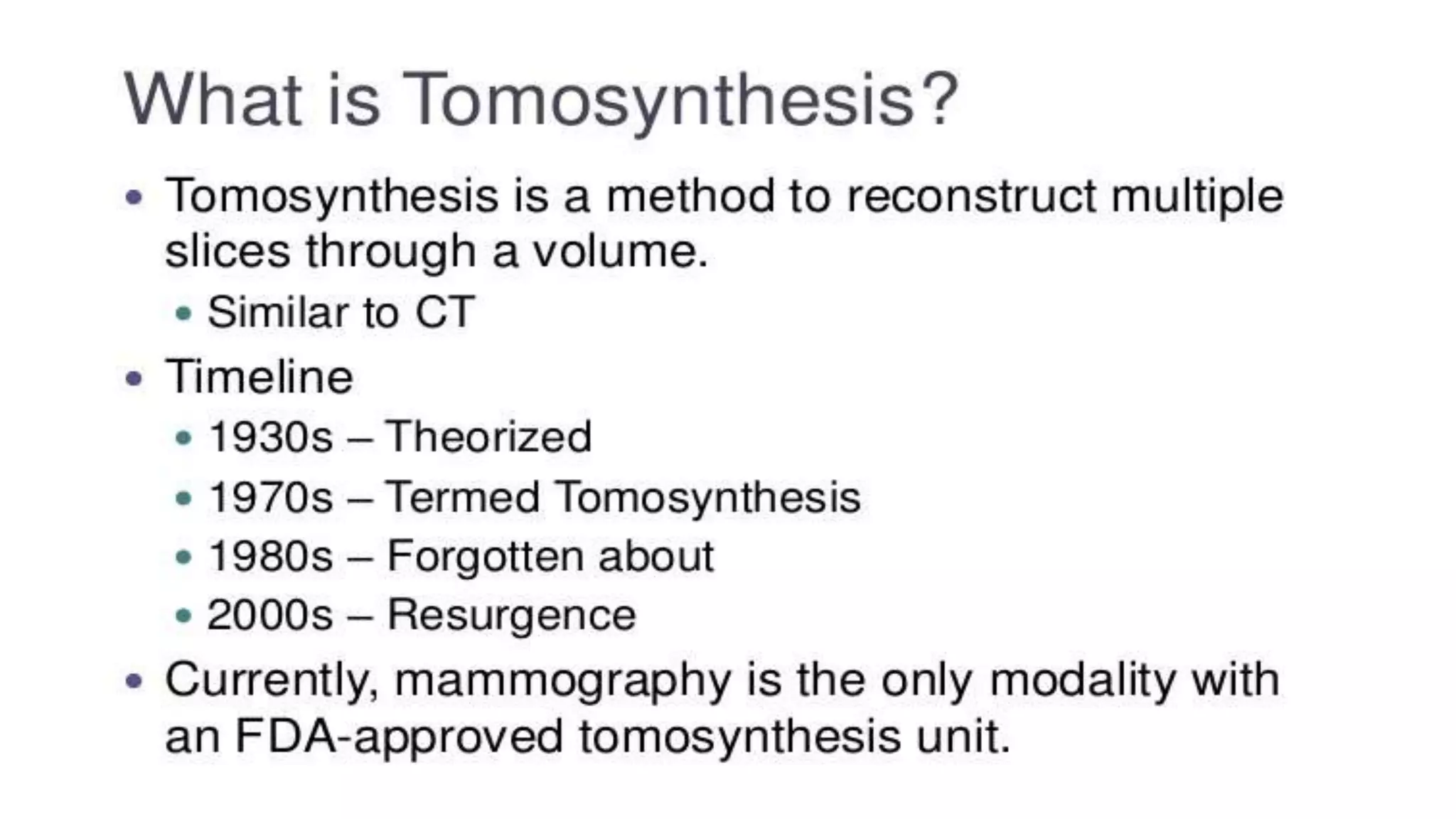
Tomosynthesis & Dexa Scan (BMD)- Its Application & Techniques
Breast tomosynthesis and DEXA bone mineral density tests provide important health information.
Breast tomosynthesis is an advanced mammography technique that uses low-dose x-rays and computer reconstruction to create 3D images of the breasts, helping detect cancers earlier. DEXA uses dual-energy x-ray beams to measure bone mineral density in the hip and spine. It identifies osteoporosis and fracture risk by comparing a patient's scores to norms for age and gender. Both tests are noninvasive, involve low radiation exposure, and provide valuable health screening and monitoring.
Introduction
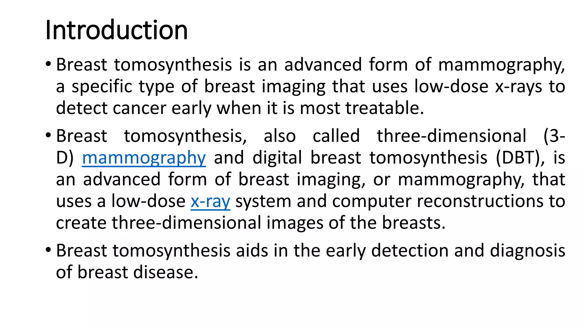
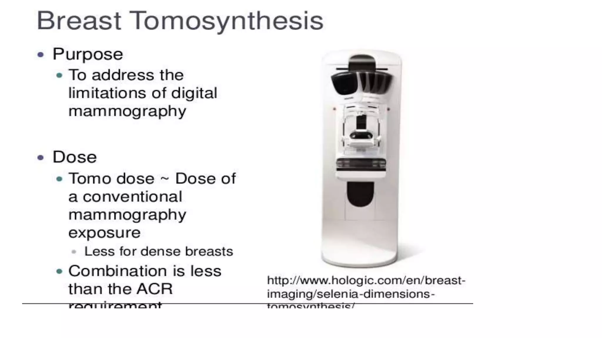
• Breast tomosynthesisis an advanced form of mammography,
a specific type of breast imaging that uses low-dose x-rays to
detect cancer early when it is most treatable.
• Breast tomosynthesis, also called three-dimensional (3-
D) mammography and digital breast tomosynthesis (DBT), is
an advanced form of breast imaging, or mammography, that
uses a low-dose x-ray system and computer reconstructions to
create three-dimensional images of the breasts.
• Breast tomosynthesis aids in the early detection and diagnosis
of breast disease.
4.
• An x-ray(radiograph) is a noninvasive medical test that helps
physicians diagnose and treat medical conditions.
• Imaging with x-rays involves exposing a part of the body to a
small dose of ionizing radiation to produce pictures of the
inside of the body.
• X-rays are the oldest and most frequently used form of
medical imaging.
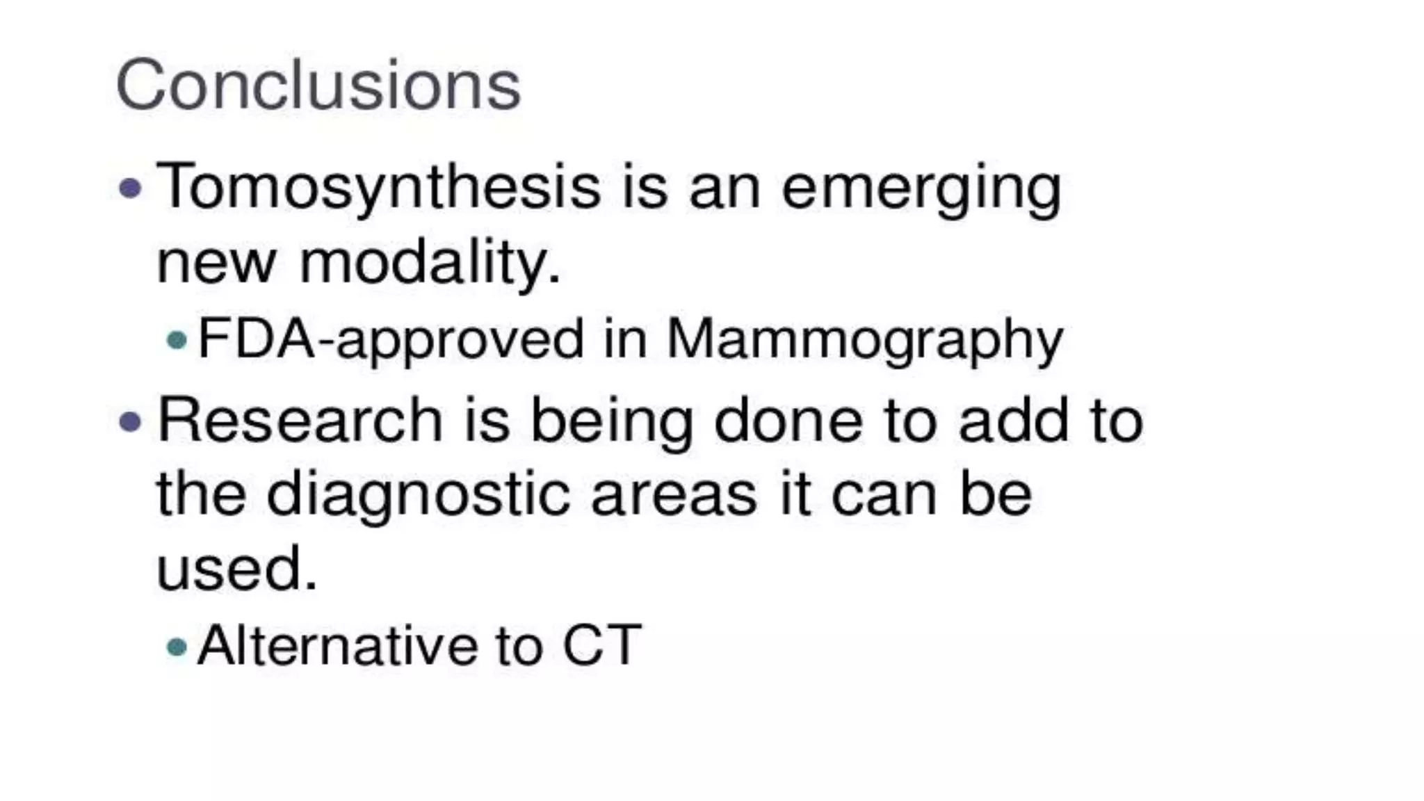
• While mammography is the best screening tool for breast
cancer available today, it does not detect all breast cancers.
Breast tomosynthesis overcomes some of the limitations of
standard mammography, but it is not yet available in all
imaging facilities.
5.
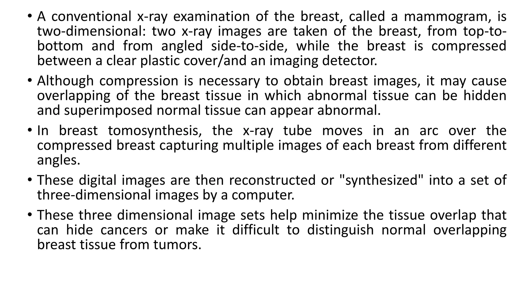
• A conventionalx-ray examination of the breast, called a mammogram, is
two-dimensional: two x-ray images are taken of the breast, from top-to-
bottom and from angled side-to-side, while the breast is compressed
between a clear plastic cover/and an imaging detector.
• Although compression is necessary to obtain breast images, it may cause
overlapping of the breast tissue in which abnormal tissue can be hidden
and superimposed normal tissue can appear abnormal.
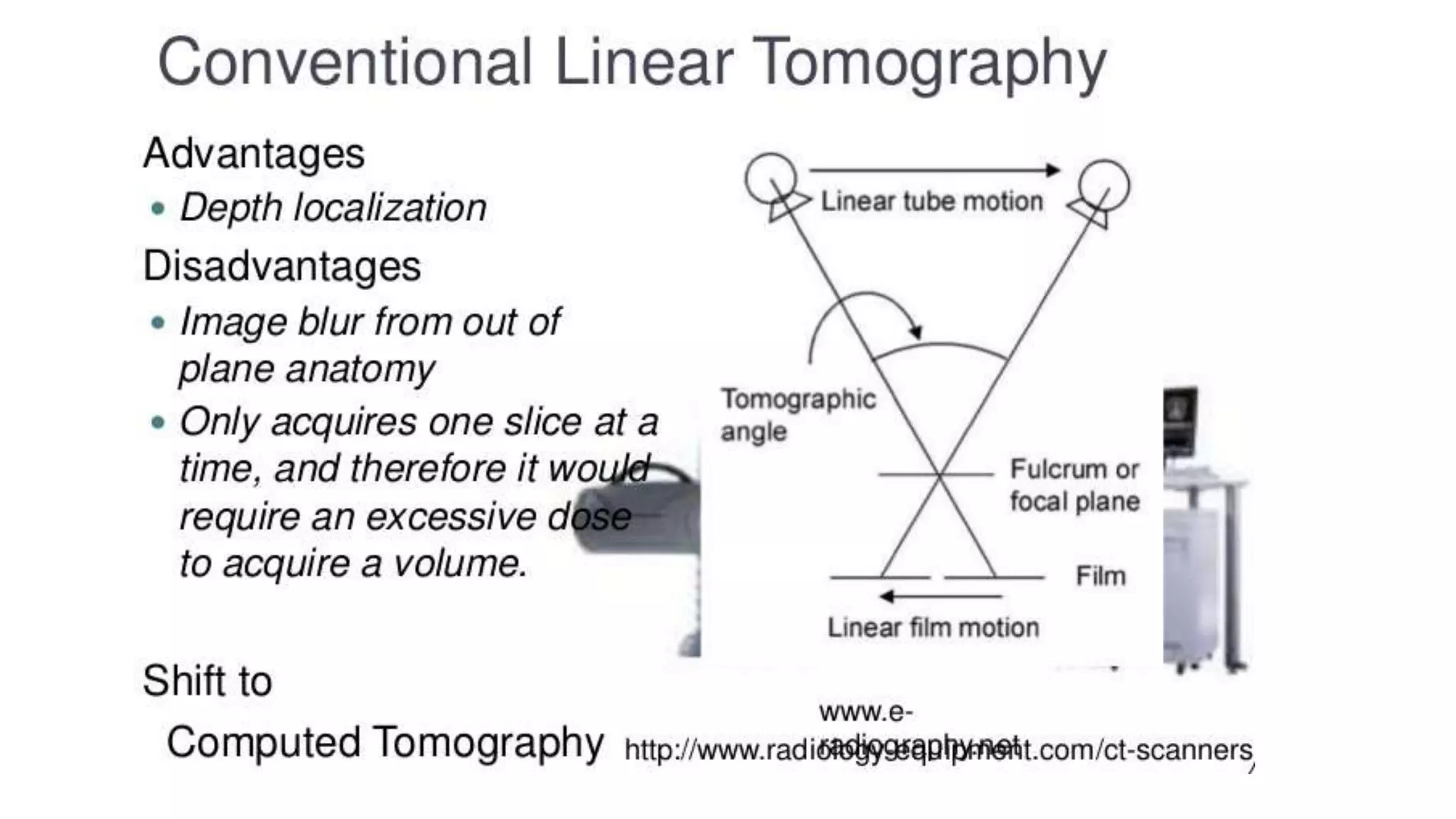
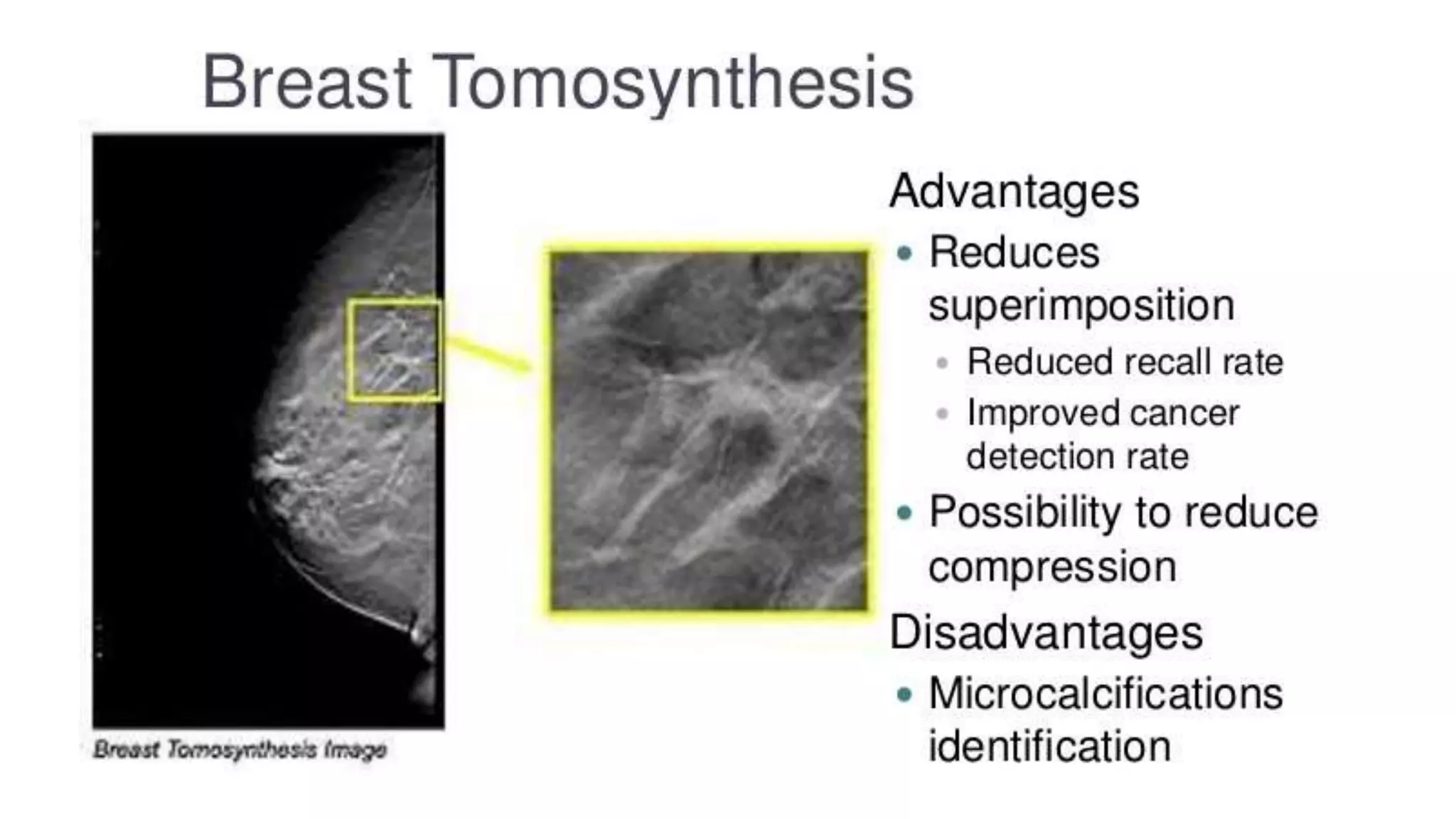
• In breast tomosynthesis, the x-ray tube moves in an arc over the
compressed breast capturing multiple images of each breast from different
angles.
• These digital images are then reconstructed or "synthesized" into a set of
three-dimensional images by a computer.
• These three dimensional image sets help minimize the tissue overlap that
can hide cancers or make it difficult to distinguish normal overlapping
breast tissue from tumors.
12.
• DBT allowsthe detection of a greater number of expansive lesions and a better
morphological analysis of masses and architectural distortions,
• the contrast of findings greater than the background, given by the more shade structures
belonging to the upper and lower layers, and then to the smaller amount of noise.
• It is thus exceeded one of the limits of two-dimensional imaging, which is the
masking of lesions caused by the superimposition of normal structures.
• The possibility of separating different layers suggests a possible reduction of false
negatives and false positives due to overlapping.
• According to early trials data, DBT is designed to offer the conspicuity of a higher
percentage of breast cancers than conventional mammography, reducing false
negative (FN) percentage at an estimated value of around 15%.
• More recent studies indicate about 30% increased DBT sensitivity and specificity
compared to FFDM (Full Field Digital Mammography) with a recalls reduction in
screening by approximately 40%.
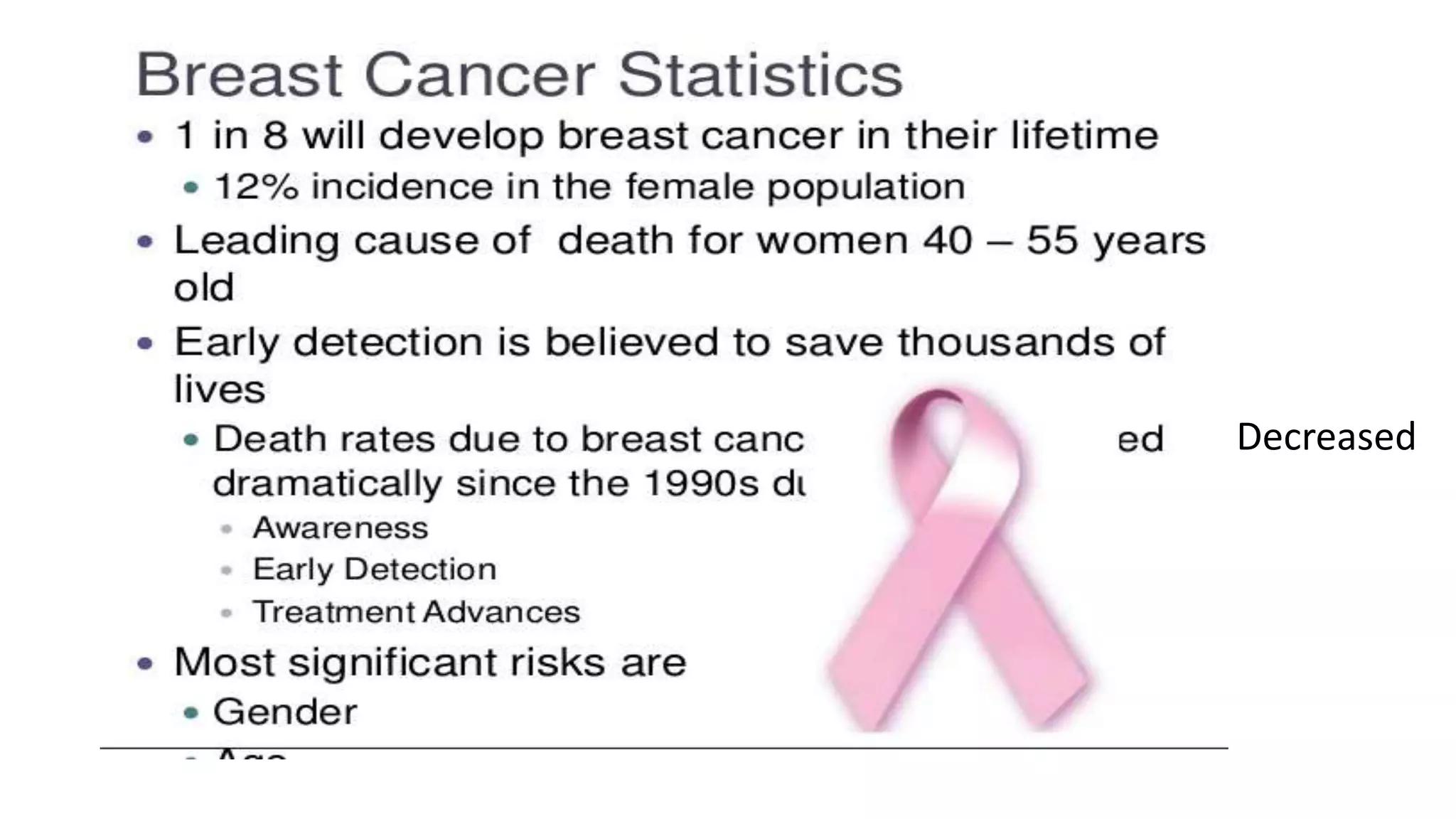
Introduction
• DEXA-DUAL ENERGYX-RAYABSORPTIOMETRY
• Most effective technique for measuring bone mineral density
(BMD)
• Osteoporosis – deficient bone matrix with normal mineralization
• Women after menopause and estrogen deficiency
• Vertebral anomalies, medications causing bone loss and thyroid
conditions
• Hip and spine
23.
Photons produced froma low dose energy source
2 X-ray beams with 2 different energy peaks are passed through the
body, one peak gets absorbed by the soft tissue and the other by the
bone
Generates a two dimensional image
Soft tissue amount is subtracted from the total area, giving the bone
mineral density.
These measurements are then compared with the normal ranges
matched for chronological age(T and Z scores)
24.
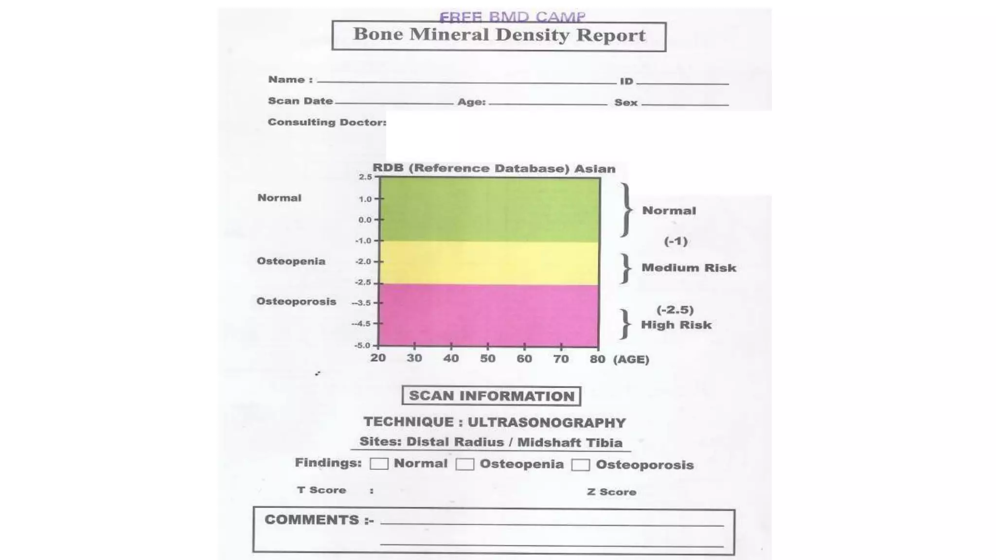
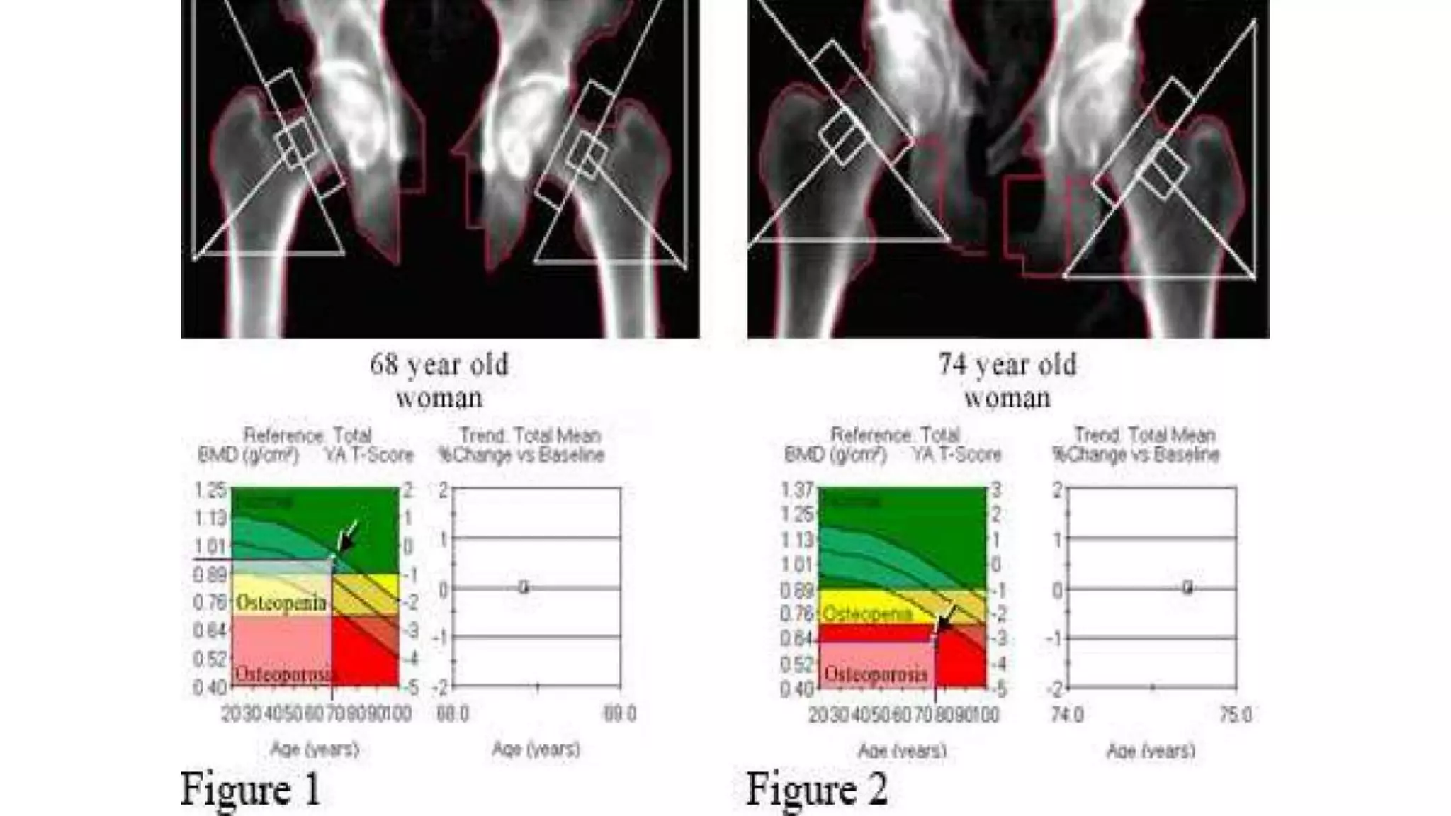
T score showsthe amount of bone that is compared with a
young adult of the same gender with peak bone mass. The T
score is used to estimate your risk of developing a fracture.
A score above -1 is considered normal
A score between -1 and -2.5 - Osteopenia(low bone mass)
A score below -2.5 is defined as osteoporosis.
Z score shows the amount of bone, compared with other
people in your age group and of the same size and gender. Z
score mainly diagnoses to the risk of having a fracture.
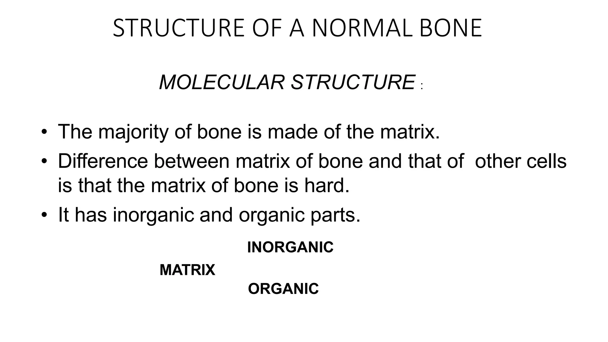
STRUCTURE OF ANORMAL BONE
MOLECULAR STRUCTURE :
• The majority of bone is made of the matrix.
• Difference between matrix of bone and that of other cells
is that the matrix of bone is hard.
• It has inorganic and organic parts.
INORGANIC
MATRIX
ORGANIC
28.
• The inorganicpart/bone mineral consists of calcium
hydroxyapatite (Ca10(PO4)6(OH)2).
• The organic part of matrix is mainly composed of Type I
collagen.
It is also composed of various growth factors
like glycosaminoglycans,osteocalcin, osteonectin,
osteopontin and Cell Attachment Factor.
29.
CELLULAR
STRUCTURE :
• Thereare several types of cells constituting the bone :
-Osteoblasts are bone-forming cells.They are the immature
bone cells which lay down the matrix as unmineralised
osteoid.They produce alkaline phosphatase which helps in
mineralisation of bone.
-Osteocytes originate from osteoblasts.Their functions incude
matrix maintenance and calcium homeostasis.They are
mature bone cells.
-Osteoclasts are the cells responsible for bone resorption, thus
they break down bone. New bone is then formed by the
osteoblasts.
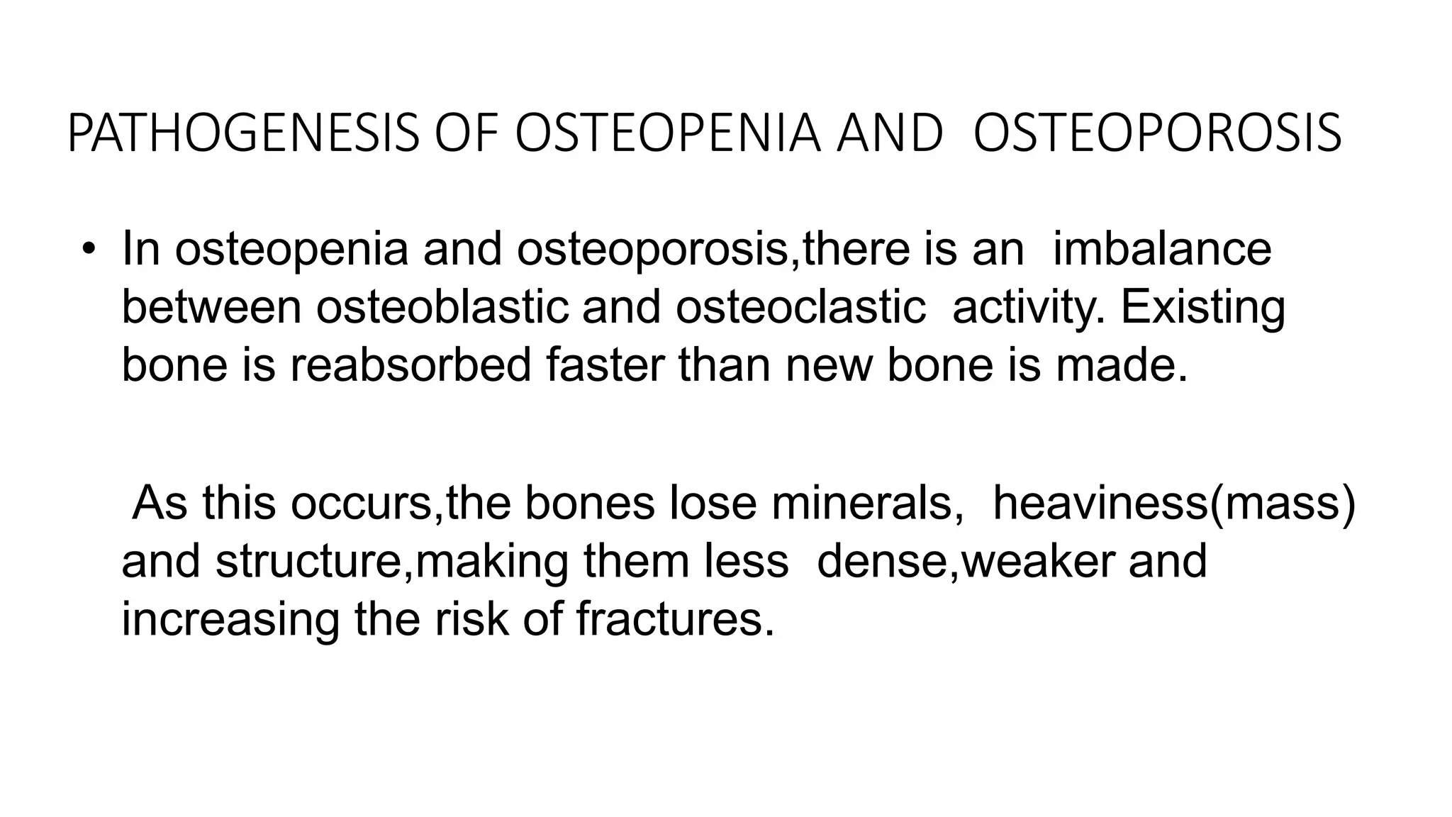
• In osteopeniaand osteoporosis,there is an imbalance
between osteoblastic and osteoclastic activity. Existing
bone is reabsorbed faster than new bone is made.
• As this occurs,the bones lose minerals, heaviness(mass)
and structure,making them less dense,weaker and
increasing the risk of fractures.
PATHOGENESIS OF OSTEOPENIA AND OSTEOPOROSIS
32.
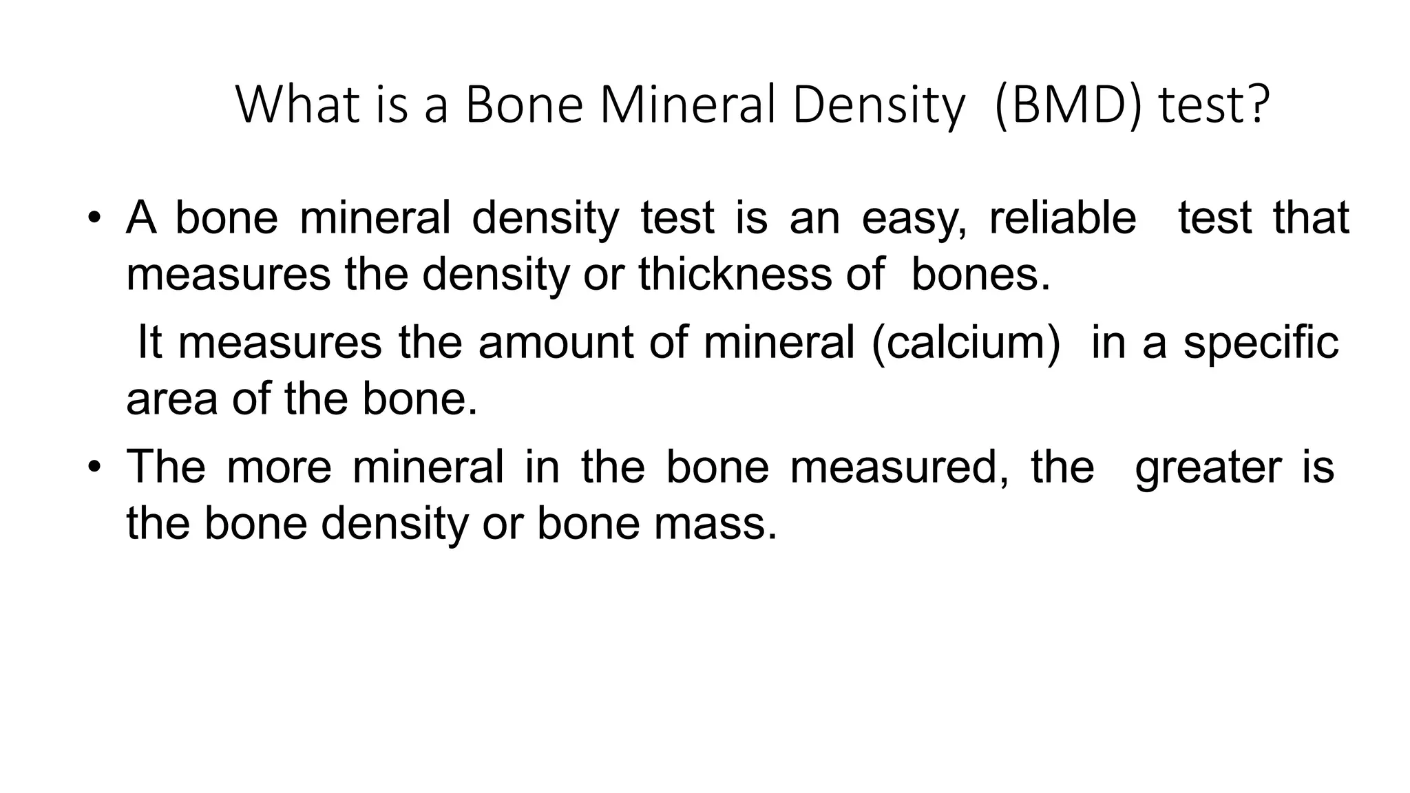
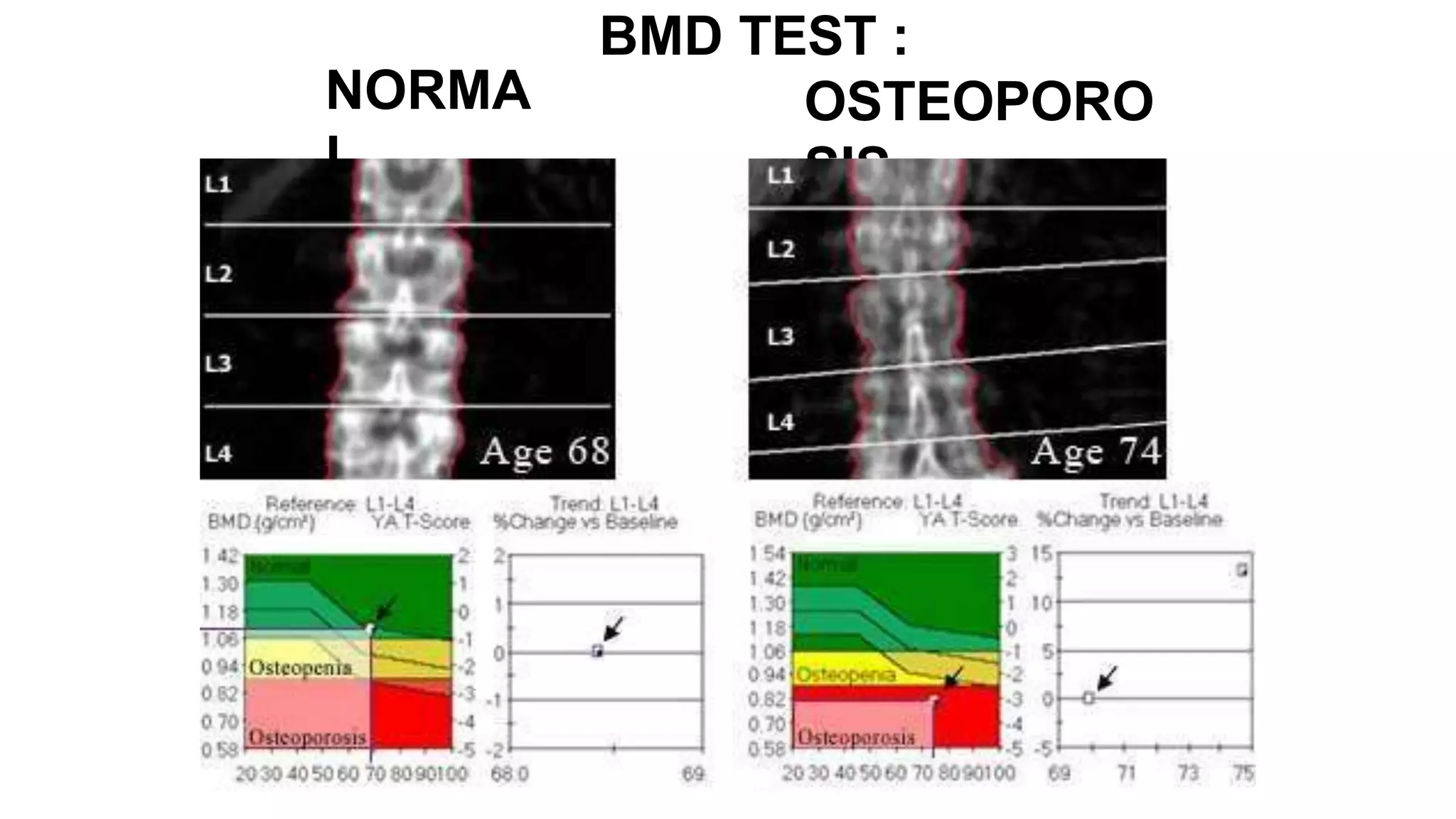
What is aBone Mineral Density (BMD) test?
• A bone mineral density test is an easy, reliable test that
measures the density or thickness of bones.
• It measures the amount of mineral (calcium) in a specific
area of the bone.
• The more mineral in the bone measured, the greater is
the bone density or bone mass.
33.
• A BMDtest can:
-Measure the density of bones
-Detect osteoporosis before a fracture occurs
-Help to predict chances of fracturing in the future
-Monitor the effectiveness of treatments for osteoporosis and
osteopenia.
•There are several different ways to measure BMD 1.Dual-energy
X-ray absorptiometry (DEXA)
2.Peripheral dual-energy X-ray absorptiometry (P-DEXA). 3.Dual
photon absorptiometry (DPA)
4.Quantitative computed tomography (QCT) 5.Quantitative
ultrasound
34.
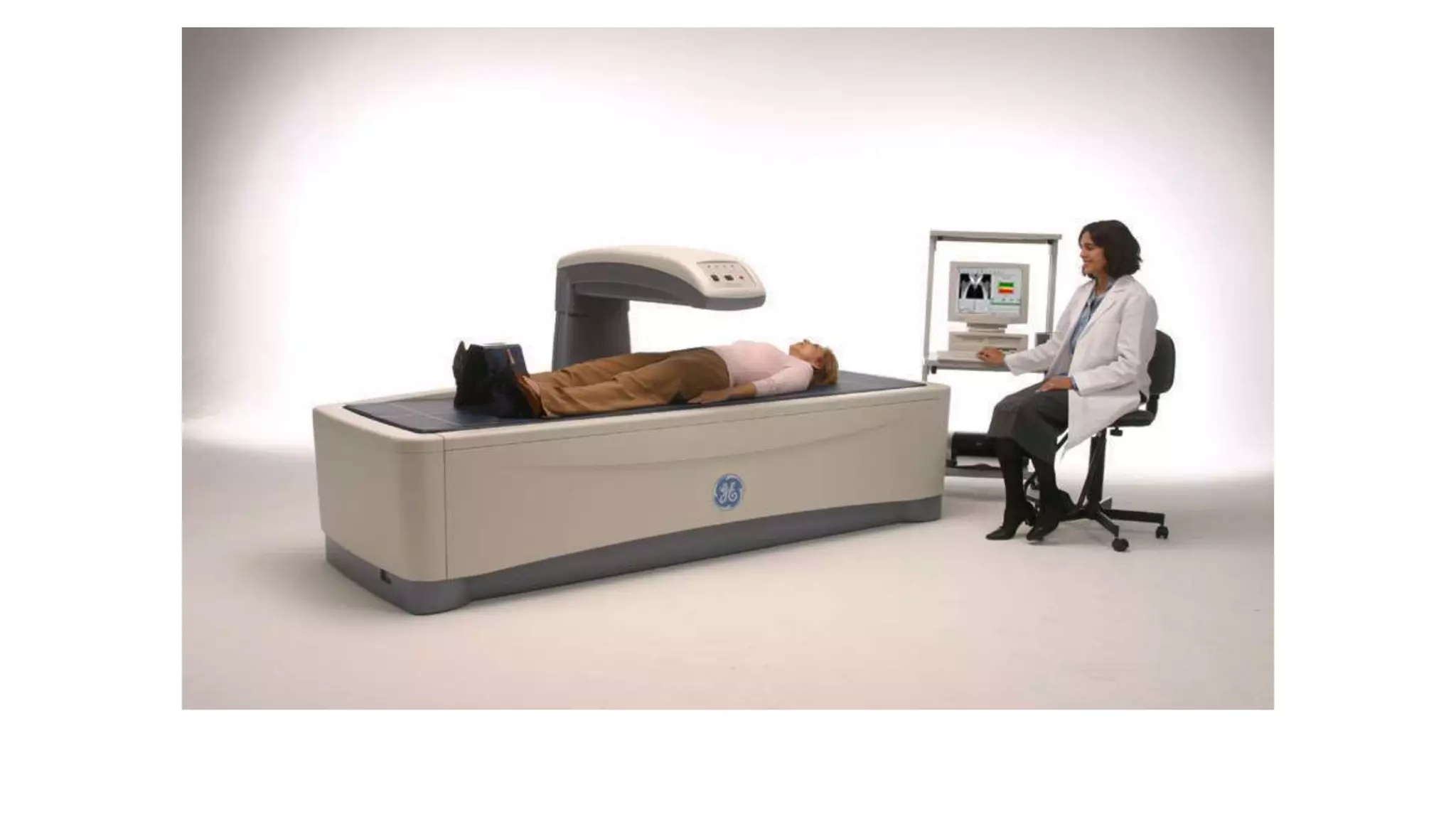
• This isthe most accurate and standardized way to
measure BMD.
• It uses two different X-ray beams to estimate bone
density in the spine and hip.
• It is a quick,easy and painless test where nothing is
injected or swallowed.
• A low-dose x-ray is taken which is is only 10% of the
radiation exposure of a chest x-ray.
Dual-energy X-ray absorptiometry
(DEXA)
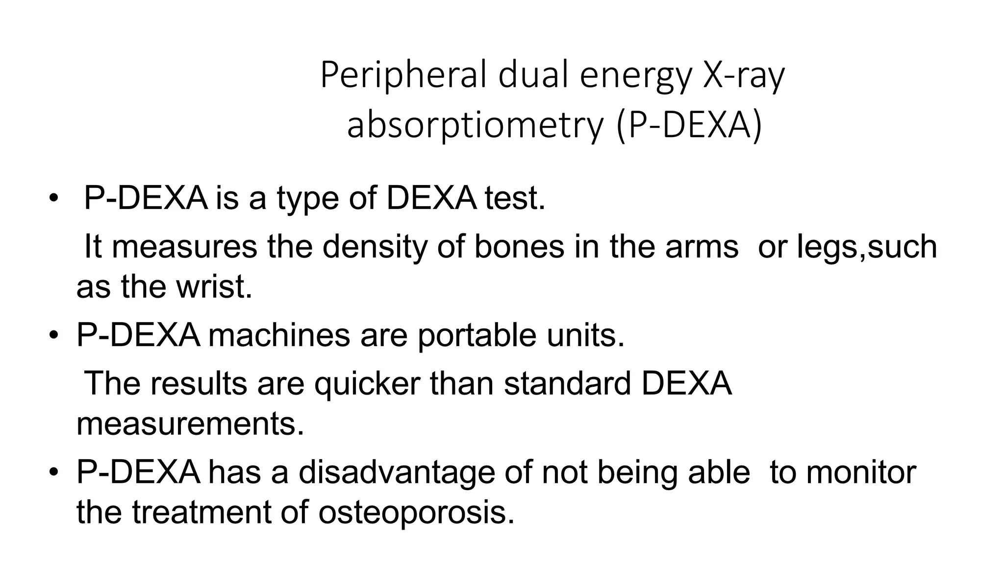
Peripheral dual energyX-ray
absorptiometry (P-DEXA)
• P-DEXA is a type of DEXA test.
• It measures the density of bones in the arms or legs,such
as the wrist.
• P-DEXA machines are portable units.
• The results are quicker than standard DEXA
measurements.
• P-DEXA has a disadvantage of not being able to monitor
the treatment of osteoporosis.
37.
Four Informative SkeletalSites
1.Radius -The distal one-third of the radius (wrist) is
efficacious in predicting fracture risk.
2.Phalanx-The proximal phalanx.
3.Metatarsus-The 5th metatarsus.Measurement at this
site is particularly important because weight-bearing
bone may lose strength at a different rate than non-
weight-bearing bone.
4.Tibia-The mid-shaft of the tibia.It is useful in the
monitoring of treatment for osteoporosis.
38.
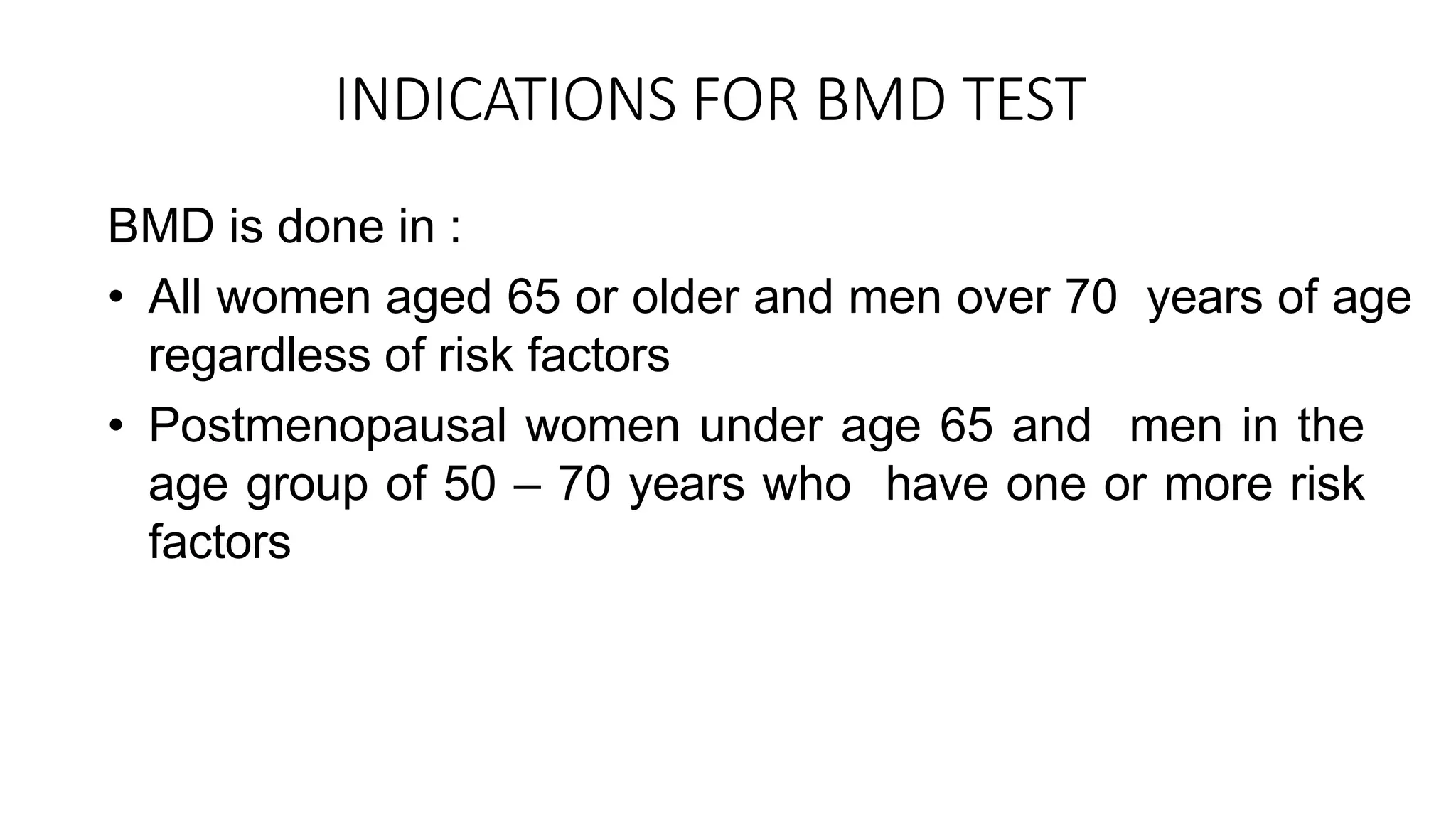
INDICATIONS FOR BMDTEST
BMD is done in :
• All women aged 65 or older and men over 70 years of age
regardless of risk factors
• Postmenopausal women under age 65 and men in the
age group of 50 – 70 years who have one or more risk
factors
39.
RISK FACTORS FOROSTEOPOROSIS
1.Having a current or previous fracture
2.On steroid medications for more than three months
3. Chronic anticonvulsant therapy
4.Men with low testosterone,alcoholism or any other secondary cause of
osteoporosis
5.Chronic rheumatoid arthritis 5.Chronic
kidney disease 6.Early menopause
6.History of hormone treatment for prostate cancer or breast cancer
6.Smoking
7.Strong family history of osteoporosis
8. Significant loss of height (vertebral compression fractures)
40.
CONTRAINDICATIONS FOR BMDTEST
• Pregnancy
• Recent gastrointestinal contrast studies(recommend
waiting for at least 72 hours before central DEXA
Scan)
• Body weight exceeding limit for DEXA
scanners(>120-130kgs)
• Bilateral hip replacements or bilateral hip pins or screws
would prevent the hip sites from being scanned.Metallic
rods or spinal fusion devices in the lumbar spine prevent
scanning at this site.
41.
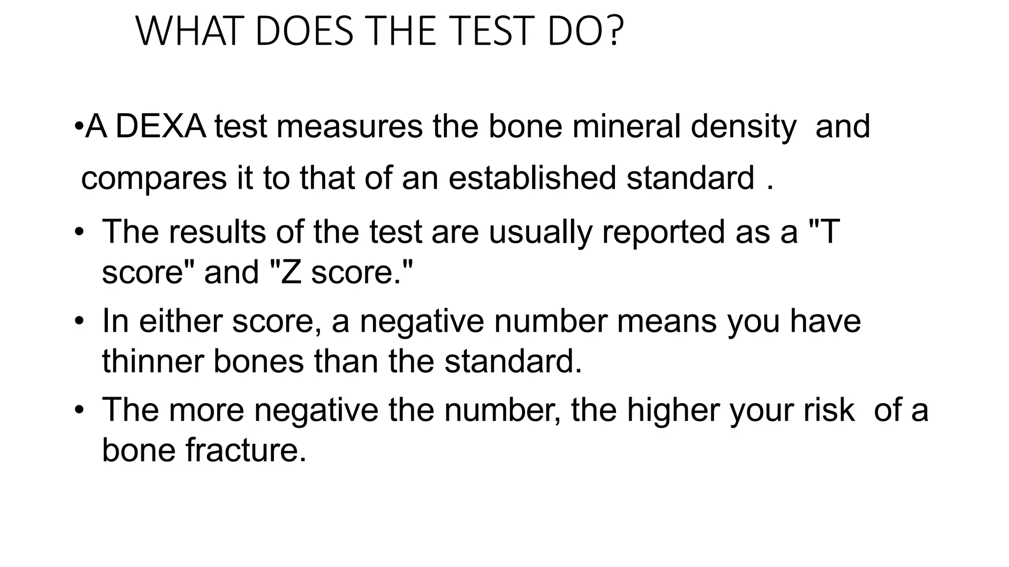
WHAT DOES THETEST DO?
•A DEXA test measures the bone mineral density and
compares it to that of an established standard .
• The results of the test are usually reported as a "T
score" and "Z score."
• In either score, a negative number means you have
thinner bones than the standard.
• The more negative the number, the higher your risk of a
bone fracture.
42.
T-SCORE
• The Tscore compares your bone density with that of
healthy young adult.
• A score of 0 means your BMD is equal to the
standard for a healthy young adult.
• Differences between your BMD and that of the healthy
young adult standard are measured in units called
standard deviations (SDs). The more standard deviations
below 0, indicated as negative numbers, the lower your
BMD and the higher your risk of fracture.
43.
• As shownin the table below, a T-score
between
+1 and −1 is considered normal or healthy.
• A T-score between −1 and −2.5 indicates that
you have low bone mass.
• A T-score of −2.5 or lower indicates that
you have osteoporosis.
• The greater the negative number, the
more severe the osteoporosis.
45.
Z-SCORE
• The Zscore compares your bone density with
that of other people of same age and gender.
• A low Z-score (below —2.0) is a warning sign
that you have less bone mass or that you are
losing bone more rapidly than expected for
someone of your age.
• Z-score= T-score - Reference T-score
48.
How is a"Bone Scan" different
from a BMD test?
• Unlike a BMD test, a bone scan is an invasive test.
• The patient is injected with a dye that allows a scanner
to look at the condition of bone tissue.
• A bone scan can diagnose inflammation, fractures, bone
lesions and cancer.
• It cannot predict the risk of osteoporosis or
diagnose the condition.
49.
Osteopenia
• Osteopenia refersto bone mineral density that is lower than
normal peak BMD (i.e,between -1.0 and
-2.5) but not low enough to be classified as
osteoporosis.
• It is a sign of normal ageing, in contrast to osteoporosis which
is a sign of pathologic ageing.
• It occurs more frequently in post-menopausal women,can be
exacerbated by lifestyle factors such as lack of exercise,
alcoholism, smoking or prolonged use of glucocorticoid
medications for asthma.
50.
• Osteopenia hasno symptoms. There is no pain as the
bone becomes thinner but the risk of fracture increases
as the bone becomes less dense.
• Diagnosis is by a bone mineral density test. A standard X-
ray is not useful in diagnosing because it is not sensitive
enough to detect small amounts of bone loss or minor
changes in bone density
52.
• Osteopenia istreated by taking steps to prevent it from
progressing to osteoporosis like –
1.Lifestyle changes
2.Dietary modifications : Foods rich in
calcium(milk,dairy products,green leafy vegetables)
and vitamin D(eggs,fish,fish oils)
3.Vitamin D and calcium supplements
53.
Osteoporosis
• Osteoporosis ="porous bones."
• Osteoporosis is defined by the WHO as a bone mineral
density that is 2.5 standard deviations or more below the
mean bone mass (average of young,healthy adults).
• The underlying mechanism in osteoporosis is an
imbalance between bone resorption and bone formation.
55.
• Osteoporosis isclassified as
• primary type 1 - postmenopausal osteoporosis
• primary type 2 - senile osteoporosis occuring after age
75
• secondary - resulting from chronic predisposing medical
conditions or prolonged use of glucocorticoids,when the
disease is called steroid- or glucocorticoid-induced
osteoporosis.
• The risk factors are same as that of osteopenia.
56.
• Osteoporosis hasno symptoms but its main
consequence is the increased risk of bone fractures.
• Osteoporotic fractures are those that occur in situations
where healthy people would not normally break a
bone;they are therefore called fragility fractures.
57.
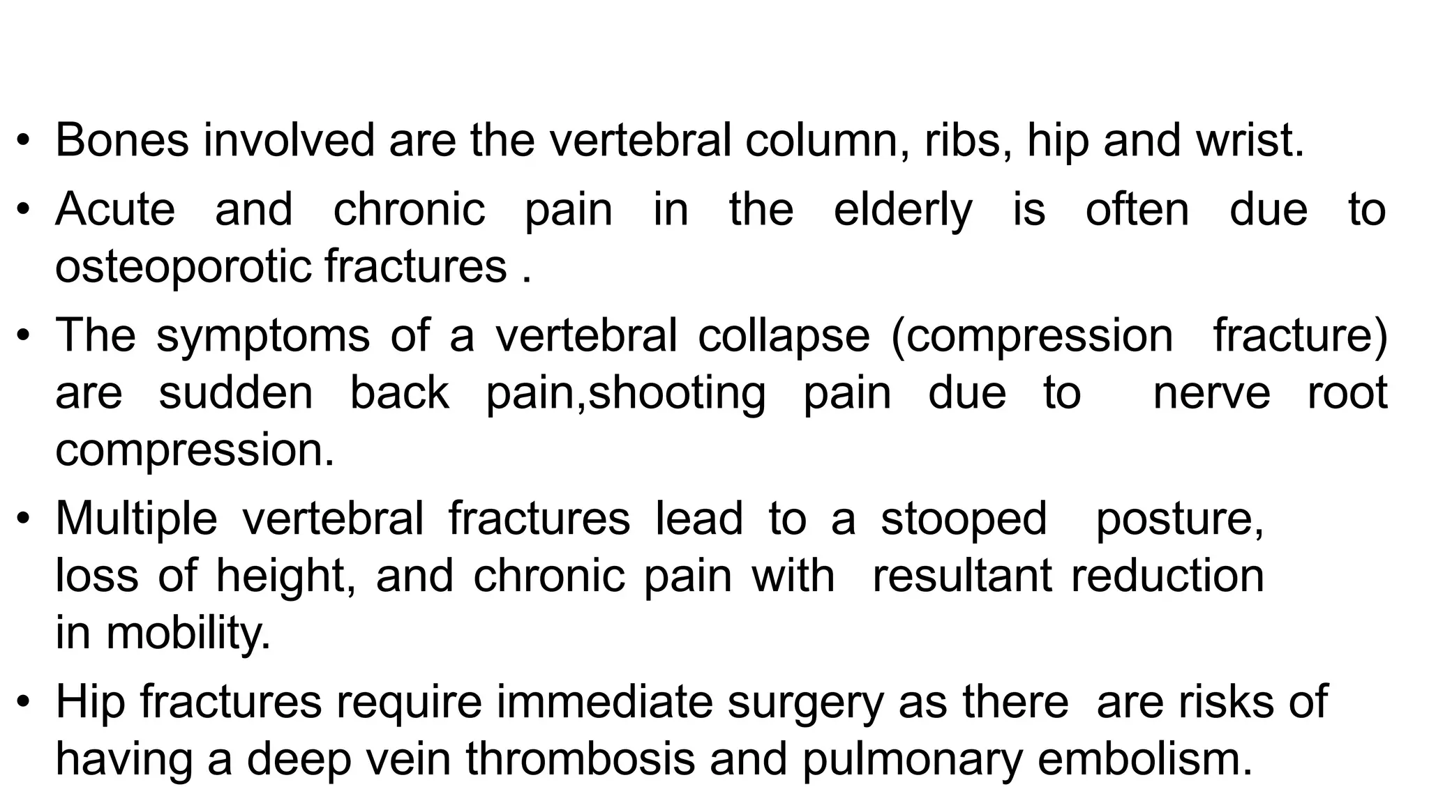
• Bones involvedare the vertebral column, ribs, hip and wrist.
• Acute and chronic pain in the elderly is often due to
osteoporotic fractures .
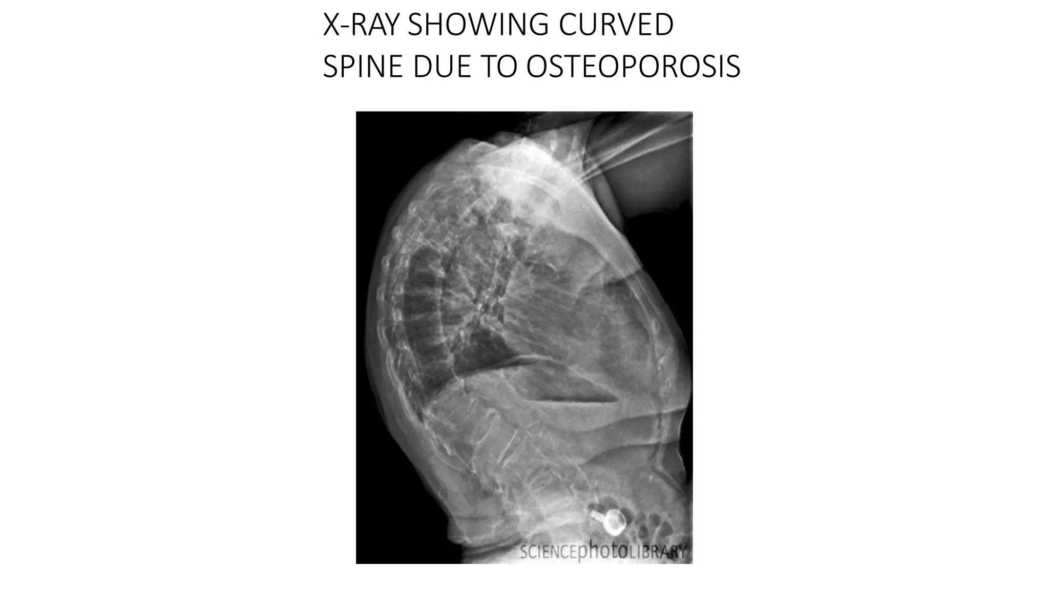
• The symptoms of a vertebral collapse (compression fracture)
are sudden back pain,shooting pain due to nerve root
compression.
• Multiple vertebral fractures lead to a stooped posture,
loss of height, and chronic pain with resultant reduction
in mobility.
• Hip fractures require immediate surgery as there are risks of
having a deep vein thrombosis and pulmonary embolism.
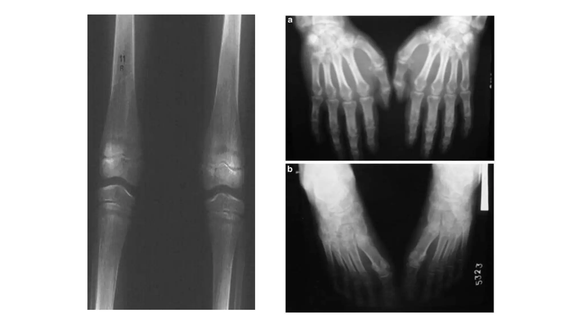
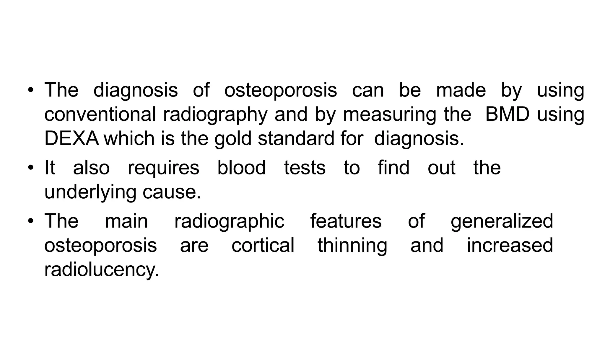
• The diagnosisof osteoporosis can be made by using
conventional radiography and by measuring the BMD using
DEXA which is the gold standard for diagnosis.
• It also requires blood tests to find out the
underlying cause.
• The main radiographic features of generalized
osteoporosis are cortical thinning and increased
radiolucency.


![Understanding Parkinson’s Disease: Causes, Symptoms, and Treatment [2025]](https://cdn.slidesharecdn.com/ss_thumbnails/understandingparkinson-251208102525-80ba3223-thumbnail.jpg?width=640&height=640&fit=bounds)


