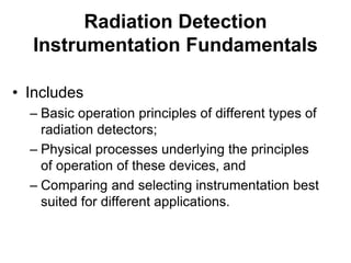
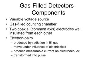
This document provides an overview of different types of radiation detection instrumentation, including gas-filled detectors, scintillation detectors, and solid state detectors. It describes the basic components and operating principles of gas-filled detectors like ionization chambers and proportional counters. It also covers scintillation detectors, explaining how organic and inorganic scintillators work and how photomultiplier tubes are used to detect the light pulses produced in scintillators. Key concepts like dead time, resolving time, and quenching effects are defined. Applications of different detector types are also briefly discussed.
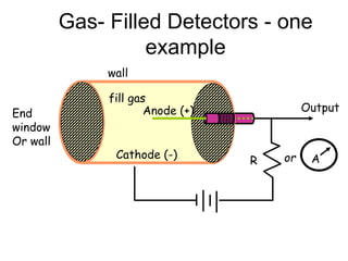
This document discusses various types of radiation detection devices, including film badges, ionization chambers, Geiger-Muller counters, proportional counters, scintillation counters, photographic plates, electroscopes, bubble chambers, solid-state detectors, cloud chambers, and spark counters. Each detection method works by using different processes like ionization, fluorescence, or track visualization to detect and sometimes quantify radiation levels or particle energy. Regular monitoring of radiation is important for safety when working with radioactive materials.
This document discusses different types of gas-filled and scintillation radiation detectors. It provides information on GM counters, proportional counters, scintillators, photomultiplier tubes, and thermoluminescent dosimeters. Key points include: how GM counters differ from proportional counters in their avalanche chain reactions; common scintillator materials like NaI(Tl) and BGO; how photomultiplier tubes convert light photons to electrical signals and amplify signals through dynode multiplication; and applications of different detector types in nuclear medicine imaging. The document is in a question-answer format where various concepts are explained in response to questions.
This document discusses different types of radiation detectors, including scintillation detectors, Geiger-Muller (GM) counters, and proportional counters. Scintillation detectors detect radiation via scintillation light produced in luminescent materials coupled to photomultiplier tubes. GM counters use gas multiplication in Geiger-Muller tubes to detect ionizing radiation via voltage pulses. Proportional counters operate at lower voltages to provide energy information, while GM counters only indicate detection.
This document discusses different types of gas-filled radiation detectors, including their operating principles and applications. It covers ionization chambers, proportional counters, and Geiger-Muller counters. Key points include:
1) Ionization chambers measure current and have a wide operating voltage range where ion pairs are collected. They are commonly used to measure exposure in radiology and nuclear medicine.
2) Proportional counters use a higher electric field to cause ion avalanches, amplifying the signal. Noble gases like argon and xenon are used to enable gas multiplication.
3) Geiger-Muller counters operate at an even higher voltage where each interaction produces the same-sized pulse, regardless of
There are three main types of radiation detectors: gas-filled detectors which use a gas between electrodes, scintillation detectors which use materials that produce light when irradiated, and semiconductor detectors made of purified crystalline materials. Detectors can also be classified by the type of information they provide, such as counting interactions, measuring energy, or indicating dose. The main challenges for detectors are dead time at high interaction rates and maintaining good energy resolution and detection efficiency.
The document discusses various types of radiation dosimetry including film dosimetry. Film dosimetry uses photoemulsions like silver bromide dispersed in gelatin that form latent images when exposed to radiation. During development, sensitized grains are converted to metallic silver making the tracks visible. The optical density of the film is proportional to the absorbed dose based on Beer's law. A characteristic H-D curve describes the film performance in terms of speed, contrast, latitude and resolution. Film dosimetry provides high spatial resolution and flexibility but response varies with energy and processing conditions.
A Geiger-Muller counter consists of a gas-filled tube that detects ionizing radiation such as alpha particles, beta particles, and gamma rays. When radiation enters the tube, it ionizes the gas and produces a pulse of current that is counted by a scaler. To prevent additional pulses from a single radiation event, a small amount of quenching gas is added which absorbs excess energy and prevents further ionization of the main gas. The Geiger-Muller counter has a dead time after each detection where it cannot detect additional radiation as it re-establishes the electric field inside the tube.
This document discusses scintillation detectors and their properties. Scintillation detectors work by emitting light when exposed to radiation, and this light output can be used to measure incident radiation. The key properties of scintillation detectors are that they must have high scintillation efficiency, light yield proportional to deposited energy, short decay time, transparency to emitted wavelengths, and be able to be made in large sizes and desired shapes. Common inorganic scintillators discussed are NaI(Tl), which is widely used due to its availability and high detection efficiency, and BGO, which has high intrinsic efficiency for high gamma energies.
The document discusses the interaction of radiation with matter. It covers topics including the atomic structure, quantities and units used in physics, production of bremsstrahlung and characteristic x-rays, photon interactions such as the photoelectric effect and Compton scattering, beam attenuation, and the principles of radiological image formation. The interaction of radiation depends on factors like the photon energy and atomic number of the absorbing material. Different interaction mechanisms dominate based on these factors and contribute to image contrast in medical imaging.
Radiation units can be divided into units of radioactivity and units of radiation dose. The curie and becquerel are units of radioactivity, with 1 curie equal to 3.7x10¹⁰ decays per second. Exposure dose is measured in roentgens or C/kg, while absorbed dose is measured in rads or grays. Equivalent dose accounts for radiation type and takes the Q factor into account, measured in rems or sieverts. Effective dose considers radiation exposure to different tissues, calculated from equivalent dose and tissue weighting factors.
The document discusses types of radiation detectors, including gas-filled detectors like ionization chambers and GM counters, and how they are used in medical applications. It covers concepts like detection methods, operating modes, dead time, and how detector performance is affected by high count rates. The goal is to classify detectors used in various departments like nuclear medicine.
Radioactive decay is the process by which an unstable atomic nucleus loses energy by emitting ionizing particles such as alpha particles, beta particles, or gamma rays. Alpha decay occurs when an atom ejects an alpha particle (helium nucleus). Beta decay is the emission of an electron. Gamma decay is the release of energy in the form of electromagnetic waves.
This document provides information about nuclear radiation detectors. It discusses three main types of gaseous ionization detectors: ionization chambers, proportional counters, and Geiger-Müller tubes. Ionization chambers detect radiation by collecting all ion pairs created through gas ionization when radiation passes through. Proportional counters can measure radiation energy by producing output proportional to radiation energy through gas amplification of ion pairs. Geiger-Müller tubes operate at very high voltages where any initial ionization causes a self-sustaining discharge and produces a standard pulse height independent of radiation type.
This document discusses various methods for detecting radiation. It outlines passive detectors like photographic film, electroscopes, dosimeters, and thermoluminescent dosimeters (TLDs) which do not require a power source. Active detectors mentioned include Geiger-Muller tubes and scintillation detectors, which need a constant energy supply. Both types detect radiation indirectly by ionizing matter and detecting the ions produced, though active detectors provide more information about the radiation type and energy.
This document discusses various topics related to radiation and its units of measurement. It defines (1) radiation oncology as the field concerned with treating cancer and other diseases using ionizing radiation, (2) the two main types of radiation as ionizing and non-ionizing, and (3) some key units used to measure radiation exposure and its effects on living tissue, including the becquerel, curie, gray, sievert, and rem. It also provides background on the scientists who discovered various types of radiation and developed these units, such as Becquerel, Curie, Roentgen, and Gray.
Radioactive Contamination and Procedures of Decontaminationmahbubul hassan
Training Course on Radiation Protection for Radiation Workers and RCOs of BAEC, Medical Facilities and Industries, TI, AERE, BAEC Savar, 27 October 2021
This document provides information about radiation detection and measurement instruments. It discusses various types of gas-filled detectors like ionization chambers, proportional counters, and Geiger-Muller counters. It also describes semiconductor detectors, scintillation counters, and instruments used for personnel dosimetry and measuring dose rates and contamination levels. The key purpose of these various instruments is to detect and measure different types of ionizing radiation like alpha, beta, gamma, and neutrons by converting radiation interactions into electrical pulses or light flashes that can be analyzed. Regular calibration of radiation monitors is emphasized to ensure accurate measurements for different radionuclides and radiation energies.
LiF:Mg,Ti (TLD-100) is a nearly tissue-equivalent thermoluminescent dosimeter material that is widely used for radiation dosimetry. It has good reproducibility and sensitivity for low doses. TLDs must be calibrated against absolute dosimeters. The TL response of TLD-100 depends on radiation quality and dose, and it exhibits fading over time. Computerized glow curve analysis can provide information on individual TL peaks. LiF:Mg,Cu,P has higher sensitivity and better tissue equivalence than TLD-100. CaF:Tm (TLD-300) can separate high and low LET radiation components based on peak heights. Various TLD materials are used for different applications
The document summarizes the interaction of radiation with matter. It discusses the different types of electromagnetic and particulate radiation. It then describes the four main processes radiation can undergo when interacting with matter - attenuation, absorption, scattering, and transmission. It provides details on the photoelectric effect, Compton effect, and pair production - the three primary physical interactions responsible for photon attenuation in matter.
This document discusses different types of solid state radiation detectors, including scintillation detectors, thermoluminescent dosimeters (TLD), and semiconductor detectors. Scintillation detectors detect radiation via light emission in inorganic crystal materials like NaI or organic crystals like anthracene. TLDs "capture" radiation dose information and release light when heated, allowing dose measurement. Common TLD materials are LiF:Mg,Ti and Li2B4O7:Mn. Semiconductor detectors like silicon and germanium act as solid state ionization chambers and are used for high resolution energy measurement of alpha and beta particles.
1. Different units are used to measure various properties of ionizing radiation, including the curie, becquerel, roentgen, rad, gray, rem, and sievert.
2. The roentgen measures exposure to gamma or X-rays, while the rad measures absorbed dose, accounting for different materials. The gray is now the SI unit for absorbed dose, replacing the rad.
3. The rem accounts for the different biological effects of various types of ionizing radiation based on their quality factor, and is used to measure equivalent dose. The sievert, replacing the rem, measures stochastic health risks from radiation exposure.
This document discusses semi-conductor detectors and their use in radiation detection. It describes the basic structure of atoms and different types of bonds between atoms. It explains that in crystalline materials like semiconductors, electrons exist in energy bands separated by gaps. In semiconductors and insulators, the valence band is full while the conduction band is empty, allowing electrons to move if given enough energy to cross the band gap. Semiconductors can be "doped" with impurities to create p-type or n-type materials and function as radiation detectors by generating electron-hole pairs when ionizing radiation transfers electrons to the conduction band.
This document discusses various radiation quantities and units used to characterize ionizing radiation. It describes key concepts such as activity, kerma, exposure, absorbed dose, equivalent dose, effective dose, annual limit intake (ALI), and derived air concentration (DAC). The International Commission on Radiation Protection (ICRP) and International Commission on Radiation Units (ICRU) help define these quantities and their relationships. Primary quantities like equivalent dose relate radiation risk, while operational quantities like exposure are used for measurements. Tissue weighting factors account for different tissue sensitivities in calculating effective dose from equivalent dose.
This document provides an overview of active methods for neutron detection, including gas filled detectors like ionization chambers and proportional counters, scintillation detectors using materials like lithium iodide and organic scintillators, and semiconductor detectors. It describes the basic detection mechanisms, advantages and disadvantages of different methods, and their typical applications in neutron dosimetry and spectrometry.
Wilhelm Roentgen discovered X-rays in 1895 while experimenting with Crookes tubes. He called them "x-rays" as the nature of the radiation was unknown. The first X-ray photograph was of his wife Bertha's hand. X-rays are a form of electromagnetic radiation with much shorter wavelengths than visible light, in the range of 0.5-2.5 angstroms. They can be considered as both waves and particles called photons. X-rays are generated in an X-ray tube when high speed electrons interact with and lose kinetic energy in the target material, usually tungsten. This produces bremsstrahlung or braking radiation and characteristic radiation when electrons change energy levels within
Neutron detection in medical industry and hsopitalmedicalHunter2
This document discusses the need for radiation surveys of new and modified radiation facilities to evaluate shielding requirements. It provides guidelines for performing surveys, including recording parameters and taking measurements at various gantry angles. Recommendations are given for survey instruments, with ionization chambers preferred for use around medical linear accelerators due to their ability to operate in pulsed radiation fields. Shielding design goals to limit equivalent dose in controlled and uncontrolled areas are provided based on NCRP recommendations.
This document provides an overview of sensors and sensor systems for an ECE 480 class taught by Professor Mason. It defines sensors and transducers, describes common sensor components and configurations, and gives examples of primary transducers including temperature, light, pressure and displacement sensors. It also discusses signal conditioning with operational amplifiers, connecting sensors to microcontrollers and networks, and sensor calibration techniques.
Liquid Sensing: Visible light absorption spectroscopy and colorimetry are two fundamental tools used in chemical analysis. Most of these light-based systems use photodiodes as the light sensor, and require similar high input impedance signal chains. This session examines the different components of a photodiode amplifier signal chain, including a programmable gain transimpedance amplifier, a hardware lock-in amplifier, and a Σ-Δ ADC that can measure a sample and reference channel to greatly reduce any measurement error due to variations in intensity of the light source.
Gas Sensing: Many industrial processes involve toxic compounds, and it is important to know when dangerous concentrations exist. Electrochemical sensors offer several advantages for instruments that detect or measure the concentration of toxic gases. This session will describe a portable toxic gas detector using an electrochemical sensor. The system presented here includes a potentiostat circuit to drive the sensor, as well as a transimpedance amplifier to take the very small output current from the sensor and translate it to a voltage that can take advantage of the full-scale input of an ADC.
This document provides an overview of various analysis tools available in EWB software for circuit simulation and analysis. It describes the following analysis types: DC operating point analysis, AC frequency analysis, transient analysis, Fourier analysis, noise analysis, distortion analysis, DC sweep analysis, sensitivity analysis, parameter sweep analysis, temperature sweep analysis, transfer function analysis, worst case analysis, pole zero analysis, and Monte Carlo analysis. For each analysis type, it provides a brief description of the analysis and an example circuit to demonstrate how to set up and interpret the results of that analysis.
Complete detail about the Radiopharmaceutical, General Introduction, Radioactive substance, Radioactive rays like alpha, beta and gamma rays. All the Measurement method to determine the radioactivity of any element and widely used instrument Geiger Muller Counter. And some Radiopharmaceutical product used in many diagnosis , treatment such like sodium iodide solution & capsule, Rose Bengal I 131 and Application of Radiopharmaceuticals.
This document provides an overview of sensors and transducers for an electrical and computer engineering course. It defines sensors and transducers as devices that convert one form of energy to another. The document describes common sensor types including temperature, light, pressure, and displacement sensors. It also discusses electronic sensor systems, signal conditioning circuits, and interfacing sensors with microcontrollers and computer systems.
This document provides an overview of sensors and transducers for an electrical and computer engineering course. It defines sensors and transducers as devices that convert one form of energy to another. The document describes common sensor types including temperature, pressure, light, and displacement sensors. It also discusses electronic sensor systems, primary and secondary transducers, microelectromechanical systems sensors, and the use of operational amplifiers and microcontrollers in sensor systems.
Principles and Practices of Traceability and CalibrationJasmin NUHIC
To learn and understand different types of measurements units, measurement constants, calibration and measurement standards as well as principles and practices of treaceability.
Mechatronics is a multidisciplinary field that refers to the skill sets needed in the contemporary, advanced automated manufacturing industry. At the intersection of mechanics, electronics, and computing, mechatronics specialists create simpler, smarter systems.
This training program provides information on fire and gas detection systems. The objectives are to ensure safety, protect the environment and plant, and provide early detection and alarms. A variety of detectors are discussed to detect different hazards like fire, toxic gases, and combustible gases. Factors in selecting and locating the detectors are addressed. Detector types for different applications are explained along with their working principles. Alarm settings and types of output devices are also summarized.
The document discusses sensors and transducers. It defines a transducer as a device that converts one form of energy to another, with sensors detecting signals from the real world and actuators generating signals. Electronic sensors typically use primary transducers to convert a parameter into an electrical signal, and secondary transducers to further process the signal. Common sensor components and configurations are described such as op-amps, instrumentation amplifiers, and connecting sensors to microcontrollers and networks. The document also covers transducer types including mechanical, thermal, optical, and chemical. Sensor calibration techniques are discussed to address non-ideal sensor effects.
The document discusses sensors and transducers. It defines a transducer as a device that converts one form of energy to another. Sensors are transducers that detect or measure a signal, while actuators generate a signal. Electronic sensors typically have a primary transducer that converts a parameter to an electrical signal and a secondary transducer that converts this to analog or digital values. Common primary transducers discussed include light, pressure, displacement, temperature, and MEMS sensors. Secondary transducers and common sensor readout circuits like Wheatstone bridges and operational amplifiers are also described.
Energy is the ability to do work and can exist in various forms. Sensors are devices that measure a physical quantity and convert it into a signal. Energy sensors specifically respond to an input quantity by generating an output signal, often electrical or optical. Common energy sensors measure mechanical energy through quantities like acceleration, force, displacement; heat/thermal energy through temperature; and light through photoresistors, photodiodes or phototransistors. Energy sensors have applications in areas like fuel cells, wind and solar energy, and nuclear systems.
This document provides information on various sensor technologies. It discusses key concepts in sensor terminology and categorization. It describes considerations for instrumentation and measurement. It also provides details on specific sensor types including their operating principles, specifications, advantages, and disadvantages. These include sensors for strain, acceleration, force, displacement, velocity, and shock.
This document provides information on various sensor technologies. It discusses key concepts related to phase linearity and distortion in sensors. It also defines transducers and categorizes sensors based on physical phenomena measured and measuring mechanisms. The document outlines considerations for instrumentation and describes common sensor specifications. It provides examples of specific sensor types for measuring strain, acceleration, force, displacement, velocity, and shock and describes their operating principles and attributes.
This document provides information on various sensor technologies. It discusses key concepts in sensor terminology and categorization. It describes common sensor types including those that measure phase linearity, mechanical properties like strain and displacement, thermal properties, acceleration, force, velocity, and shock. For each sensor type, it outlines important specifications and considerations in sensor design and measurement.
The document discusses various sensor technologies and considerations for sensing systems. It describes different types of sensors categorized by the physical phenomena they measure such as mechanical, thermal, and optical sensors. It also discusses sensor specifications including accuracy, resolution, sensitivity, bandwidth, and noise. The document covers various sensor technologies for measuring strain, acceleration, force, displacement, velocity, and angular motion. It describes MEMS sensors and their fabrication techniques. Finally, it discusses considerations for designing sensing systems such as sensor selection, data collection, communication, power, and environmental influence.
Similar to Radiation detection instrumentation fundamentals (20)
Role of artificial intellegence (a.i) in radiology department nitish virmaniNitish Virmani
Artificial intelligence can help radiologists make more accurate diagnoses by recognizing patterns in medical images. AI systems trained on large datasets of images can identify abnormalities and make recommendations to radiologists rapidly. PACS systems that incorporate AI may be the future of radiology to help analyze images and optimize workflow. While AI shows great promise, its decisions are based on algorithms and data, unlike human judgment, so radiologists will still be needed to apply clinical expertise.
Breast tomosynthesis and DEXA bone mineral density tests provide important health information.
Breast tomosynthesis is an advanced mammography technique that uses low-dose x-rays and computer reconstruction to create 3D images of the breasts, helping detect cancers earlier. DEXA uses dual-energy x-ray beams to measure bone mineral density in the hip and spine. It identifies osteoporosis and fracture risk by comparing a patient's scores to norms for age and gender. Both tests are noninvasive, involve low radiation exposure, and provide valuable health screening and monitoring.
MRI spectroscopy- Its Application, Principle & Techniques Nitish Virmani
Magnetic resonance spectroscopy (MRS) is a noninvasive imaging technique that measures biochemical and metabolic processes in tissues without using ionizing radiation. MRS provides additional clinical information for diseases such as brain tumors, metabolic disorders, and systemic diseases. MRS can distinguish between neoplastic and non-neoplastic brain masses, identify tumor recurrence versus radiation necrosis, and help monitor treatment response. Key metabolites that MRS evaluates include NAA, creatine, choline, lactate, lipids, and myo-inositol. Metabolite ratios are used for interpretation, with abnormal ratios indicating various conditions like tumors, demyelination, ischemia, and infection. MRS has wide clinical applications and provides valuable physiological information to
MRI shoulder and knee- Anatomy, Scan Planning & Its Techniques Nitish Virmani
The document provides information about MRI procedures for the shoulder and knee joints. It describes the anatomy and indications for MRI of each joint. Suggested imaging protocols including sequences, slice thickness, and field of view are outlined for the shoulder and knee. Patient positioning and equipment used are also discussed. Common findings addressed in reports on the shoulder include rotator cuff tears and labral injuries, while meniscal tears and ligament injuries are highlighted for the knee.
Magnetic Resonance Cholangiopancreatography- MRCPNitish Virmani
Magnetic resonance cholangiopancreatography (MRCP) is a noninvasive MRI technique used to visualize the biliary and pancreatic ducts. It utilizes heavily T2-weighted sequences to provide high signal from fluid-filled structures like the biliary tree and pancreatic ducts. MRCP has largely replaced diagnostic ERCP as it is less invasive and avoids risks of radiation exposure. It is useful for evaluating biliary diseases like cysts, stones, strictures, and pancreatic conditions such as divisum and cancer. With refinements in techniques such as secretin administration, MRCP provides accurate assessment of pancreaticobiliary anatomy and pathology in a safe, noninvasive manner.
This document provides information about MRI contrast agents. It discusses that MRI contrast agents are used to improve visibility of internal structures during MRI scans. The most commonly used contrast agents contain gadolinium and work by shortening the T1 relaxation time of protons. This causes enhanced contrast between tissues. Contrast agents must have unpaired electrons to generate a magnetic field and interact with proton spins in tissues. The document discusses various types of contrast agents classified by their composition, effects, and applications. It also covers administration routes, side effects like allergic reactions and nephrogenic systemic fibrosis (NSF), and contraindications for use.
Trauma radiography involves specialized procedures to image patients with traumatic injuries. Trauma centers are classified by level depending on available resources and specialties. Level I centers have the most comprehensive services available 24/7. Radiographers in emergency departments must be prepared to perform a variety of trauma imaging procedures on patients of all ages using specialized equipment. Common trauma projections include lateral cervical spine, cervicothoracic, and thoracic and lumbar spine views. Quality, speed, accuracy and attention to detail are important principles for optimal trauma radiography.
This document discusses various concepts related to radiographic image quality and measurements. It defines terms like radiographic contrast, spatial resolution, contrast resolution, noise, and artifacts. It describes how factors like the film, geometry, and subject can impact radiographic quality. It also discusses optical density, sensitometry, and how the characteristic curve relates exposure to density. The modulation transfer function and how it relates to spatial frequencies is explained. Overall, the document provides an overview of key technical factors and measurements that influence the quality of radiographic images.
Radiation has benefits for medical imaging but can harm healthy cells. Proper protection techniques like time, distance and shielding can avoid overexposure, especially for pregnant technicians. Technicians are at risk of radiation exposure and need protection including lead aprons, gloves, glasses and shields to block scatter radiation and stay under exposure limits. New suspended shielding systems aim to improve radiation protection.
The document provides guidance on radiography for pediatric patients. It discusses preparing the patient and environment, immobilization techniques, common conditions seen in pediatric radiography, and appropriate exams and exposure adjustments. Successful pediatric radiography requires effective communication, building trust, and immobilizing the patient to minimize motion during exposures while avoiding unnecessary radiation.
Chandrima Spa Ajman is one of the leading Massage Center in Ajman, which is open 24 hours exclusively for men. Being one of the most affordable Spa in Ajman, we offer Body to Body massage, Kerala Massage, Malayali Massage, Indian Massage, Pakistani Massage Russian massage, Thai massage, Swedish massage, Hot Stone Massage, Deep Tissue Massage, and many more. Indulge in the ultimate massage experience and book your appointment today. We are confident that you will leave our Massage spa feeling refreshed, rejuvenated, and ready to take on the world.
Visit : https://massagespaajman.com/
Call : 052 987 1315
R3 Stem Cell Therapy: A New Hope for Women with Ovarian FailureR3 Stem Cell
Discover the groundbreaking advancements in stem cell therapy by R3 Stem Cell, offering new hope for women with ovarian failure. This innovative treatment aims to restore ovarian function, improve fertility, and enhance overall well-being, revolutionizing reproductive health for women worldwide.
Healthy Eating Habits:
Understanding Nutrition Labels: Teaches how to read and interpret food labels, focusing on serving sizes, calorie intake, and nutrients to limit or include.
Tips for Healthy Eating: Offers practical advice such as incorporating a variety of foods, practicing moderation, staying hydrated, and eating mindfully.
Benefits of Regular Exercise:
Physical Benefits: Discusses how exercise aids in weight management, muscle and bone health, cardiovascular health, and flexibility.
Mental Benefits: Explains the psychological advantages, including stress reduction, improved mood, and better sleep.
Tips for Staying Active:
Encourages consistency, variety in exercises, setting realistic goals, and finding enjoyable activities to maintain motivation.
Maintaining a Balanced Lifestyle:
Integrating Nutrition and Exercise: Suggests meal planning and incorporating physical activity into daily routines.
Monitoring Progress: Recommends tracking food intake and exercise, regular health check-ups, and provides tips for achieving balance, such as getting sufficient sleep, managing stress, and staying socially active.
At Apollo Hospital, Lucknow, U.P., we provide specialized care for children experiencing dehydration and other symptoms. We also offer NICU & PICU Ambulance Facility Services. Consult our expert today for the best pediatric emergency care.
For More Details:
Map: https://cutt.ly/BwCeflYo
Name: Apollo Hospital
Address: Singar Nagar, LDA Colony, Lucknow, Uttar Pradesh 226012
Phone: 08429021957
Opening Hours: 24X7
Michigan HealthTech Market Map 2024. Includes 7 categories: Policy Makers, Academic Innovation Centers, Digital Health Providers, Healthcare Providers, Payers / Insurance, Device Companies, Life Science Companies, Innovation Accelerators. Developed by the Michigan-Israel Business Accelerator
Can Allopathy and Homeopathy Be Used Together in India.pdfDharma Homoeopathy
This article explores the potential for combining allopathy and homeopathy in India, examining the benefits, challenges, and the emerging field of integrative medicine.
Exploring the Benefits of Binaural Hearing: Why Two Hearing Aids Are Better T...Ear Solutions (ESPL)
Binaural hearing using two hearing aids instead of one offers numerous advantages, including improved sound localization, enhanced sound quality, better speech understanding in noise, reduced listening effort, and greater overall satisfaction. By leveraging the brain’s natural ability to process sound from both ears, binaural hearing aids provide a more balanced, clear, and comfortable hearing experience. If you or a loved one is considering hearing aids, consult with a hearing care professional at Ear Solutions hearing aid clinic in Mumbai to explore the benefits of binaural hearing and determine the best solution for your hearing needs. Embracing binaural hearing can lead to a richer, more engaging auditory experience and significantly improve your quality of life.
We are one of the top Massage Spa Ajman Our highly skilled, experienced, and certified massage therapists from different corners of the world are committed to serving you with a soothing and relaxing experience. Luxuriate yourself at our spas in Sharjah and Ajman, which are indeed enriched with an ambiance of relaxation and tranquility. We could confidently claim that we are one of the most affordable Spa Ajman and Sharjah as well, where you can book the massage session of your choice for just 99 AED at any time as we are open 24 hours a day, 7 days a week.
Visit : https://massagespaajman.com/
Call : 052 987 1315
Unlocking the Secrets to Safe Patient Handling.pdfLift Ability
Furthermore, the time constraints and workload in healthcare settings can make it challenging for caregivers to prioritise safe patient handling Australia practices, leading to shortcuts and increased risks.
Hypertension and it's role of physiotherapy in it.Vishal kr Thakur
This particular slides consist of- what is hypertension,what are it's causes and it's effect on body, risk factors, symptoms,complications, diagnosis and role of physiotherapy in it.
This slide is very helpful for physiotherapy students and also for other medical and healthcare students.
Here is summary of hypertension -
Hypertension, also known as high blood pressure, is a serious medical condition that occurs when blood pressure in the body's arteries is consistently too high. Blood pressure is the force of blood pushing against the walls of blood vessels as the heart pumps it. Hypertension can increase the risk of heart disease, brain disease, kidney disease, and premature death.
Can coffee help me lose weight? Yes, 25,422 users in the USA use it for that ...nirahealhty
The South Beach Coffee Java Diet is a variation of the popular South Beach Diet, which was developed by cardiologist Dr. Arthur Agatston. The original South Beach Diet focuses on consuming lean proteins, healthy fats, and low-glycemic index carbohydrates. The South Beach Coffee Java Diet adds the element of coffee, specifically caffeine, to enhance weight loss and improve energy levels.
2. Radiation Detection
Instrumentation Fundamentals
• Includes
– Basic operation principles of different types of
radiation detectors;
– Physical processes underlying the principles
of operation of these devices, and
– Comparing and selecting instrumentation best
suited for different applications.
5. Gas-Filled Detectors -
Components
• Variable voltage source
• Gas-filled counting chamber
• Two coaxial (common axis) electrodes well
insulated from each other
• Electron-pairs
– produced by radiation in fill gas
– move under influence of electric field
– produce measurable current on electrodes, or
– transformed into pulse
6. Gas- Filled Detectors - one
example
wall
fill gas
R
Output
Aor
Anode (+)
Cathode (-)
End
window
Or wall
10. Voltage versus Ions Collected
Voltage
Number
of Ion
Pairs
collected
Ionization region
Saturation Voltage
100 % of initial
ions are collected
Recom-
bination
region
11. Saturation Current
• The point at which 100% of ions begin to be
collected
• All ion chambers operate at a voltage that
produces a saturation current
• The region over which the saturation current is
produced is called the ionization region
• It levels the voltage range because all charges
are already collected and rate of formation is
constant
12. Observed Output: Pulse Height
• Ions collected
• Number of ionizations relate to specific
ionization value of radiation
• Gas filled detectors operate in either
– current mode
• Output is an average value resulting from detection
of many values
– pulse mode
• One pulse per particle
14. Ionization Region Recap
• Pulse size depends on # ions produced in
detector.
• No multiplication of ions due to secondary
ionization (gas amplification is unity)
• Voltage produced (V) = Q/C
• Where
– Q is total charge collected
– C is capacitance of the ion chamber
15. Ionization Chambers, continued
• Chamber’s construction determines its operating
characteristics
• Physical size, geometry, and materials define its
ability to maintain a charge
• Operates at a specific voltage
• When operating, the charge collected due to
ionizing events is
Q = CΔV
16. Ionization Chambers, continued
• The number of ions (N) collected can
be obtained once the charge is
determined:
N = Q / k
• Where k is a conversion factor
– (1.6 x 10⁻¹⁹C/e)
17. Other Aspects of Gas-Filled
Detectors
• Accuracy of measurement
– Detector Walls composed of air equivalent material or
– tissue equivalent
• Wall thickness
– must allow radiation to enter/ cause interactions
– alpha radiation requires thin wall (allowed to pass)
– gammas require thicker walls (interactions needed)
• Sensitivity
– Air or Fill gas Pressure
18. Current vs. Voltage for Fill Gases in
a Cylindrical Ion Chamber
Applied Voltage (volts)
Relative
Current
(%)
Helium at low pressure
Air at low pressure
Helium at high pressure
Air at high pressure
0.1
1.0
10
100
19. Correcting Ion Chambers for T-
Temperature, P- Pressure
• Ion chambers operate in pressurized
mode which varies with ambient conditions
• Detector current (I) and exposure rate (X)
are functions of gas temperature and
pressure as well as physical size of
detector.
20. Correcting Ion Chambers for T, P
• Detector current (I) and exposure rate (X)
related by:
• k, conversion factor
• ρ detector gas density
• V detector volume
• STP standard temp and pressure (273K, 760
torr (1 atm)
X
P
P
T
T
VρkI
stp
stp
21. Operating Regions of Gas-Filled
DetectorsRecombinationRegion
IonizationRegion
ProportionalRegion
LimitedProportional
Region
Geiger-MuellerRegion
ContinuousDischargeRegion
PulseHeight
Voltage
23. Proportional Counters
• Operates at higher voltage than ionization
chamber
• Initial electrons produced by ionization
– are accelerated with enough speed to cause
additional ionizations
– cause additional free electrons
– produces more electrons than initial event
• Process is termed: gas amplification
25. Distinguishing Alpha & Beta
• Proportional counters
– can distinguish between different radiation types
– specifically alpha and beta-gamma
• Differential detection capability
– due to size of pulses produced by initial ionizing
events
– requires voltage setting in range of 900 to 1,300 volts
• alpha pulses above discriminator
• beta/gamma pulses too small
26. Gas Flow Proportional Counters
• Common type of proportional counter
• Fixed radiation detection instrument used in
counting rooms
• Windowed or windowless
• Both employ 2 geometry
– essentially all radiation emitted from the surface of the
source enters active volume of detector
• Windowless
– used for alpha detection
29. Gas Flow Proportional,
continued
• Fill gas
– selected to enhance gas multiplication
– no appreciable electron attachment
– most common is P-10 (90% Argon and 10%
methane)
30. Geiger Mueller Detectors
• Operate at voltages above proportional
detectors
• Each primary ionization
– produces a complete avalanche of ions throughout
the detector volume
– called a Townsend Avalanche
– continues until maximum number of ion pairs are
produced
– avalanche may be propagated by photoelectrons
– quenching is used to prevent process
31. • No proportional relationship between
energy of incident radiation and number of
ionizations detected
• A level pulse height occurs throughout the
entire voltage range
Geiger Mueller Detectors,
continued
32. Advantages/Disadvantages of Gas-
Filled Detectors
• Ion Chamber: simple, accurate, wide range,
sensitivity is function of chamber size, no dead
time
• Proportional Counter: discriminate hi/lo LET,
higher sensitivity than ion chamber
• GM Tube: cheap, little/no amplification, thin
window for low energy; limited life
33. Points to Remember for Gas-filled
Detectors
• Know operating principles of your detector
– Contamination only?
– High range?
– Alpha / beta detection?
– Dose rate?
– Alpha/beta shield?
34. Points to Remember for Gas-filled
Detectors
• Power supply requirements
– Stable?
– Batteries ok?
• Temperature, pressure correction
requirements
• Calibration
– Frequency
– Nuclides
35. Issues with Gas Filled Detectors: Dead
Time
• Minimum time at which detector recovers
enough to start another avalanche (pulse)
• The dead time may be set by:
– limiting processes in the detector, or
– associated electronics
• “Dead time losses”
– can become severe in high counting rates
– corrections must be made to measurements
• Term is used loosely - beware!
36. Issues with Gas Filled Detectors:
Recovery Time
• Time interval between dead time and full
recovery
• Recovery Time = Resolving time- dead
time
37. Issues with Gas Filled Detectors:
Resolving Time
• Minimum time interval that must elapse
after detection of an ionizing particle
before a second particle can be detected.
38. Correcting for Dead Time
• For some systems (GMs) dead time may be
large.
• A correction to the observed count rate can
be calculated as:
• Where
– T is the dead time
– R0 is the observed count rate and
– RC is the corrected count rate
TR1
R
R
0
o
c
39. Relationship among dead time,
recovery time, and resolving time
Pulse
Height
Time, microseconds
100 200 300 400 5000
Recovery timeDead
Time
Resolving time
40. Geiger Tube as Exposure Meter
• “Exposure” is the parameter measuring
the ionization of air.
• Geiger tube measures ionization pulses
per second - a “count rate”.
• The number of ionizations in the Geiger
tube is a constant for a particular energy
but is energy dependent.
41. COMPENSATED GEIGER DOSE
RATE METERS
• GMs have a high sensitivity but are very
dependent upon the energy of photon radiations.
• The next graph illustrates the relative response
(R) of a typical GM vs photon energy (E).
• At about 60 keV the response reaches a
maximum which may be thirty times higher than
the detector’s response at other radiation
energies.
43. RadEye
• Pocket meter
– low power components
– automatic self checks
– essential functions accessed while wearing protective
gloves.
– Alarm-LED can be seen while the instrument is worn
in a belt-holster.
– Instrument also equipped with a built in vibrator and
an earphone-output for silent alarming or use in very
noisy environment.
• Number of optional components
44. RadEye
• Options
– RadEye PRD - High Sensitivity Personal
Radiation Detector
• The RadEye PRD is 5000 - 100000 times more
sensitive than typical electronic dosimeter.
• The RadEye PRD uses Natural Background
Rejection (NBR) technology. It is the only
instrument of its type and size to achieve this.
• Probably a plastic scintillator – more about this
later
45. RadEye
• Options
– RadEye G - Wide Range Gamma Survey Meter for
Personal Radiation Protection
• linearity over 6 decades of radiation intensity: from
background level to 5 R/h
• overrange indication up to 1000 R/h.
• RadEye G incorporates a large energy compensated GM-
tube for dose rate measurement for gamma and x-ray.
– NBR = Natural Background Rejection
The NBR measurement technology has been
developed by Thermo Electron for the supression of
alarms caused by variations of the natural
background.
47. Scintillators
• Emit light when irradiated
– promptly (<10-8s)
• fluorescence
– delayed (>10-8s)
• phosphorescence
• Can be
– liquid
– solid
– gas
– organic
– inorganic
48. Basis of Scintillation - Energy
Structure in an Atom
Excited state
Ground state, last filled
(outer) orbital
Energy
49. Basis of Scintillation - Energy
Structure in a Molecule
Excited state
Ground state
Interatomic distance
Energy
Ao
A1
Bo
B1
EA0
EA1
EB1
EB0
50. Scintillator Properties
• A large number of different scintillation crystals
exist for a variety of applications.
• Some important characteristics of scintillators
are:
– Density and atomic number (Z)
– Light output (wavelength + intensity)
– Decay time (duration of the scintillation light pulse)
– Mechanical and optical properties
– Cost
http://www.scionixusa.com/pages/navbar/scin_crystals.html
51. Liquid scintillation counting
• Standard laboratory method for measuring radiation from beta-
emitting nuclides.
• Samples are dissolved or suspended in a "cocktail" containing an
aromatic solvent (historically benzene or toluene, and small amounts
of other additives known as fluors.
– Beta particles transfer energy to the solvent molecules, which in turn
transfer their energy to the fluors;
– Excited fluor molecules dissipate the energy by emitting light.
– Each beta emission (ideally) results in a pulse of light.
– Scintillation cocktails may contain additives to shift the wavelength of
the emitted light to make it more easily detected.
• Samples are placed in small transparent or translucent (often glass)
vials that are loaded into an instrument known as a liquid scintillation
counter.
52. Organic Scintillators
• Examples
• Differences
Anthracene
CH3
Excited state
Ground state
Interatomic distance
Energy
Ao
A1
Bo
B1
EA0
EA1
EB1
EB0
Toluene
53. Inorganic (Crystal) Scintillators
• Most are crystals of alkali metals (iodides)
– NaI(Tl)
– CsI(Tl)
– CaI(Na)
– LiI(Eu)
– CaF2(Eu)
• Impurity in trace amounts
– “activator” causes luminescence
– e.g., (Eu) is 10-3 of crystal
54. Organic vs. Inorganic
Scintillators
• Inorganic scintillators have greater:
– light output
– longer delayed light emission
– higher atomic numbers
– than organic scintillators
• Inorganic scintillators also
– linear energy response (light output is
energy absorbed)
59. Detecting Scintillator Output:-
PhotoCathode & Photomultiplier
Tubes
• Radiation interaction in scintillator produces
light (may be in visible range)
• Quantification of output requires light
amplification and detection device(s)
• This is accomplished with the:
– Photocathode
– Photomultiplier tube
• Both components are
– placed together as one unit
– optically coupled to the scintillator
61. Cutaway diagram of solid-fluor
scintillation detector
Gamma ray
Scintillation
event
Reflector housing
Fluor crystal NaI (Tl)
Photocathode
Photoelectrons
Dynodes
Photomultiplier tube
62. Major components of PM Tube
• Photocathode material
• Dynodes
– electrodes which eject additional electrons after being
struck by an electron
– Multiple dynodes result in 106 or more signal
enhancement
• Collector
– accumulates all electrons produced from final dynode
• Resistor
– collected current passed through resistor to generate
voltage pulse
63. Generalized Detection System
using a Scintillator
Oscilloscope
Scaler
Multi-
Channel
Analyzer
DiscriminatorAmplifier
Pre-
Amp
High
Voltage
Detector
(Crystal &
Photomultiplier)
64. Liquid Scintillation Systems
• Used to detect low energy (ie., low range)
radiations
– beta
– alpha
• Sample is immersed in scintillant
• Provides 4 geometry
• Quenching can limit output
– chemical
– color quenching
– optical quenching
65. Chemical Quenching
• Dissipation of energy prior to transfer from
organic solvent to scintillator
• Reduces total light output
• Common chemical quenching agents
– Dissolved oxygen is most common
– Acids
– Excessive concentration of one component (e.g.,
primary fluor)
– Too little scintillation media
– halogenated hydrocarbons
66. Optical Quenching
• Absorption of light photons after they are
emitted from the scintillator liquid and
before they reach the PMT
• Reduces total light output
• Common optical quenching agents:
– fingerprints
– condensation
– dirt on the LS vials
67. Circuitry in LSC systems
• Shielded counting well
• Two (or more) PMT’s optically coupled to
sample well
• Coincidence circuitry to compare PMT
pulses
• Pulse Summation Circuit
– adds signals from PMTs
– gates single pulse to amplifier
– summation circuit doubles height of signal
68. Field Applications for Liquid and Solid
Scintillation Counters
• Solid Scintillators
– in-situ measurement of low to high energy gammas
– laboratory systems
• spectroscopy
• SCA or MCA mode
• Liquid Scintillators
– wipe tests
– contaminants in solids (concrete)
– contaminants in aqueous/organic liquids
69. Selecting Scintillators - Density and
Atomic number
• Efficient detection of gamma-rays requires
material with a high density and high Z
• Inorganic scintillation crystals meet the
requirements of stopping power and
optical transparency,
– Densities range from roughly 3 to 9 g/cm3
– Very suitable to absorb gamma rays.
– Materials with high Z-values are used for
spectroscopy at high energies (>1 MeV).
70. Relative Importance of Three Major
Interaction Mechanisms
• The lines show the values of Z and hv for which the two neighboring
effects are just equal
71. Light output of Scintillators
• Scintillation material with a high light
output is preferred for all spectroscopic
applications.
• Emission wavelength should be matched
to the sensitivity of the light detection
device that is used (PMT of photodiode).
72. Decay time
• Scintillation light pulses (flashes) are usually
characterized by a fast increase of the intensity
in time (pulse rise time) followed by an
exponential decrease.
• Decay time of a scintillator is defined by the time
after which the intensity of the light pulse has
returned to 1/e of its maximum value.
• Most scintillators are characterized by more than
one decay time and usually, the effective
average decay time is given
• The decay time is of importance for fast counting
and/or timing applications
73. Mechanical and Optical Properties
• NaI(Tl) is one of the most important scintillants.
– Hygroscopic
– Can only be used in hermetically sealed metal containers
• Some scintillation crystals may easily crack or cleave under
mechanical pressure
• CsI is “plastic” and will deform.
• Important aspects of commonly used scintillation materials are listed
on the next 2 slides.
• The list is not exhaustive, and each scintillation crystal has its own
specific application.
– For high resolution spectroscopy, NaI(Tl), or CsI(Na) (high light
output) are normally used.
– For high energy physics applications, the use of bismuth
germanate Bi4Ge3O12 (BGO) crystals (high density and Z)
improves the lateral confinement of the shower.
– For the detection of beta-particles, CaF2(Eu) can be used
instead of plastic scintillators (higher density).
74. Material
Density
[g/cm3]
Emission
Max [nm]
Decay
Constant
(1)
Refractive
Index (2)
Conversion
Efficiency
(3)
Hygro-
scopic
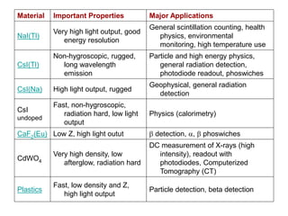
NaI(Tl) 3.67 415 0.23 ms 1.85 100 yes
CsI(Tl) 4.51 550 0.6/3.4 ms 1.79 45 no
CsI(Na) 4.51 420 0.63 ms 1.84 85 slightly
CsI
undoped
4.51 315 16 ns 1.95 4 - 6 no
CaF2
(Eu)
3.18 435 0.84 ms 1.47 50 no
6LiI (Eu) 4.08 470 1.4 ms 1.96 35 yes
6Li -
glass
2.6 390 - 430 60 ns 1.56 4 - 6 no
CsF 4.64 390 3 - 5 ns 1.48 5 - 7 yes
(1) Effective average decay time For -rays.
(2) At the wavelength of the emission maximum.
(3) Relative scintillation signal at room temperature for -rays when coupled to a photomultiplier tube
with a Bi-Alkalai photocathode.
Commonly Used Scintillators
75. Material
Density
[g/cm3]
Emission
Maximum
[nm]
Decay
Constant
(1)
Refractive
Index (2)
Conversion
Efficiency
(3)
Hygros
copic
BaF2 4.88
315
220
0.63 ms
0.8 ns
1.50
1.54
16
5
no
YAP (Ce) 5.55 350 27 ns 1.94 35 - 40 no
GSO (Ce) 6.71 440 30 - 60 ns 1.85 20 - 25 no
BGO 7.13 480 0.3 ms 2.15 15 - 20 no
CdWO4 7.90 470 / 540 20 / 5 ms 2.3 25 - 30 no
Plastics 1.03 375 - 600 1 - 3 ms 1.58 25 - 30 no
(1) Effective agerage decay time For -rays.
(2) At the wavelength of the emission maximum.
(3) Relative scintillation signal at room temperature for -rays when coupled to a photomultiplier tube
with a Bi-Alkalai photocathode.
Commonly Used Scintillators
76. Afterglow
• Defined as the fraction of scintillation light still present for
a certain time after the X-ray excitation stops.
– Originates from the presence of millisecond to even hour long
decay time components.
– Can be as high as a few % after 3 ms in most halide scintillation
crystals .
– CsI(Tl) long duration afterglow can be a problem for many
applications.
– Afterglow in halides is believed to be intrinsic and correlated to
certain lattice defects.
• BGO and Cadmium Tungstate (CdWO4) crystals are
examples of low afterglow scintillation materials
77. Scintillators - Neutron Detection
• Neutrons do not produce ionization directly
in scintillation crystals
• Can be detected through their interaction
with the nuclei of a suitable element.
– 6LiI(Eu) crystal -neutrons interact with 6Li
nuclei to produce an alpha particle and 3H
which both produce scintillation light that can
be detected.
– Enriched 6Li containing glasses doped with
Ce as activator can also be used.
79. Neutron Detection
• Conventional neutron meters surround a
thermal neutron detector with a large and
heavy (20 lb) polyethylene neutron
moderator.
• Other meters utilizes multiple windows
formed of a fast neutron scintillator (ZnS in
an epoxy matrix), with both a thermal
neutron detector and a photomultiplier
tube.
80. Radiation Damage in Scintillators
• Radiation damage results in change in
scintillation characteristics caused by prolonged
exposure to intense radiation.
• Manifests as decrease of optical transmission of
a crystal
– decreased pulse height
– deterioration of the energy resolution
• Radiation damage other than activation may be
partially reversible; i.e. the absorption bands
disappear slowly in time.
81. Radiation Damage in Scintillators
• Doped alkali halide scintillators such as NaI(Tl)
and CsI(Tl) are rather susceptible to radiation
damage.
• All known scintillation materials show more or
less damage when exposed to large radiation
doses.
• Effects usually observed in thick (> 5 cm)
crystals.
• A material is usually called radiation hard if no
measurable effects occur at a dose of 10,000
Gray. Examples of radiation hard materials are
CdWO4 and GSO.
82. Emission Spectra of Scintillation
Crystals
• Each scintillation material has characteristic
emission spectrum.
• Spectrum shape is sometimes dependent on the
type of excitation (photons / particles).
• Emission spectrum is important when choosing
the optimum readout device (PMT /photodiode)
and the required window material.
• Emission spectrum of some common scintillation
materials shown in next two slides.
85. Temperature Influence on the
Scintillation Response
• Light output (photons per MeV gamma) of most
scintillators is a function of temperature.
– Radiative transitions, responsible for the production of
scintillation light compete with non-radiative
transitions (no light production).
– In most light output is quenched (decreased) at higher
temperatures.
– An exception is the fast component of BaF2 where
intensity is essentially temperature independent.
87. Choosing a Scintillator
• Following table lists characteristics such as high density,
fast decay etc.
• Choice of a certain scintillation crystal in a radiation
detector depends strongly on the application.
• Questions such as :
– What is the energy of the radiation to measure ?
– What is the expected count rate ?
– What are the experimental conditions (temperature, shock)?
88. Material Important Properties Major Applications
NaI(Tl)
Very high light output, good
energy resolution
General scintillation counting, health
physics, environmental
monitoring, high temperature use
CsI(Tl)
Non-hygroscopic, rugged,
long wavelength
emission
Particle and high energy physics,
general radiation detection,
photodiode readout, phoswiches
CsI(Na) High light output, rugged
Geophysical, general radiation
detection
CsI
undoped
Fast, non-hygroscopic,
radiation hard, low light
output
Physics (calorimetry)
CaF2(Eu) Low Z, high light outut detection, , phoswiches
CdWO4
Very high density, low
afterglow, radiation hard
DC measurement of X-rays (high
intensity), readout with
photodiodes, Computerized
Tomography (CT)
Plastics
Fast, low density and Z,
high light output
Particle detection, beta detection
89. Material Important Properties Major Applications
6LiI(Eu)
High neutron cross-section,
high light output
Thermal neutron detection and
spectroscopy
6Li -
glass
High neutron cross-section,
non-hygroscopic
Thermal neutron detection
BaF2 Ultra-fast sub-ns UV emission
Positron life time studies, physics
research, fast timing
YAP(Ce
)
High light output, low Z, fast
MHz X-ray spectroscopy, synchrotron
physics
GSO(Ce
)
High density and Z, fast,
radiation hard
Physics research
BGO High density and Z
Particle physics, geophysical
research, PET, anti-Compton
spectrometers
CdWO4
Very high density, low
afterglow, radiation hard
DC measurement of X-rays (high
intensity), readout with
photodiodes, Computerized
Tomography (CT)
Plastics
Fast, low density and Z, high
light output
Particle detection, beta detection
90. PRACTICAL SCINTILLATION
COUNTERS
• Highly sensitive surface contamination probes
incorporate a range phosphors
• Examples include:
– zinc sulphide (ZnS(Ag)) powder coatings (5–10
mg·cm–2) on glass or plastic substrates or coated
directly onto the photomultiplier window for detecting
alpha and other heavy particles;
– cesium iodide (CsI(Tl)) that is thinly machined (0.25
mm) and that may be bent into various shapes;
– and plastic phosphors in thin sheets or powders fixed
to a glass base for beta detection.
91.
92. PRACTICAL SCINTILLATION
COUNTERS
• Probes (A and B previous slide) and their associated
ratemeters (C) tend not to be robust.
• Photomultipliers are sensitive to shock damage and are
affected by localized magnetic fields.
• Minor damage to the thin foil through which radiation
enters the detector allows ambient light to enter and
swamp the photomultiplier.
• Cables connecting ratemeters and probes are also a
common problem.
• Very low energy beta emitters (for example 3H) can be
dissolved in liquid phosphors in order to be detected.
93. 43-93 Alpha/Beta Scintillator
• The Model 43-93 is a 100 cm² dual
phosphor alpha/beta scintillator that is
designed to be used for simultaneously
counting alpha and beta contamination
94. 43-93 Alpha/Beta Scintillator
• INDICATED USE: Alpha beta survey
• SCINTILLATOR: ZnS(Ag) adhered to 0.010" thick plastic scintillation
material
• WINDOW: 1.2 mg/cm² recommended for outdoor use
• WINDOW AREA:
– Active - 100 cm²
– Open - 89 cm²
• EFFICIENCY (4pi geometry): Typically 15% - Tc-99; 20% - Pu-239; 20% -
S-90/Y-90
• NON-UNIFORMITY: Less than 10%
• BACKGROUND: Alpha - 3 cpm or less
• Beta - Typically 300 cpm or less (10 µR/hr field )
• CROSS TALK:
– Alpha to beta - less than 10%
– Beta to alpha - less than 1%
95. 43-93 Alpha/Beta Scintillator
• COMPATIBLE INSTRUMENTS: Models 2224, 2360
• TUBE: 1.125"(2.9cm) diameter magnetically shielded
photomultiplier
• OPERATING VOLTAGE: Typically 500 - 1200 volts
• DYNODE STRING RESISTANCE: 100 megohm
• CONNECTOR: Series “C” (others available )
• CONSTRUCTION: Aluminum housing with beige
polyurethane enamel paint
• TEMPERATURE RANGE: 5°F(-15°C) to 122°F(50°C)
May be certified to operate from -40°F(-40°C) to
150°F(65°C)
• SIZE: 3.2"(8.1 cm)H X 3.5"(8.9 cm)W X 12.2"(31 cm)L
• WEIGHT: 1 lb (0.5kg)
96. 44-2 Gamma Scintillator
• The Model 44-2 is a 1" X 1" NaI(Tl)
Gamma Scintillator that can be used with
several different instruments including
survey meters, scalers, ratemeters, and
alarm ratemeters
97. • INDICATED USE: High energy gamma detection
• SCINTILLATOR: 1" (2.5 cm) diameter X 1" (2.5 cm) thick sodium iodide
(NaI)Tl scintillator
• SENSITIVITY: Typically 175 cpm/microR/hr (Cs-137)
• COMPATIBLE INSTRUMENTS: General purpose survey meters,
ratemeters, and scalers
• TUBE: 1.5:(3.8cm) diameter magnetically shielded photomultiplier
• OPERATING VOLTAGE: Typically 500 - 1200 volts
• DYNODE STRING RESISTANCE: 100 megohm
• CONNECTOR: Series "C" (others available )
• CONSTRUCTION: Aluminum housing with beige polyurethane enamel
paint
• TEMPERATURE RANGE: -4° F(-20° C) to 122° F(50° C)
May be certified for operation from -40° F(-40° C) to 150° F(65° C)
• SIZE: 2" (5.1 cm) diameter X 7.3" (18.5 cm)L
• WEIGHT: 1 lb (0.5kg)
44-2 Gamma Scintillator
98. Scintillation Detectors
• Best:
– Measure low gamma dose rates
• Also:
– Measure beta dose rates (with corrections)
• However:
– Somewhat fragile and expensive
• CANNOT:
– Not intended for detecting contamination, only
radiation fields
104. Semiconductor Detectors
• Solids have
– lattice structure (molecular level)
– quantized energy levels
– valence bands
– conduction bands
• Semiconductors have lattice structure
– similar to inorganic scintillators
– composed of Group IVB elements
– ability to easily share electrons with adjoining atoms
106. Basic Nature of Semiconductors
• Schematic view of lattice of Group IV-B (Group 4B)
element Si
• Dots represent electron pair bonds between the Si
atoms
Si
Si
Si
Si
Si
Si
107. Basic Nature, cont’d
• Schematic diagram of energy levels of crystalline
Si.
• Pure Si is a poor conductor of electricity
Conduction Band
Valence Band
Energy
1.08 eV Forbidden Gap
108. Basic Nature, cont’d
• Schematic view of lattice of Group IV element
Si, doped with P (Group VB) as an impurity –
note extra electron
Si
Si
Si
Si
Si
P
109. Basic Nature, cont’d
• Schematic diagram of disturbed energy levels of
crystalline Si.
• Si with Group V impurities like P is said to be an n-
type silicon because of the negative charge carriers
(the electrons)
Conduction Band
Valence Band
Energy
0.05 eV Donor level
110. Basic Nature, cont’d
• Schematic view of lattice of Group IV element
Si, doped with B (Group IIIB) as an impurity –
note hole in electron orbital
Si
Si
Si
Si
Si
B
111. Basic Nature, cont’d
• Schematic diagram of disturbed energy levels of
crystalline Si with B impurity.
• Si with Group III impurities is said to be a p-type silicon
because of the positive charge carriers (the holes)
Conduction Band
Valence Band
Energy
0.08 eV Acceptor level
112. Occupation of energy states for
n and p-type semiconductors
Conduction Band
Valence Band
After Turner
0.67 eV
As donor
impurity
levels
Ga acceptor
impurity
levels
0.013 eV
0.011eV
113. Operating Principles of
Semiconductor detectors
• Si semiconductor is a layer of p-type Si in contact with n-
type Si.
• What happens when this junction is created?
– Electrons from n-type migrate across junction to fill holes
in p-type
– Creates an area around the p-n junction with no excess of
holes or electrons
– Called a “depletion region”
• Apply (+) voltage to n-type and (-) to p-type:
– Depletion region made thicker
– Called a “reverse bias”
114. Energy-level diagram for n-p
junction
Conduction
Band
Valence
Band
After Turner
n-type
p-type
Junction
region
115. Detector specifics
• Depletion region acts as sensitive volume of
the detector
• Passage of ionizing radiation through the
region
– Creates holes in valence band
– Electrons in conduction band
– Electrons migrate to positive charge on n side
– Holes migrate to negative voltage on p side
– Creates electrical output
• Requires about 3.6 eV to create an electron
hole pair in Si
116. Detector Specifics, cont’d
• Reverse bias n-p junction is good detector
– Depletion region
• Has high resistivity
• Can be varied by changing bias voltage
– Ions produced can be quickly collected
– Number of ion pairs collected is proportional to
energy deposited in detector
• Junction can be used as a spectrometer
• Types of detectors:
– HPGe
– GeLi (lithium drifted detectors)
– Surface barrier detectors
– Electronic dosimeters
117.
118. SOLID STATE DETECTORS RECAP
• Solid state detectors utilize semiconductor
materials.
• Intrinsic semiconductors are of very high purity
and extrinsic semiconductors are formed by
adding trace quantities (impurities) such as
phosphorus (P) and lithium (Li) to materials such
as germanium (Ge) and silicon (Si).
• There are two groups of detectors:
– junction detectors and bulk conductivity detectors.
119. SOLID STATE DETECTORS
• Junction detectors are of either
– diffused junction or
– surface barrier type:
– an impurity is either diffused into, or spontaneously oxidized
onto, a prepared surface of intrinsic material to change a layer of
‘p-type’ semiconductor from or to ‘n-type’.
• When a voltage (reverse bias) is applied to the surface barrier
detector it behaves like a solid ionization chamber.
• Bulk conductivity detectors are formed from intrinsic semiconductors
of very high bulk resistivity (for example CdS and CdSe).
• They also operate like ionization counters but with a higher density
than gases and a ten-fold greater ionization per unit absorbed dose.
• Further amplification by the detector creates outputs of about one
microampere at 10 mSv·h–1
120. Solid State Counters
– A - very thin metal
(gold) electrode.
– P - thin layer of p-
type semiconductor.
– D - depletion region,
3–10 mm thick
formed by the
voltage, is free of
charge in the absence
of ionizing radiations.
– N - n-type
semiconductor.
– B - thin metal
electrode which
provides a positive
potential at the n-type
semiconductor.
121. PRACTICAL SOLID STATE
DETECTORS
• The main applications for semiconductor detectors are in
the laboratory for the spectrometry of both heavy
charged (alpha) particle and gamma radiations.
• However, energy compensated PIN diodes and special
photodiodes are used as pocket electronic (active)
dosimeters.
– PIN diode: Acronym for positive-intrinsic-negative diode.
– A photodiode with a large, neutrally doped intrinsic region
sandwiched between p-doped and n-doped semiconducting
regions.
– A PIN diode exhibits an increase in its electrical conductivity as a
function of the intensity, wavelength, and modulation rate of the
incident radiation. Synonym PIN photodiode.
122. PIN Diodes
• Ordinary Silicon PIN photodiodes can serve as detectors for X-ray
and gamma ray photons. The detection efficiency is a function of the
thickness of the silicon wafer. For a wafer thickness of 300 microns
(ignoring attenuation in the diode window and/or package) the
detection efficiency is close to 100% at 10 KeV, falling to
approximately 1% at 150 KeV(3).
• For energies above approximately 60 KeV, photons interact almost
entirely through Compton scattering. Moreover, the active region of
the diode is in electronic equilibrium with the surrounding medium--
the diode package, substrate, window and outer coating, etc., so
that Compton recoil electrons which are produced near--and close
enough to penetrate--the active volume of the diode, are also
detected.
• For this reason the overall detection efficiency at 150 KeV and
above is maintained fairly constant (approximately 1%) over a wide
range of photon energies.
• Thus, a silicon PIN diode can be thought of as a solid-state
equivalent to an ionization-chamber radiation detector.
123. PRACTICAL SOLID STATE
DETECTORS
• Specially combined thin and thick detectors provide the
means to identify charged particles.
– used to monitor for plutonium in air, discriminating against alpha
particles arising from natural radioactivity, and for monitoring for
radon daughter products in air.
– Small physical size and insensitivity to gamma radiation have
found novel applications: inside nuclear fuel flasks monitoring for
alpha contamination and checking sealed radium sources for
leakage.
• Bulk conductivity detectors can measure high dose rates
but with minute-long response times. A Ge(Li) detector
operated at –170°C is capable of a very high gamma
resolution of 0.5%. The temperature dependence and
high cost add to their impracticality.
124. Another type of Solid State /
Scintillation system
Thermoluminescent Dosimeters
125. Thermoluminescence
• (TL) is the ability to convert energy from
radiation to a radiation of a different wavelength,
normally in the visible light range.
• Two categories
– Fluorescence - emission of light during or immediately
after irradiation
– Not a particularly useful reaction for TLD use
– Phosphorescence - emission of light after the
irradiation period. Delay can be seconds to months.
• TLDs use phosphorescence to detect radiation.
126. Thermoluminescence
• Radiation moves electrons into “traps”
• Heating moves them out
• Energy released is proportional to
radiation
• Response is ~ linear
• High energy trap data is stored in TLD for
a long time
127. TL Process
Valence Band (outermost electron shell)
Conduction Band (unfilled shell)
Phosphor atom
Incident
radiation
Electron trap
(metastable state)
-
128. TL Process, continued
Valence Band (outermost electron shell)
Conduction Band
Phosphor atom
Thermoluminescent
photon Heat Applied-
129. Output – Glow Curves
• A glow curve is obtained from heating
• Light output from TLis not easily interpreted
• Multiple peaks result from electrons in "shallow" traps
• Peak results as traps are emptied.
• Light output drops off as these traps are depleted.
• Heating continues
• Electrons in deeper traps are released.
• Highest peak is typically used to calculate dose
• Area under represents the radiation energy deposited in
the TLD
130. Trap Depths - Equate to LongTerm
Stability of Information
Time or temperature
131. TLD Reader Construction
Power Supply
PMT
DC Amp
Filter
Heated Cup
TL material
To High
Voltage To ground
Recorder or meter
132. Advantages
• Advantages (as compared to film dosimeter
badges) includes:
– Able to measure a greater range of doses
– Doses may be easily obtained
– They can be read on site instead of being sent away
for developing
– Quicker turnaround time for readout
– Reusable
– Small size
– Low cost
134. NON-TL Dosimeters
• LUXEL DOSIMETER
• "Optically Stimulated Luminescence"
(OSL) technology
• Minimum detectable dose
– 1 mRem for gamma and x-ray radiation,
– 10 mRem for beta radiation.
135. Non TL Dosimeters, continued
• Uses thin layer of Al2O3:C
• Has a TL sensitivity 50 times greater than
TLD-100 (LiF:Mg,Ti)
• Almost tissue equivalent.
• Strong sensitivity to light
• Thermal quenching.
• Readout stimulated using laser
• Dosimeter luminesces in proportion to
radiation dose.
136. Summary
• Wide range of detection equipment
available
• Understand strengths and weaknesses of
each
• No single detector will do everything
• We’ll get to selection issues in the next
two days
137. Suggested Reading
• Glenn F. Knoll, Radiation Detection and
Measurement, John Wiley & Sons.
• Hernam Cember, Introduction to Health
Physics, McGraw Hill.
• Nicholas Tsoulfanidis, Measurement and
Detection of Radiation, Taylor & Francis.
• C.H. Wang, D.L.Willis, W.D. Loveland,
Radiotracer Methodology in the Biological,
Environmental and Physical Sciences,
Prentice-Hall