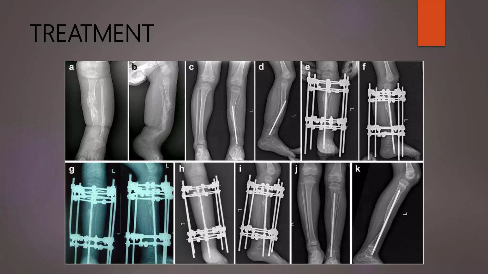
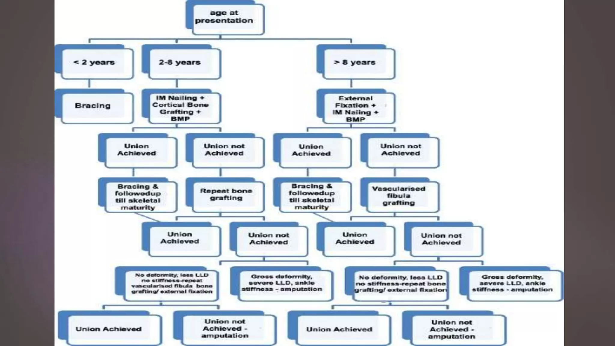
CONGENITAL
PSEUDOARTHROSIS
OF TIBIA
PRESENTED BY: DR. SACHIN. M.
2ND YEAR PGT, DEPT OF ORTHOPAEDICS, SMCH
MODERATOR: DR. BIPUL BORTHAKUR
PROFESSOR, DEPT OF ORTHOPAEDICS, SMCH
LEARNING OBJECTIVES
 INTRODUCTION
 ETIOPATHOGENESIS
 CLASSIFICATION
 INVESTIGATIONS
 TREATMENT
 COMPLICATIONS
INTRODUCTION
 Congenital pseudarthrosis is a specific type of a non-union
 Pseudarthrosis – “false joint”
 Either present or incipient at birth
 Most commonly involves distal half of the tibia and ipsilateral fibula
CONGENITAL PSEUDARTHROSIS OF
TIBIA
 Rare disorder with an incidence of 1 in 2,50,000 live births
 Non-union of a tibial fracture that occurs spontaneously or after a minor
trauma
 Usually develops within first 2 years of life; can be seen before birth also
ETIOPATHOGENESIS
 Exact cause is not known, but closely related to patients with
 Neurofibromatosis or related stigmata in 50% cases (NF1 gene, 17q11)
 Fibrous dysplasia in 10% of cases
 Others are – constriction band syndromes and Osteofibrous dysplasia
 Non-union occurs due to poor healing following fracture of the bone
ETIOPATHOGENESIS
 Most accepted theory is linked to the periosteum
 An accumulation of nerve cells (NF1 haplopotent MSCs) around the periosteal
vasculature, that destroy its blood supply.
 Hamartomatous thickened fibrous tissue with limited vascular ingrowth is seen
at the site of pseudarthrosis
CLASSIFICATION
 ANDERSEN’S CLASSIFICATION SYSTEM - Based on morphology
 Dysplastic
 Cystic (no association with NF)
 Sclerotic
 Clubfoot type that arises due to associated abnormalities
CLASSIFICATION
 CRAWFORD’S CLASSIFICATION SYSTEM –Radiological description of lesions
NON-DYSPLASTIC DYSPLASTIC DYSPLASTIC DYSPLASTIC
Hourglass
constriction
Cysts
Frank
pseudarthrosis
CLASSIFICATION – BOYD’S
 BOYD’S CLASSIFICATION OF CPT
 TYPE I: Anterior bowing and a defect in the tibia present at birth
 TYPE II: Anterior bowing + hourglass constriction of the tibia present at birth
(Crawford’s type IIA)
 Most common type
 High risk tibia with poorest prognosis
 Tibia is tapered, rounded and sclerotic with obliterated medullary canal
 Re-fracture is common during the growth period, ceases after skeletal maturity
CLASSIFICATION
 TYPE III: Anterior bowing + congenital cysts in the bone
 Near the junction of middle and distal thirds of the tibia
 Less chances of re-fracture
 TYPE IV: occurs in a sclerotic segment of the bone without narrowing of the
tibia
 Partial or complete obliteration of the medullary canal
 Starts as stress or insufficiency fracture – complete fracture – non-union
 Good prognosis if treatment is started before completion of the insufficiency
fracture
CLASSIFICATION
 TYPE V: pseudarthrosis of the tibia occurs with a dysplastic fibula
 Dysplastic fibula may end up in pseudarthrosis – poor prognosis
 TYPE VI: Intra-osseous neurofibroma or schwannoma that results in the
pseudarthrosis
 Extremely rare
 Prognosis – depends on the aggressiveness of intra-osseous lesions and its
treatment
CLASSIFICATION – PALEY’S
 EL ROSASSY – PALEY – HERZENBERG’S CLASSIFICATION: based on
 Geometry and mobility of the bone ends: thick & stiff OR thin & mobile
 Previous, unsuccessful surgery
 TYPE I:
 Atrophic (narrow) bone ends
 Mobile pseudarthrosis
 No previous surgery
CLASSIFICATION – PALEY’S
 TYPE II:
 Atrophic (narrow) bone ends
 Mobile pseudarthrosis
 Previous unsuccessful surgery
 TYPE III:
 Hypertrophic (wide) bone ends
 Stiff pseudarthrosis
 With or without previous surgery
CLASSIFICATION – JOHNSON ET AL
 Can be used to guide the treatment and prognosis
 Based on
 Presence or absence of fracture
 Age of first fracture
 Fractured tibia – require surgical treatment
 Intact tibia – Observation and splinting
INVESTIGATIONS
 Conventional radiography
INVESTIGATIONS
 MRI - provides valuable information on the extent of the disease and is
helpful for the preoperative planning in that the borders for resection can
be defined precisely
 CT scan - confirm radiographic findings, showing osteolytic lesions
containing solid tissue
 Total bone scintigraphy - a slight uptake at the beginning of the dynamic
venous phase and a high uptake during terminal phase
TREATMENT
 Primary goals are:
 Union
 No re-fractures
 Correction of diaphyseal deformity
 Secondary goals are:
 Correction of ankle-knee valgus
 Treatment of limb length discrepancy
 Correction of foot deformity
TREATMENT
 Depends on the age of the patient and presence or absence of the fracture
 Before walking age – little treatment (casting)
 Once ambulation begins – casting or bracing (Clamshell orthosis or PTB
orthosis), if there is no fracture; can be continued till skeletal maturity
 Once fracture occurs – surgical management
TREATMENT
 SURGICAL MANAGEMENT
 Basic principles include:
 Resection of the entire pseudarthrosis and surrounding hamartomatous tissue
 Restoration of mechanical alignment
 Intramedullary fixation
 Augmentation techniques:
 Primary shortening, bone transport, supplemental bone grafting and BMPs
TREATMENT
 SURGICAL MANAGEMENT
 Amputation – rarely required in initial stages
 Anticipated shortening of > 2-3 inches
 History of multiple failed surgical procedures
 Stiffness and decreased function of a limb that would be more useful after an
amputation and prosthetic fitting
TREATMENT
 INTRAMEDULLARY FIXATION – William’s IM rod (Anderson et al)
 Most commonly used technique
 In very distal tibia CPT – may require to cross the ankle joint to provide
additional stability
 Rod can migrate with growth or can be surgically reposition the rod above the
ankle to restore the ankle motion
 In proximal tibia CPT – possible to avoid crossing the ankle joint
 Larger diameter rod or the one with interlocking option aid in stability
TREATMENT
 INTRAMEDULLARY FIXATION
Fassier Duval telescopic nailWilliam’s IM rod
TREATMENT
 VASCULARIZED BONE GRAFTS
 Reconstruction of the resected pseudarthrosis with vascularized bone graft
– can be harvested by fibula or iliac crest
 Requires experience in microvascular techniques
 Two surgical teams – one for harvesting the graft and the other for
preparing the pseudarthrosis site, would be helpful
 Vascularized fibular grafts – CPT with gap > 3cm and CPT with multiple
failed surgeries
TREATMENT
 BONE MORPHOGENIC PROTEINS
 Can be used as an adjunctive to the surgical stabilization of the
pseudarthrosis
 Both rhBMP-2 and rhBMP-7 have been used
 Early union rates have been reported
 Long term follow-up is needed to understand the long term efficacy and
safety of these treatments
TREATMENT
 ILIZAROV FIXATION
 Offer the advantage of maintaining or gaining tibial length
 Disadvantages include –
 Difficulty transporting the proximal tibia
 Docking malalignment
 Poor quality of regenerated bone
TREATMENT
 PALEY’S INSTITUTE TREATMENT STRATEGY FOR CPT
 It’s a culmination of 28 years of Dr. Paley’s treatment technique
 Comprises of 4 papers, recent one being published in 2012
 Ilizarov fixator + bone graft + IM nailing (FD telescopic nail) + periosteal graft +
BMP insertion + Zolidronic acid infusion + tibio-fibular cross union
 Locking plates can also be used instead of Ilizarov fixator
 100% union rate with 0% re-fracture rate
TREATMENT
PROGNOSIS
 Factors leading to poor prognosis are:
 Lower age at treatment
 Previous failed treatment
 Neurofibromatosis
 Increased follow up
COMPLICATIONS
 Stiffness of the ankle and hindfoot: usually disappears once the IM nail is
proximal to the ankle joint
 If persists, rarely hampers the functional results
 Re-fracture: most common complication after surgical treatment of CPT
(50% to 75%)
 Can be managed with casting or exchange nailing
 IM nail should be left in place till skeletal maturity
COMPLICATIONS
 Valgus ankle deformity: occurs when the distal tibia fragment is not fixed
during insertion of IM nail
 More frequently seen when the fibula is left intact than when fibular osteotomy
is done
 Long term bracing during growth years to prevent progressive deformity
 Langenskiold procedure may be indicated
COMPLICATIONS
 Tibial shortening:
 Can be treated by a well-timed contralateral epiphysiodesis or limb lengthening
of the proximal tibia
 Ilizarov technique in patients with significant shortening and a wide non-union;
in patients with failed IM nail and bone grafting procedures
Congenital pseudarthrosis of tibia - Dr. Sachin M

Congenital pseudarthrosis of tibia - Dr. Sachin M

  • 1.
    CONGENITAL PSEUDOARTHROSIS OF TIBIA PRESENTED BY:DR. SACHIN. M. 2ND YEAR PGT, DEPT OF ORTHOPAEDICS, SMCH MODERATOR: DR. BIPUL BORTHAKUR PROFESSOR, DEPT OF ORTHOPAEDICS, SMCH
  • 2.
    LEARNING OBJECTIVES  INTRODUCTION ETIOPATHOGENESIS  CLASSIFICATION  INVESTIGATIONS  TREATMENT  COMPLICATIONS
  • 3.
    INTRODUCTION  Congenital pseudarthrosisis a specific type of a non-union  Pseudarthrosis – “false joint”  Either present or incipient at birth  Most commonly involves distal half of the tibia and ipsilateral fibula
  • 4.
    CONGENITAL PSEUDARTHROSIS OF TIBIA Rare disorder with an incidence of 1 in 2,50,000 live births  Non-union of a tibial fracture that occurs spontaneously or after a minor trauma  Usually develops within first 2 years of life; can be seen before birth also
  • 5.
    ETIOPATHOGENESIS  Exact causeis not known, but closely related to patients with  Neurofibromatosis or related stigmata in 50% cases (NF1 gene, 17q11)  Fibrous dysplasia in 10% of cases  Others are – constriction band syndromes and Osteofibrous dysplasia  Non-union occurs due to poor healing following fracture of the bone
  • 6.
    ETIOPATHOGENESIS  Most acceptedtheory is linked to the periosteum  An accumulation of nerve cells (NF1 haplopotent MSCs) around the periosteal vasculature, that destroy its blood supply.  Hamartomatous thickened fibrous tissue with limited vascular ingrowth is seen at the site of pseudarthrosis
  • 7.
    CLASSIFICATION  ANDERSEN’S CLASSIFICATIONSYSTEM - Based on morphology  Dysplastic  Cystic (no association with NF)  Sclerotic  Clubfoot type that arises due to associated abnormalities
  • 8.
    CLASSIFICATION  CRAWFORD’S CLASSIFICATIONSYSTEM –Radiological description of lesions NON-DYSPLASTIC DYSPLASTIC DYSPLASTIC DYSPLASTIC Hourglass constriction Cysts Frank pseudarthrosis
  • 9.
    CLASSIFICATION – BOYD’S BOYD’S CLASSIFICATION OF CPT  TYPE I: Anterior bowing and a defect in the tibia present at birth  TYPE II: Anterior bowing + hourglass constriction of the tibia present at birth (Crawford’s type IIA)  Most common type  High risk tibia with poorest prognosis  Tibia is tapered, rounded and sclerotic with obliterated medullary canal  Re-fracture is common during the growth period, ceases after skeletal maturity
  • 10.
    CLASSIFICATION  TYPE III:Anterior bowing + congenital cysts in the bone  Near the junction of middle and distal thirds of the tibia  Less chances of re-fracture  TYPE IV: occurs in a sclerotic segment of the bone without narrowing of the tibia  Partial or complete obliteration of the medullary canal  Starts as stress or insufficiency fracture – complete fracture – non-union  Good prognosis if treatment is started before completion of the insufficiency fracture
  • 11.
    CLASSIFICATION  TYPE V:pseudarthrosis of the tibia occurs with a dysplastic fibula  Dysplastic fibula may end up in pseudarthrosis – poor prognosis  TYPE VI: Intra-osseous neurofibroma or schwannoma that results in the pseudarthrosis  Extremely rare  Prognosis – depends on the aggressiveness of intra-osseous lesions and its treatment
  • 12.
    CLASSIFICATION – PALEY’S EL ROSASSY – PALEY – HERZENBERG’S CLASSIFICATION: based on  Geometry and mobility of the bone ends: thick & stiff OR thin & mobile  Previous, unsuccessful surgery  TYPE I:  Atrophic (narrow) bone ends  Mobile pseudarthrosis  No previous surgery
  • 13.
    CLASSIFICATION – PALEY’S TYPE II:  Atrophic (narrow) bone ends  Mobile pseudarthrosis  Previous unsuccessful surgery  TYPE III:  Hypertrophic (wide) bone ends  Stiff pseudarthrosis  With or without previous surgery
  • 14.
    CLASSIFICATION – JOHNSONET AL  Can be used to guide the treatment and prognosis  Based on  Presence or absence of fracture  Age of first fracture  Fractured tibia – require surgical treatment  Intact tibia – Observation and splinting
  • 15.
  • 16.
    INVESTIGATIONS  MRI -provides valuable information on the extent of the disease and is helpful for the preoperative planning in that the borders for resection can be defined precisely  CT scan - confirm radiographic findings, showing osteolytic lesions containing solid tissue  Total bone scintigraphy - a slight uptake at the beginning of the dynamic venous phase and a high uptake during terminal phase
  • 17.
    TREATMENT  Primary goalsare:  Union  No re-fractures  Correction of diaphyseal deformity  Secondary goals are:  Correction of ankle-knee valgus  Treatment of limb length discrepancy  Correction of foot deformity
  • 18.
    TREATMENT  Depends onthe age of the patient and presence or absence of the fracture  Before walking age – little treatment (casting)  Once ambulation begins – casting or bracing (Clamshell orthosis or PTB orthosis), if there is no fracture; can be continued till skeletal maturity  Once fracture occurs – surgical management
  • 19.
    TREATMENT  SURGICAL MANAGEMENT Basic principles include:  Resection of the entire pseudarthrosis and surrounding hamartomatous tissue  Restoration of mechanical alignment  Intramedullary fixation  Augmentation techniques:  Primary shortening, bone transport, supplemental bone grafting and BMPs
  • 20.
    TREATMENT  SURGICAL MANAGEMENT Amputation – rarely required in initial stages  Anticipated shortening of > 2-3 inches  History of multiple failed surgical procedures  Stiffness and decreased function of a limb that would be more useful after an amputation and prosthetic fitting
  • 21.
    TREATMENT  INTRAMEDULLARY FIXATION– William’s IM rod (Anderson et al)  Most commonly used technique  In very distal tibia CPT – may require to cross the ankle joint to provide additional stability  Rod can migrate with growth or can be surgically reposition the rod above the ankle to restore the ankle motion  In proximal tibia CPT – possible to avoid crossing the ankle joint  Larger diameter rod or the one with interlocking option aid in stability
  • 22.
    TREATMENT  INTRAMEDULLARY FIXATION FassierDuval telescopic nailWilliam’s IM rod
  • 23.
    TREATMENT  VASCULARIZED BONEGRAFTS  Reconstruction of the resected pseudarthrosis with vascularized bone graft – can be harvested by fibula or iliac crest  Requires experience in microvascular techniques  Two surgical teams – one for harvesting the graft and the other for preparing the pseudarthrosis site, would be helpful  Vascularized fibular grafts – CPT with gap > 3cm and CPT with multiple failed surgeries
  • 24.
    TREATMENT  BONE MORPHOGENICPROTEINS  Can be used as an adjunctive to the surgical stabilization of the pseudarthrosis  Both rhBMP-2 and rhBMP-7 have been used  Early union rates have been reported  Long term follow-up is needed to understand the long term efficacy and safety of these treatments
  • 25.
    TREATMENT  ILIZAROV FIXATION Offer the advantage of maintaining or gaining tibial length  Disadvantages include –  Difficulty transporting the proximal tibia  Docking malalignment  Poor quality of regenerated bone
  • 26.
    TREATMENT  PALEY’S INSTITUTETREATMENT STRATEGY FOR CPT  It’s a culmination of 28 years of Dr. Paley’s treatment technique  Comprises of 4 papers, recent one being published in 2012  Ilizarov fixator + bone graft + IM nailing (FD telescopic nail) + periosteal graft + BMP insertion + Zolidronic acid infusion + tibio-fibular cross union  Locking plates can also be used instead of Ilizarov fixator  100% union rate with 0% re-fracture rate
  • 27.
  • 29.
    PROGNOSIS  Factors leadingto poor prognosis are:  Lower age at treatment  Previous failed treatment  Neurofibromatosis  Increased follow up
  • 30.
    COMPLICATIONS  Stiffness ofthe ankle and hindfoot: usually disappears once the IM nail is proximal to the ankle joint  If persists, rarely hampers the functional results  Re-fracture: most common complication after surgical treatment of CPT (50% to 75%)  Can be managed with casting or exchange nailing  IM nail should be left in place till skeletal maturity
  • 31.
    COMPLICATIONS  Valgus ankledeformity: occurs when the distal tibia fragment is not fixed during insertion of IM nail  More frequently seen when the fibula is left intact than when fibular osteotomy is done  Long term bracing during growth years to prevent progressive deformity  Langenskiold procedure may be indicated
  • 32.
    COMPLICATIONS  Tibial shortening: Can be treated by a well-timed contralateral epiphysiodesis or limb lengthening of the proximal tibia  Ilizarov technique in patients with significant shortening and a wide non-union; in patients with failed IM nail and bone grafting procedures