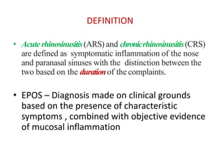
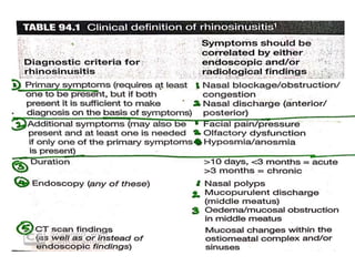
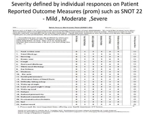
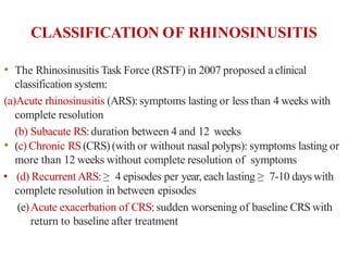
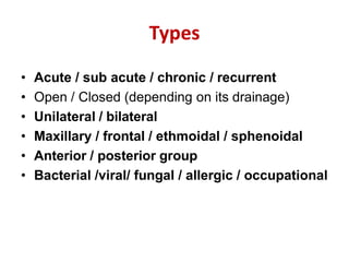
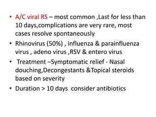
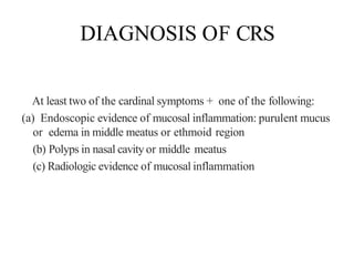
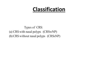
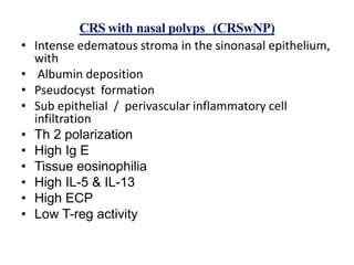
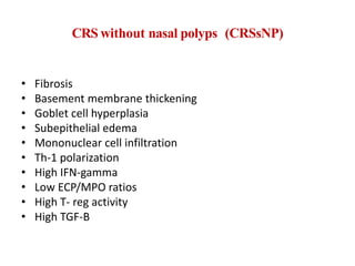
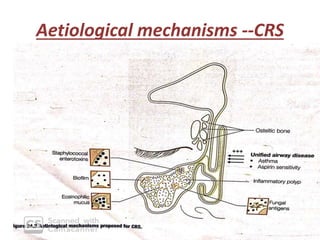
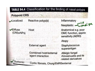
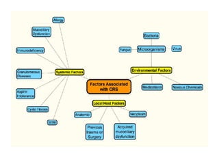
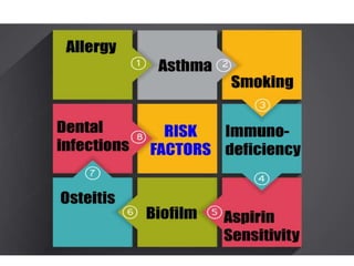
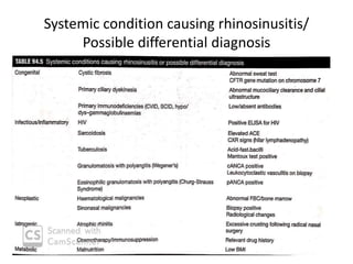
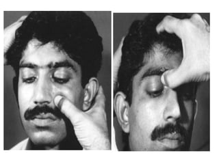
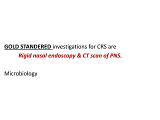
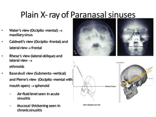
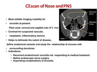
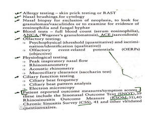
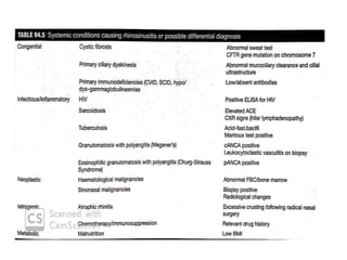
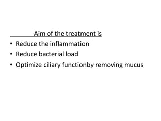
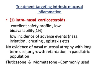
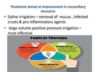
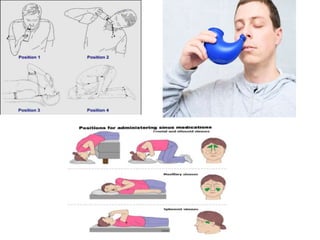
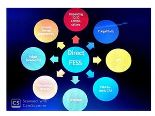
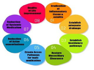
Rhinosinusitis
Dr Mohammed Nishad N
DEFINITION
• Acute rhinosinusitis(ARS) and chronicrhinosinusitis(CRS)
are defined as symptomatic inflammation of the nose
and paranasal sinuses with the distinction between the
two based on the durationof the complaints.
• EPOS – Diagnosis made on clinical grounds
based on the presence of characteristic
symptoms , combined with objective evidence
of mucosal inflammation
Severity defined by individual responces on Patient
Reported Outcome Measures (prom) such as SNOT 22
- Mild , Moderate ,Severe
CLASSIFICATION OF RHINOSINUSITIS
• The Rhinosinusitis Task Force (RSTF) in 2007 proposed a clinical
classification system:
(a)Acute rhinosinusitis (ARS):symptoms lasting or less than 4 weeks with
complete resolution
(b) Subacute RS:duration between 4 and 12 weeks
• (c) Chronic RS(CRS) (with or without nasal polyps): symptoms lasting or
more than 12 weeks without complete resolution of symptoms
• (d) Recurrent ARS:≥ 4 episodes per year, each lasting ≥ 7-10 days with
complete resolution in between episodes
(e)Acute exacerbation of CRS:sudden worsening of baseline CRSwith
return to baseline after treatment
Types
• Acute / sub acute / chronic / recurrent
• Open / Closed (depending on its drainage)
• Unilateral / bilateral
• Maxillary / frontal / ethmoidal / sphenoidal
• Anterior / posterior group
• Bacterial /viral/ fungal / allergic / occupational
• A/C viral RS – most common ,Last for less than
10 days,complications are very rare, most
cases resolve spontaneously
• Rhinovirus (50%) , influenza & parainfluenza
virus , adeno virus ,RSV & entero virus
• Treatment –Symptomatic relief - Nasal
douching,Decongestants &Topical steroids
based on severity
• Duration > 10 days consider antibiotics
• ABRS --0.5-2%
• S pneumoniae(27%) , H.Influenzae(44%) ,
M.catarrhalis(14%) , S . pyogenes ,S.aureus
• BACTERIOLOGY
− Streptococcus
pneumoniae
− Hemophilus
influenzae
− Moraxella catarrhalis
− Staphylococcus
aureus
− Neisseria
• Acute sinusitis • Chronicsinusitis
− Staph.Aureus
− Streptococcus
− H. influenzae
− Bacteroides
− Pseudomonas
Predisposing factors to ARS
• Anatomical factors (DNS, Haller cell ,Concha
bullosa , etc
• Allergy
• Smoking
• Poor mental health
• Pathophysiology relating to viral agents – Cell
invasion of respiratory epithelium –
inflammatory changes including mechanical
changes ,epithelial damage and activation of
humoral and cellular defences
• In ABRS – superinfection following an initial
viral insult –Epithelial disruption has already
occurred & associated decrease in ciliated
cells and increase in goblet cells –sinus osteal
obstruction
• The accumulating mucus causes an initial
increase in the intra sinus pressure followed
quickly by negative pressure due to lack of
ventilation . This then set up a vicious cycle of
further congestion ,mucus retention ,
impaired gas exchange and PH balance –
prevents clearance of inflammatory products
& debris ----ideal medium for bacteria to
flourish
Chronic Rhinosinusitis
• Four cardinal symptoms of CRS
(a) Anterior or posterior purulent nasal discharge
(b) Nasal obstruction
(c) Face pain or pressure
(d) Hyposmia or anosmia
DIAGNOSIS OF CRS
At least two of the cardinal symptoms + one of the following:
(a) Endoscopic evidence of mucosal inflammation: purulent mucus
or edema in middle meatus or ethmoid region
(b) Polyps in nasal cavity or middle meatus
(c) Radiologic evidence of mucosal inflammation
Classification
Types of CRS:
(a) CRSwith nasal polyps (CRSwNP)
(b) CRSwithout nasal polyps (CRSsNP)
CRS with nasal polyps (CRSwNP)
• Intense edematous stroma in the sinonasal epithelium,
with
• Albumin deposition
• Pseudocyst formation
• Sub epithelial / perivascular inflammatory cell
infiltration
• Th 2 polarization
• High Ig E
• Tissue eosinophilia
• High IL-5 & IL-13
• High ECP
• Low T-reg activity
CRS without nasal polyps (CRSsNP)
• Fibrosis
• Basement membrane thickening
• Goblet cell hyperplasia
• Subepithelial edema
• Mononuclear cell infiltration
• Th-1 polarization
• High IFN-gamma
• Low ECP/MPO ratios
• High T- reg activity
• High TGF-B
Aetiological mechanisms --CRS
FACTORSASSOCIATED WITH CRS
• Anatomic abnormalities: Septal deviation and spur, turbinate
hypertrophy, middle turbinate concha bullosa, prominent agger
nasi cell, Haller cells, prominent ethmoidal bulla, pneumatization
and inversion of uncinate process.
• Ostiomeatal complex compromise: The common drainage pathway for
frontal, anterior ethmoid, and maxillary sinuses; blockage by inflammation
or infection can lead to obstruction of sinus drainage, resulting in sinusitis.
Mucociliary impairment: Ciliaryfunction plays important role in clearance
of sinuses; loss of ciliary function may result from infection, inflammation,
or toxin; Kartagener syndrome (situs inversus, CRS,and bronchiectasis) may
be associated with CRS.
Asthma: Up to 50% o CRSpatients have asthma.
• Bacterial infection: Staphylococcus aureus, coagulase-negative
Staphylococcus, Pseudomonas aeruginosa, Klebsiella pneumoniae,
Proteus mirabilis, Enterobacter, Escherichi coli; with chronicity,
anaerobes develop Fusobacterium, Peptostreptococcus, and
Prevotella.
• Fungal infection: Maycause a range of diseases, from noninvasive
fungus balls to invasive pathologies.
• Allergy:Acontributing factor to CRS;there is increased prevalence
of allergic rhinitis in patients with CRS.
• Staphylococcal superantigen: Exotoxins secreted by certain S.
aureus strains; they activate cells by linking -cell receptors with
MHC II surface molecule on antigen presenting cells (APCs)-T
cell activation , Ig E ,IL-5 activation-- which produce localized
eosinophilic inflammatory response
• Osteitis: Area of increased bone density and thickening may be a
marker of chronic inflammation.
• Marker of severity
• Occur more often in revision cases
• cases are resistant to standered oral treatment regimens
Biofilms: 3D structures of living bacteria encased in polysaccharide;
have been found on sinus mucosa in CRS patients.
• Multiple other species have been implicated in biofilm production
in CRS patients as well,including Haemophilusinfluenzae,Streptococcus
pneumoniae, Pseudomonasaeruginosa,and Moraxellacatarrhalis,although P.
aeruginosaand S. aureusappear to convey a worse prognosis
• Thick granular(eosinophilic ) mucin –
AFRS/AERD
• Less viscous mucus s/o bacterial super
antigen etiology
• ASAor Samter triad: Nasal polyposis, aspirin (ASA)sensitivity,
and asthma; mediated by production of proinflammatory
mediators, mainly leukotrienes.
Systemic condition causing rhinosinusitis/
Possible differential diagnosis
Signs of Rhinosinusitis
• Congested and edematous nasal mucosa
• Nasal discharge (anterior and posterior
rhinoscopy)
• Tenderness over the paranasal sinuses
• Postnasal drip, granular pharyngitis
• Cheek swelling in maxillary sinusitis
• Lid edema in ethmoid & frontal sinusitis
• and edematous nasal mucosa
• Location of
facial pain in
Rhinosinusitis
Maxillary sinusitis
• Cheek, upper jaw,
forehead that
increases on bending
forward
Anterior Ethmoid:
nasal bridge and
peri-orbital, more on
eye movement
Sphenoid : vertex,
occipital, retro-
orbital pain
Frontal sinusitis
• Forehead that increases
during morning and
decreases by late
afternoon (office
headache)
Posterior
Ethmoid: deep
seated retro-
orbital
Palpation to elicitparanasal sinus
tenderness
• Maxillary: over the
canine fossa
• Anterior ethmoid: medial
to medial canthus
• Frontal: Floor of sinus
at the superomedial
aspect of the orbit or
tap over its anterior
wall on the forehead
GOLD STANDERED investigations for CRS are
Rigid nasal endoscopy & CT scan of PNS.
Microbiology
Plain X-rayof Paranasal sinuses
• Plain X-rayofParanasalsinuses• Water’s view (Occipito -mental) 
maxillarysinus
• Caldwell’s view (Occipito -frontal) and
lateralviewfrontal
• Rhese’s view (lateral oblique) and
lateral view 
ethmoids
• Baseskull view (Submento -vertical)
and Pierre’s view (Occipito -mental with
mouth open) sphenoid
− Air-fluid level seen in acute
sinusitis
− Mucosal thickening seen in
chronicsinusitis
• Most reliable imaging modality for
• sinusitis at present
• Plain axial, coronal and sagittal cuts of 3 mm
• Contrast for suspected vascular,
• neoplastic, inflammatory lesions
• Helps to delineate the extent of disease,
define anatomical variants and study the relationship of sinuses with
• surrounding structures
• Indications:
• Recurrent acute/chronic sinusitis not responding to medical treatment
• Before endoscopic sinus surgery
• Impending complications of sinusitis
C.T.scan ofNose andPNS
Plain C.T.scan NoseandPNS: Maxillaryand
ethmoidsinusitis
C.T.scan: frontalsinusitis
C.T.scan: sphenoidsinusitis
M.R.I.ofP.N.S.
• Indications
• −To assess the intracranial
extension of sinonasal disease,
brain abscess due to sinusitis
and meningocele or
encephalocele
• −Malignant neoplasms of
sinonasaltract
• −To evaluate the orbital
complicationsof sinusitis
Aim of the treatment is
• Reduce the inflammation
• Reduce bacterial load
• Optimize ciliary functionby removing mucus
Treatment targeting intrinsic mucosal
inflammation
• (1) intra- nasal corticosteroids
excellent safety profile , low
bioavailability(1%)
low incidence of adverse events (nasal
irritation , crusting , epistaxis etc)
No evidence of nasal mucosal atrophy with long
term use ,or growth retardation in paediatric
population
Fluticasone & Mometasone –Commonly used
(2) Systemic corticosteroids
short course of oral steroids
(3) Immunomodulatory antibiotics
Macrolide (roxithromycin)
anti inflammatory effect – target markers including
IL-8,IL-4,IFN-Gamma,TNF-Alpha and have
predominant effect on neutrophil mediated
inflammation
Doxycyclin –Reduce levels of MPO,ECP,MMP-9
(4) Leukotriene receptor antagonists
Montelukast
(5) Novel immunoregulation
Monoclonal antibodies
Omlizumab -- Anti-Ig E
Mepolizumab– Anti IL-5
(6) Aspirin desensitization
sampters triad pts –immune tolerance –regular
administration of aspirin (orally/intranasally) –
incrementally increasing dosage of oral aspirin
until several hundred mgs /day are tolerated
Treatment aimed at reducing microbial load
• Reducing microbial load or eradicating
pathogen from sinuses
• Culture directed short term antibiotics – used
in a/c exacerbation of CRS
• Biofilm –systemic antibiotics are ineffective
topical antibiotics and surfactants are
beneficial
Treatment aimed at improvement in mucociliary
clearance
• Saline irrigation – removal of mucus , infected
crusts & pro inflammatory agents
• large volume positive pressure irrigation –
most effective
• THANK YOU

Rhinosinusitis

  • 1.
  • 3.
    DEFINITION • Acute rhinosinusitis(ARS)and chronicrhinosinusitis(CRS) are defined as symptomatic inflammation of the nose and paranasal sinuses with the distinction between the two based on the durationof the complaints. • EPOS – Diagnosis made on clinical grounds based on the presence of characteristic symptoms , combined with objective evidence of mucosal inflammation
  • 5.
    Severity defined byindividual responces on Patient Reported Outcome Measures (prom) such as SNOT 22 - Mild , Moderate ,Severe
  • 6.
    CLASSIFICATION OF RHINOSINUSITIS •The Rhinosinusitis Task Force (RSTF) in 2007 proposed a clinical classification system: (a)Acute rhinosinusitis (ARS):symptoms lasting or less than 4 weeks with complete resolution (b) Subacute RS:duration between 4 and 12 weeks • (c) Chronic RS(CRS) (with or without nasal polyps): symptoms lasting or more than 12 weeks without complete resolution of symptoms • (d) Recurrent ARS:≥ 4 episodes per year, each lasting ≥ 7-10 days with complete resolution in between episodes (e)Acute exacerbation of CRS:sudden worsening of baseline CRSwith return to baseline after treatment
  • 7.
    Types • Acute /sub acute / chronic / recurrent • Open / Closed (depending on its drainage) • Unilateral / bilateral • Maxillary / frontal / ethmoidal / sphenoidal • Anterior / posterior group • Bacterial /viral/ fungal / allergic / occupational
  • 8.
    • A/C viralRS – most common ,Last for less than 10 days,complications are very rare, most cases resolve spontaneously • Rhinovirus (50%) , influenza & parainfluenza virus , adeno virus ,RSV & entero virus • Treatment –Symptomatic relief - Nasal douching,Decongestants &Topical steroids based on severity • Duration > 10 days consider antibiotics
  • 9.
    • ABRS --0.5-2% •S pneumoniae(27%) , H.Influenzae(44%) , M.catarrhalis(14%) , S . pyogenes ,S.aureus
  • 10.
    • BACTERIOLOGY − Streptococcus pneumoniae −Hemophilus influenzae − Moraxella catarrhalis − Staphylococcus aureus − Neisseria • Acute sinusitis • Chronicsinusitis − Staph.Aureus − Streptococcus − H. influenzae − Bacteroides − Pseudomonas
  • 11.
    Predisposing factors toARS • Anatomical factors (DNS, Haller cell ,Concha bullosa , etc • Allergy • Smoking • Poor mental health
  • 12.
    • Pathophysiology relatingto viral agents – Cell invasion of respiratory epithelium – inflammatory changes including mechanical changes ,epithelial damage and activation of humoral and cellular defences • In ABRS – superinfection following an initial viral insult –Epithelial disruption has already occurred & associated decrease in ciliated cells and increase in goblet cells –sinus osteal obstruction
  • 13.
    • The accumulatingmucus causes an initial increase in the intra sinus pressure followed quickly by negative pressure due to lack of ventilation . This then set up a vicious cycle of further congestion ,mucus retention , impaired gas exchange and PH balance – prevents clearance of inflammatory products & debris ----ideal medium for bacteria to flourish
  • 16.
    Chronic Rhinosinusitis • Fourcardinal symptoms of CRS (a) Anterior or posterior purulent nasal discharge (b) Nasal obstruction (c) Face pain or pressure (d) Hyposmia or anosmia
  • 17.
    DIAGNOSIS OF CRS Atleast two of the cardinal symptoms + one of the following: (a) Endoscopic evidence of mucosal inflammation: purulent mucus or edema in middle meatus or ethmoid region (b) Polyps in nasal cavity or middle meatus (c) Radiologic evidence of mucosal inflammation
  • 18.
    Classification Types of CRS: (a)CRSwith nasal polyps (CRSwNP) (b) CRSwithout nasal polyps (CRSsNP)
  • 19.
    CRS with nasalpolyps (CRSwNP) • Intense edematous stroma in the sinonasal epithelium, with • Albumin deposition • Pseudocyst formation • Sub epithelial / perivascular inflammatory cell infiltration • Th 2 polarization • High Ig E • Tissue eosinophilia • High IL-5 & IL-13 • High ECP • Low T-reg activity
  • 20.
    CRS without nasalpolyps (CRSsNP) • Fibrosis • Basement membrane thickening • Goblet cell hyperplasia • Subepithelial edema • Mononuclear cell infiltration • Th-1 polarization • High IFN-gamma • Low ECP/MPO ratios • High T- reg activity • High TGF-B
  • 21.
  • 25.
    FACTORSASSOCIATED WITH CRS •Anatomic abnormalities: Septal deviation and spur, turbinate hypertrophy, middle turbinate concha bullosa, prominent agger nasi cell, Haller cells, prominent ethmoidal bulla, pneumatization and inversion of uncinate process. • Ostiomeatal complex compromise: The common drainage pathway for frontal, anterior ethmoid, and maxillary sinuses; blockage by inflammation or infection can lead to obstruction of sinus drainage, resulting in sinusitis. Mucociliary impairment: Ciliaryfunction plays important role in clearance of sinuses; loss of ciliary function may result from infection, inflammation, or toxin; Kartagener syndrome (situs inversus, CRS,and bronchiectasis) may be associated with CRS. Asthma: Up to 50% o CRSpatients have asthma.
  • 26.
    • Bacterial infection:Staphylococcus aureus, coagulase-negative Staphylococcus, Pseudomonas aeruginosa, Klebsiella pneumoniae, Proteus mirabilis, Enterobacter, Escherichi coli; with chronicity, anaerobes develop Fusobacterium, Peptostreptococcus, and Prevotella. • Fungal infection: Maycause a range of diseases, from noninvasive fungus balls to invasive pathologies. • Allergy:Acontributing factor to CRS;there is increased prevalence of allergic rhinitis in patients with CRS.
  • 27.
    • Staphylococcal superantigen:Exotoxins secreted by certain S. aureus strains; they activate cells by linking -cell receptors with MHC II surface molecule on antigen presenting cells (APCs)-T cell activation , Ig E ,IL-5 activation-- which produce localized eosinophilic inflammatory response • Osteitis: Area of increased bone density and thickening may be a marker of chronic inflammation. • Marker of severity • Occur more often in revision cases • cases are resistant to standered oral treatment regimens Biofilms: 3D structures of living bacteria encased in polysaccharide; have been found on sinus mucosa in CRS patients. • Multiple other species have been implicated in biofilm production in CRS patients as well,including Haemophilusinfluenzae,Streptococcus pneumoniae, Pseudomonasaeruginosa,and Moraxellacatarrhalis,although P. aeruginosaand S. aureusappear to convey a worse prognosis
  • 28.
    • Thick granular(eosinophilic) mucin – AFRS/AERD • Less viscous mucus s/o bacterial super antigen etiology • ASAor Samter triad: Nasal polyposis, aspirin (ASA)sensitivity, and asthma; mediated by production of proinflammatory mediators, mainly leukotrienes.
  • 29.
    Systemic condition causingrhinosinusitis/ Possible differential diagnosis
  • 30.
    Signs of Rhinosinusitis •Congested and edematous nasal mucosa • Nasal discharge (anterior and posterior rhinoscopy) • Tenderness over the paranasal sinuses • Postnasal drip, granular pharyngitis • Cheek swelling in maxillary sinusitis • Lid edema in ethmoid & frontal sinusitis • and edematous nasal mucosa
  • 31.
    • Location of facialpain in Rhinosinusitis Maxillary sinusitis • Cheek, upper jaw, forehead that increases on bending forward Anterior Ethmoid: nasal bridge and peri-orbital, more on eye movement Sphenoid : vertex, occipital, retro- orbital pain Frontal sinusitis • Forehead that increases during morning and decreases by late afternoon (office headache) Posterior Ethmoid: deep seated retro- orbital
  • 32.
    Palpation to elicitparanasalsinus tenderness • Maxillary: over the canine fossa • Anterior ethmoid: medial to medial canthus • Frontal: Floor of sinus at the superomedial aspect of the orbit or tap over its anterior wall on the forehead
  • 34.
    GOLD STANDERED investigationsfor CRS are Rigid nasal endoscopy & CT scan of PNS. Microbiology
  • 35.
    Plain X-rayof Paranasalsinuses • Plain X-rayofParanasalsinuses• Water’s view (Occipito -mental)  maxillarysinus • Caldwell’s view (Occipito -frontal) and lateralviewfrontal • Rhese’s view (lateral oblique) and lateral view  ethmoids • Baseskull view (Submento -vertical) and Pierre’s view (Occipito -mental with mouth open) sphenoid − Air-fluid level seen in acute sinusitis − Mucosal thickening seen in chronicsinusitis
  • 36.
    • Most reliableimaging modality for • sinusitis at present • Plain axial, coronal and sagittal cuts of 3 mm • Contrast for suspected vascular, • neoplastic, inflammatory lesions • Helps to delineate the extent of disease, define anatomical variants and study the relationship of sinuses with • surrounding structures • Indications: • Recurrent acute/chronic sinusitis not responding to medical treatment • Before endoscopic sinus surgery • Impending complications of sinusitis C.T.scan ofNose andPNS
  • 37.
    Plain C.T.scan NoseandPNS:Maxillaryand ethmoidsinusitis
  • 38.
  • 39.
  • 40.
    M.R.I.ofP.N.S. • Indications • −Toassess the intracranial extension of sinonasal disease, brain abscess due to sinusitis and meningocele or encephalocele • −Malignant neoplasms of sinonasaltract • −To evaluate the orbital complicationsof sinusitis
  • 45.
    Aim of thetreatment is • Reduce the inflammation • Reduce bacterial load • Optimize ciliary functionby removing mucus
  • 46.
    Treatment targeting intrinsicmucosal inflammation • (1) intra- nasal corticosteroids excellent safety profile , low bioavailability(1%) low incidence of adverse events (nasal irritation , crusting , epistaxis etc) No evidence of nasal mucosal atrophy with long term use ,or growth retardation in paediatric population Fluticasone & Mometasone –Commonly used
  • 47.
    (2) Systemic corticosteroids shortcourse of oral steroids (3) Immunomodulatory antibiotics Macrolide (roxithromycin) anti inflammatory effect – target markers including IL-8,IL-4,IFN-Gamma,TNF-Alpha and have predominant effect on neutrophil mediated inflammation Doxycyclin –Reduce levels of MPO,ECP,MMP-9
  • 48.
    (4) Leukotriene receptorantagonists Montelukast (5) Novel immunoregulation Monoclonal antibodies Omlizumab -- Anti-Ig E Mepolizumab– Anti IL-5 (6) Aspirin desensitization sampters triad pts –immune tolerance –regular administration of aspirin (orally/intranasally) – incrementally increasing dosage of oral aspirin until several hundred mgs /day are tolerated
  • 50.
    Treatment aimed atreducing microbial load • Reducing microbial load or eradicating pathogen from sinuses • Culture directed short term antibiotics – used in a/c exacerbation of CRS • Biofilm –systemic antibiotics are ineffective topical antibiotics and surfactants are beneficial
  • 52.
    Treatment aimed atimprovement in mucociliary clearance • Saline irrigation – removal of mucus , infected crusts & pro inflammatory agents • large volume positive pressure irrigation – most effective
  • 56.