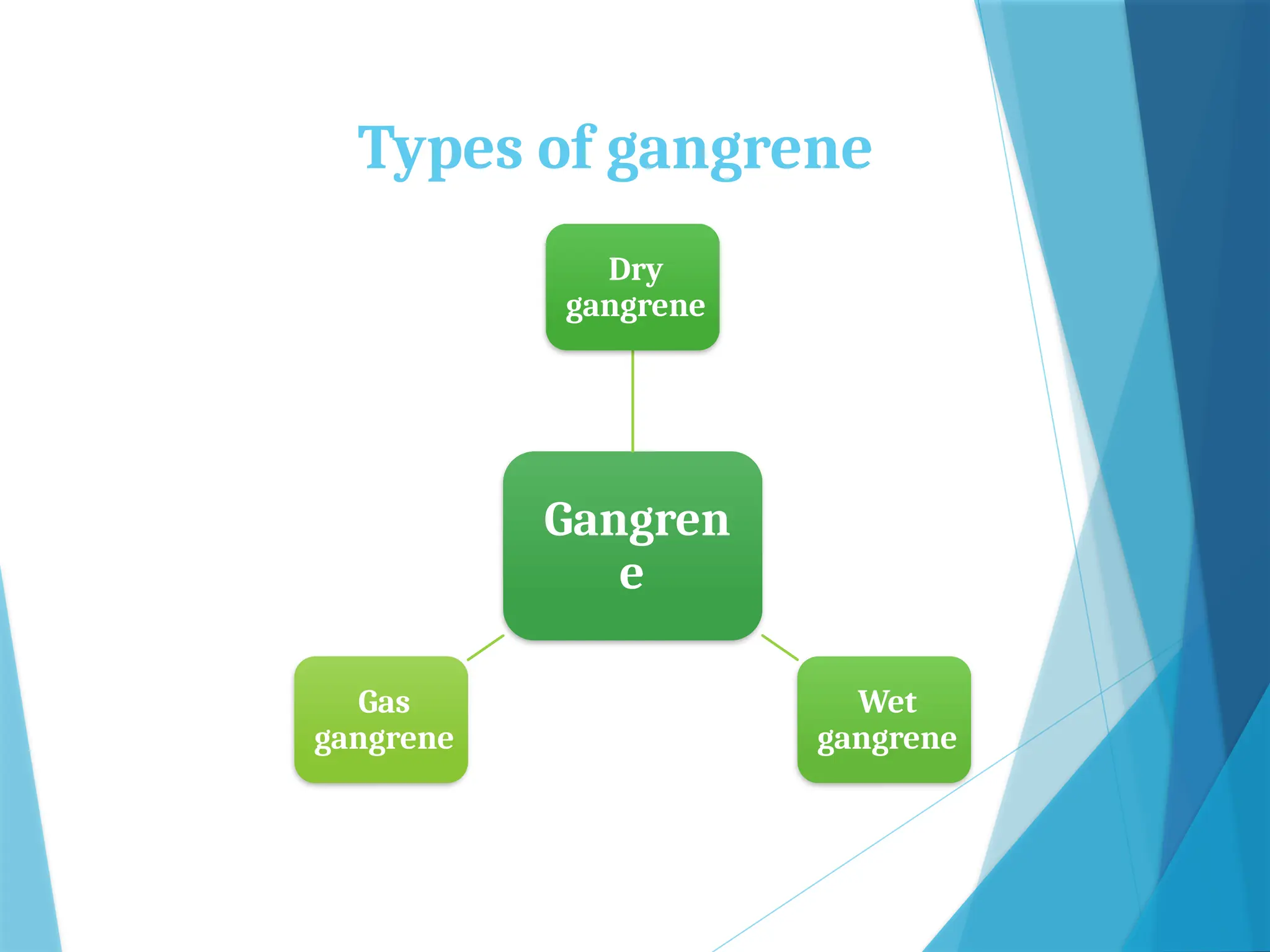
PATHOLOGY FOR NURSES
CELL INJURY
Dr. Binu Babu
Ph.D (N)
Professor
Mrs. Jincy Ealias
M.Sc (N)
Professor
 Cell injury refers to a state in which a cell's normal function is
disrupted due to various harmful stimuli. This disruption can
range from mild and reversible to severe and irreversible,
potentially leading to cell death.
 Etiology
 Cell injury can be caused by a variety of factors, including:
 Physical Agents: Trauma, extreme temperatures, radiation.
 Chemical Agents: Toxins, Drugs poisons.
 Biological Agents: Bacteria
 Nutritional Imbalances: deficiencies or excess nutrition
 Genetic factors: Mutations, inherited diseases
 Hypoxia: Insufficient oxygen supply to the cells.
 The response of a cell to injury can vary, and it may include
changes such as:
 Adaptation: Cells may adapt to mild injury by changing their
function or structure through hypertrophy or atrophy.
 Reversible Injury: If the injury is mild or short-term, cells may
recover fully once the harmful stimulus is removed.
 Irreversible Injury: Severe or prolonged injury can lead to
irreversible damage and cell death, either through necrosis or
apoptosis.
Pathogenesis of reversible
and irreversible cell injury
 The pathogenesis of cell injury involves the mechanisms by
which various harmful stimuli cause changes in cell structure
and function.
 Cell injury can be categorized into reversible and irreversible
injury, based on the cell's ability to recover once the harmful
stimulus is removed.
Pathogenesis of cell injury
 Reversible Cell Injury
 Pathogenesis:
 ATP Depletion: Lack of oxygen or nutrient supply, or mitochondrial
damage, leads to decreased ATP production.
 Reduced ATP levels impair cellular functions such as ion transport,
protein synthesis, and cell signaling.
 Cell Swelling:
 ATP depletion affects the function of the sodium-potassium pump,
leading to sodium and water accumulation in the cell.
 This causes cellular swelling, known as hydropic change or vascular
degeneration.
 Cells can recover if the damaging stimulus is removed in time and
homeostasis is restored.
 Cellular functions return to normal once ATP levels are replenished,
swelling is reduced, and metabolic processes are normalized.
Pathogenesis of cell injury
 Irreversible Cell Injury
 Severe Mitochondrial Damage:
 Persistent or severe injury leads to irreversible mitochondrial
damage.
 Mitochondrial permeability transition pores open, leading to loss
of membrane potential and cessation of ATP production.
 The release of pro-apoptotic proteins triggers cell death
pathways.
 Massive Calcium Influx
 Loss of calcium homeostasis results in excessive intracellular calcium
 Calcium activates destructive enzymes such as phospholipase,
proteases, endonucleases, and ATPases.
 Membrane Damage:
 Increased phospholipid degradation and reduced synthesis weaken
cellular membranes.
 Lysosomal membrane damage releases hydrolytic enzymes, leading to
autolysis.
 Plasma membrane damage results in loss of cellular contents and
influx of extracellular substances.
 Oxidative Stress:
 Generation of reactive oxygen species (ROS) and free radicals causes
widespread damage to lipids, proteins, and DNA
 DNA Damage:
 Severe injury can cause irreversible damage to nuclear DNA.
 Accumulation of misfolded prot DNA damage triggers apoptotic pathways.
 Outcomes:
 Cells undergo necrosis apoptosis, depending on the type and extent of
damage
 Necrosis leads to cell lysis and inflammation, whereas apoptosis involves
programmed cell death without inflammation.
 Reversible Injury: Characterized by cellular swelling,
reduced ATP production and disrupted metabolism. Recovery
is possible if the stimulus is removed.
 Irreversible Injury: Involves severe mitochondrial damage,
massive calcium influx, membrane damage, oxidative stress,
and DNA damage. This leads to cell death via necrosis or
apoptosis
Necrosis
 Necrosis is a form of cell injury that results in the premature
death of cells in living tissue, characterized by the
uncontrolled breakdown of cellular structures. It typically
elicits an inflammatory response in the surrounding tissue.
Definition and General
Features
 Irreversible cell injury leads to cell death and subsequent
degradation of cellular components.
 Causes: Ischemia (lack of blood supply), toxins, infections,
physical trauma, and other damaging stimuli.
 Morphological changes: Cell swelling, loss of membrane
integrity, leakage of cellular contents, and inflammation.
 Inflammatory Response: Unlike apoptosis, necrosis often
triggers an inflammatory response due to the release of
intracellular contents into the extracellular space.
Types of Necrosis
Necrosis
Coagulative
Necrosis
Liquefactive
Necrosis
Caseous
Necrosis
Fat Necrosis
Fibrinoid
Necrosis
Gangrenous
Necrosis
Coagulative Necrosis:
 Characteristics: Preservation of basic cell outlines for a few
days.
 Cause: Typically due to ischemia or infarction.
 Location: Common in solid organs like the heart, kidneys,
spleen.
 Appearance: Tissue appears firm and pale.
Liquefactive Necrosis:
 Characteristics: Complete digestion of dead cells resulting in a
liquid viscous mass.
 Cause: Often due to bacterial or fungal infections; also seen in
 brain infarcts.
 Location: Common in the brain
 and abscesses.
 Appearance: Tissue becomes soft and liquefied.
Caseous Necrosis
 Characteristics: combination of coagulative and liquefactive
necrosis
 Causes: Typically associated with tuberculosis and certain
fungal infections.
 Appearance: Tissue appears soft, friable, and white,
resembling cheese.
Fat Necrosis:
 Characteristics: Focal areas of fat destruction.
 Cause: Often due to trauma to fatty tissue or pancreatitis.
 Location: Common in breast tissue and abdominal fat.
 Appearance: Chalky white areas due to the formation of
calcium soap (saponification).
Fibrinoid Necrosis:
 Characteristics: Immune complexes and fibrin are deposited
in the walls of arteries.
 Cause: Immune-mediated diseases such vasculitis.
 Appearance: Bright pink and morphous appearance on H&E
staining.
Gangrenous Necrosis
 Characteristics: not a specific pattern of cell death but a
clinical term.
 Types
 Dry gangrene: Coagulative necrosis due to ischemia, often in limbs.
 Wet Gangrene: Liquefactive necrosis due to bacterial infection, often
in the intestines or limbs.
Gangrene
 Gangrene is a serious and potentially life-threatening
condition characterized by the death of body tissue due to a
lack of blood supply, infection, or both. It can affect any body
part but is most commonly seen in the extremities.
Definition and General
Features
 Gangrene: Death of tissue caused by an insufficient blood
supply, often compounded by bacterial infection.
Types of gangrene
Gangren
e
Dry
gangrene
Wet
gangrene
Gas
gangrene
Dry Gangrene
 Cause: Typically results from chronic ischemia without
infection.
 Common in conditions like peripheral artery disease and
diabetes.
 Pathogenesis: Reduced blood flow causes tissue to die
gradually. The dead issue then dries out and shrinks.
 Clinical Features: The affected area becomes dry, shrunken,
and dark brown or black. There is a clear line of demarcation
between healthy and dead tissue.
 Complications: If not managed, it can progress to wet
gangrene if infection occurs.
Wet Gangrene
 Cause: Usually results from a sudden loss of blood supply
followed by bacterial infection. Often seen in severe burns,
frostbite, or traumatic injuries.
 Pathogenesis: Rapid tissue death due to ischemia is followed
by bacterial invasion. The presence of infection leads to more
rapid and extensive tissue destruction.
 Clinical Features: The affected area becomes swollen blistered,
 and wet. The tissue appears soft, putrid, and black, often emits
a foul odor.
 Complications: High risk of sepsis and systemic infection,
which can be fatal if not treated promtly .
Gas Gangrene
 Cause: Caused by infection with Clostridium bacteria,
particularly Clostridiym peringens, which are anaerobic and
thrive in low- oxygen environments.
 Pathogenesis: The bacteria produce toxins and gas within
tissue causing rapid tissue necrosis and gas bubble formation.
 Clinical features: Severe pain, swelling, pale-to-dark red skin
color, and crepitus (crackling sound) due to gas in the tissues.
The affected area may exude a foul-smelling, brownish fluid.
 Complications: Extremely rapid progression with high risk of
septic shock and death if not treated immediately.
References
 Suresh K Sharma. Textbook of Pharmacology, Pathology and
Genetics for Nurses (Vol-I) Jaypee Brothers Medical Publishers.
 Swaminathan K, Pathology and Genetics for Nurses, Jaypee
Brothers Medical Publishers.
 Ramdas Nayak, Sharada Rai & Astha Gupta. Textbook of
Pathology and Genetics for Nurses, Jaypee Brothers Medical
Publishers.
 Mandal AK & Shramana Choudhary. Comprehensive textbook
of Pathology for Nursing, Avichal Publishers.
 Vinay Kumar, Abbas A K, Aster C J, Robbins & Cotran Pathologic
Basis of Disease, ELSEVIER.

PATHOLOGY FOR NURSES - CELL INJURY......

  • 1.
    PATHOLOGY FOR NURSES CELLINJURY Dr. Binu Babu Ph.D (N) Professor Mrs. Jincy Ealias M.Sc (N) Professor
  • 2.
     Cell injuryrefers to a state in which a cell's normal function is disrupted due to various harmful stimuli. This disruption can range from mild and reversible to severe and irreversible, potentially leading to cell death.  Etiology  Cell injury can be caused by a variety of factors, including:  Physical Agents: Trauma, extreme temperatures, radiation.  Chemical Agents: Toxins, Drugs poisons.  Biological Agents: Bacteria  Nutritional Imbalances: deficiencies or excess nutrition  Genetic factors: Mutations, inherited diseases  Hypoxia: Insufficient oxygen supply to the cells.
  • 3.
     The responseof a cell to injury can vary, and it may include changes such as:  Adaptation: Cells may adapt to mild injury by changing their function or structure through hypertrophy or atrophy.  Reversible Injury: If the injury is mild or short-term, cells may recover fully once the harmful stimulus is removed.  Irreversible Injury: Severe or prolonged injury can lead to irreversible damage and cell death, either through necrosis or apoptosis.
  • 4.
    Pathogenesis of reversible andirreversible cell injury  The pathogenesis of cell injury involves the mechanisms by which various harmful stimuli cause changes in cell structure and function.  Cell injury can be categorized into reversible and irreversible injury, based on the cell's ability to recover once the harmful stimulus is removed.
  • 5.
    Pathogenesis of cellinjury  Reversible Cell Injury  Pathogenesis:  ATP Depletion: Lack of oxygen or nutrient supply, or mitochondrial damage, leads to decreased ATP production.  Reduced ATP levels impair cellular functions such as ion transport, protein synthesis, and cell signaling.  Cell Swelling:  ATP depletion affects the function of the sodium-potassium pump, leading to sodium and water accumulation in the cell.  This causes cellular swelling, known as hydropic change or vascular degeneration.  Cells can recover if the damaging stimulus is removed in time and homeostasis is restored.  Cellular functions return to normal once ATP levels are replenished, swelling is reduced, and metabolic processes are normalized.
  • 6.
    Pathogenesis of cellinjury  Irreversible Cell Injury  Severe Mitochondrial Damage:  Persistent or severe injury leads to irreversible mitochondrial damage.  Mitochondrial permeability transition pores open, leading to loss of membrane potential and cessation of ATP production.  The release of pro-apoptotic proteins triggers cell death pathways.
  • 7.
     Massive CalciumInflux  Loss of calcium homeostasis results in excessive intracellular calcium  Calcium activates destructive enzymes such as phospholipase, proteases, endonucleases, and ATPases.  Membrane Damage:  Increased phospholipid degradation and reduced synthesis weaken cellular membranes.  Lysosomal membrane damage releases hydrolytic enzymes, leading to autolysis.  Plasma membrane damage results in loss of cellular contents and influx of extracellular substances.
  • 8.
     Oxidative Stress: Generation of reactive oxygen species (ROS) and free radicals causes widespread damage to lipids, proteins, and DNA  DNA Damage:  Severe injury can cause irreversible damage to nuclear DNA.  Accumulation of misfolded prot DNA damage triggers apoptotic pathways.  Outcomes:  Cells undergo necrosis apoptosis, depending on the type and extent of damage  Necrosis leads to cell lysis and inflammation, whereas apoptosis involves programmed cell death without inflammation.
  • 9.
     Reversible Injury:Characterized by cellular swelling, reduced ATP production and disrupted metabolism. Recovery is possible if the stimulus is removed.  Irreversible Injury: Involves severe mitochondrial damage, massive calcium influx, membrane damage, oxidative stress, and DNA damage. This leads to cell death via necrosis or apoptosis
  • 10.
    Necrosis  Necrosis isa form of cell injury that results in the premature death of cells in living tissue, characterized by the uncontrolled breakdown of cellular structures. It typically elicits an inflammatory response in the surrounding tissue.
  • 11.
    Definition and General Features Irreversible cell injury leads to cell death and subsequent degradation of cellular components.  Causes: Ischemia (lack of blood supply), toxins, infections, physical trauma, and other damaging stimuli.  Morphological changes: Cell swelling, loss of membrane integrity, leakage of cellular contents, and inflammation.  Inflammatory Response: Unlike apoptosis, necrosis often triggers an inflammatory response due to the release of intracellular contents into the extracellular space.
  • 12.
  • 13.
    Coagulative Necrosis:  Characteristics:Preservation of basic cell outlines for a few days.  Cause: Typically due to ischemia or infarction.  Location: Common in solid organs like the heart, kidneys, spleen.  Appearance: Tissue appears firm and pale.
  • 14.
    Liquefactive Necrosis:  Characteristics:Complete digestion of dead cells resulting in a liquid viscous mass.  Cause: Often due to bacterial or fungal infections; also seen in  brain infarcts.  Location: Common in the brain  and abscesses.  Appearance: Tissue becomes soft and liquefied.
  • 15.
    Caseous Necrosis  Characteristics:combination of coagulative and liquefactive necrosis  Causes: Typically associated with tuberculosis and certain fungal infections.  Appearance: Tissue appears soft, friable, and white, resembling cheese.
  • 16.
    Fat Necrosis:  Characteristics:Focal areas of fat destruction.  Cause: Often due to trauma to fatty tissue or pancreatitis.  Location: Common in breast tissue and abdominal fat.  Appearance: Chalky white areas due to the formation of calcium soap (saponification).
  • 17.
    Fibrinoid Necrosis:  Characteristics:Immune complexes and fibrin are deposited in the walls of arteries.  Cause: Immune-mediated diseases such vasculitis.  Appearance: Bright pink and morphous appearance on H&E staining.
  • 18.
    Gangrenous Necrosis  Characteristics:not a specific pattern of cell death but a clinical term.  Types  Dry gangrene: Coagulative necrosis due to ischemia, often in limbs.  Wet Gangrene: Liquefactive necrosis due to bacterial infection, often in the intestines or limbs.
  • 19.
    Gangrene  Gangrene isa serious and potentially life-threatening condition characterized by the death of body tissue due to a lack of blood supply, infection, or both. It can affect any body part but is most commonly seen in the extremities.
  • 20.
    Definition and General Features Gangrene: Death of tissue caused by an insufficient blood supply, often compounded by bacterial infection.
  • 21.
  • 22.
    Dry Gangrene  Cause:Typically results from chronic ischemia without infection.  Common in conditions like peripheral artery disease and diabetes.  Pathogenesis: Reduced blood flow causes tissue to die gradually. The dead issue then dries out and shrinks.  Clinical Features: The affected area becomes dry, shrunken, and dark brown or black. There is a clear line of demarcation between healthy and dead tissue.  Complications: If not managed, it can progress to wet gangrene if infection occurs.
  • 23.
    Wet Gangrene  Cause:Usually results from a sudden loss of blood supply followed by bacterial infection. Often seen in severe burns, frostbite, or traumatic injuries.  Pathogenesis: Rapid tissue death due to ischemia is followed by bacterial invasion. The presence of infection leads to more rapid and extensive tissue destruction.  Clinical Features: The affected area becomes swollen blistered,  and wet. The tissue appears soft, putrid, and black, often emits a foul odor.  Complications: High risk of sepsis and systemic infection, which can be fatal if not treated promtly .
  • 24.
    Gas Gangrene  Cause:Caused by infection with Clostridium bacteria, particularly Clostridiym peringens, which are anaerobic and thrive in low- oxygen environments.  Pathogenesis: The bacteria produce toxins and gas within tissue causing rapid tissue necrosis and gas bubble formation.  Clinical features: Severe pain, swelling, pale-to-dark red skin color, and crepitus (crackling sound) due to gas in the tissues. The affected area may exude a foul-smelling, brownish fluid.  Complications: Extremely rapid progression with high risk of septic shock and death if not treated immediately.
  • 25.
    References  Suresh KSharma. Textbook of Pharmacology, Pathology and Genetics for Nurses (Vol-I) Jaypee Brothers Medical Publishers.  Swaminathan K, Pathology and Genetics for Nurses, Jaypee Brothers Medical Publishers.  Ramdas Nayak, Sharada Rai & Astha Gupta. Textbook of Pathology and Genetics for Nurses, Jaypee Brothers Medical Publishers.  Mandal AK & Shramana Choudhary. Comprehensive textbook of Pathology for Nursing, Avichal Publishers.  Vinay Kumar, Abbas A K, Aster C J, Robbins & Cotran Pathologic Basis of Disease, ELSEVIER.