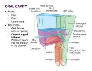
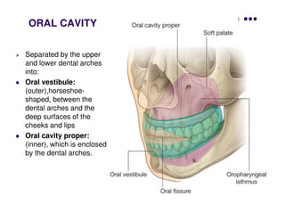
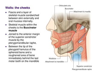
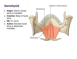
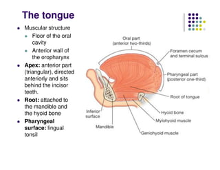
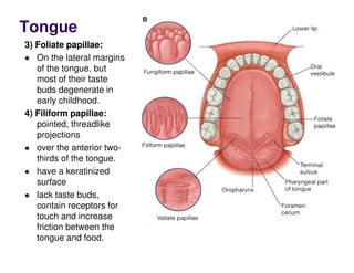
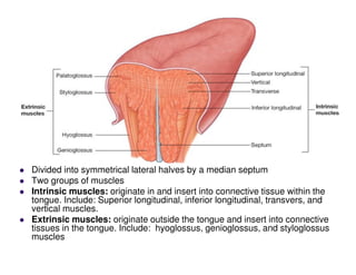
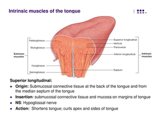
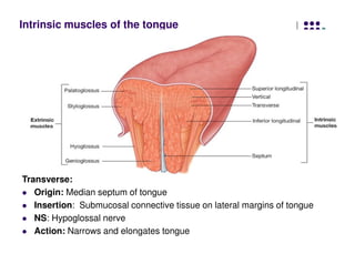
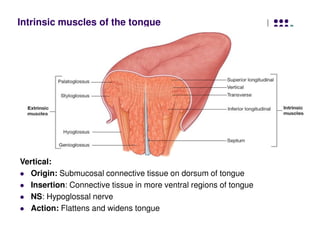
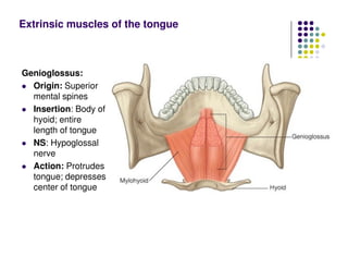
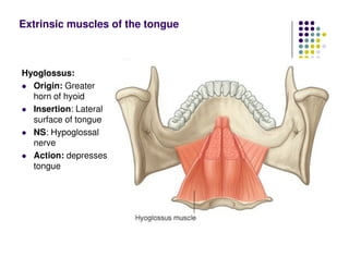
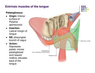
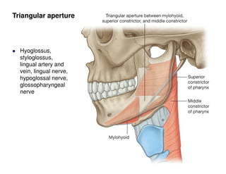
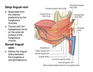
ORAL CAVITY
Walls:
Roof
Floor
Lateral walls
Opennings
Oral fissure:
anterior opening
Oropharyngeal
isthmus:
Posterior, opens
into the oral part
of the pharynx
Separated by the upper
and lower dental arches
into:
Oral vestibule:
(outer),horseshoe-
shaped, between the
dental arches and the
deep surfaces of the
cheeks and lips
Oral cavity proper:
(inner), which is enclosed
by the dental arches.
ORAL CAVITY
Walls: the cheeks
Fascia and a layer of
skeletal muscle sandwiched
between skin externally and
oral mucosa internally.
Skeletal muscle within the
cheeks is the Buccinator
muscle
Joined to the anterior margin
of the superior constrictor
muscle by the
pterygomandibular raphe,
Between the tip of the
pterygoid hamulus of the
sphenoid bone and a
roughened area of bone
immediately behind the last
molar tooth on the mandible
Floor
Formed by three
structures:
Muscular diaphragm:
fills the U-shaped gap
between the left and
right sides of the body
of the mandible
composed
Mylohyoid
muscles
Geniohyoid muscles
Tongue, which is
superior to the
geniohyoid muscles.
Origin: Mylohyoid line of body of mandible
Insertion: Body of hyoid bone
NS: Inferior alveolar nerve
Action: Elevates floor of mouth and hyoid bone or depresses
mandible
Mylohyoid
Geniohyoid
Origin: Inferior mental
spine of mandible
Insertion: Body of hyoid
bone
NS: C1 nerve
Action: Elevates hyoid
bone or depresses
mandible
The tongue
Muscular structure
Floor of the oral
cavity
Anterior wall of
the oropharynx
Apex: anterior part
(triangular), directed
anteriorly and sits
behind the incisor
teeth.
Root: attached to
the mandible and
the hyoid bone
Pharyngeal
surface: lingual
tonsil
Tongue
Papillae: projections of
the lamina propria
covered with stratified
squamous epithelium.
(The dorsal and lateral
surfaces of the tongue).
1) Fungiform papillae:
mushroom like
elevations (red dots)
Dorsum of the tongue,
concentrated mainly
around the margins and
tip of the tongue
most of them contain
taste buds
2) Vallate (circumvallate) papillae: About 12 in number
Arranged in an inverted V-shape on the posterior surface of the tongue.
All of them contain taste buds.
Tongue
3) Foliate papillae:
On the lateral margins
of the tongue, but
most of their taste
buds degenerate in
early childhood.
4) Filiform papillae:
pointed, threadlike
projections
over the anterior two-
thirds of the tongue.
have a keratinized
surface
lack taste buds,
contain receptors for
touch and increase
friction between the
tongue and food.
Inferior surface of
tongue
Lacks papillae
Frenulum of
tongue: single
median fold
continuous with the
mucosa covering the
floor of the oral
cavity,
On each side of the
frenulum is a lingual
vein, and lateral to
each vein is a rough
fimbriated fold.
Ankyloglossia?
Tongue
Divided into symmetrical lateral halves by a median septum
Two groups of muscles
Intrinsic muscles: originate in and insert into connective tissue within the
tongue. Include: Superior longitudinal, inferior longitudinal, transvers, and
vertical muscles.
Extrinsic muscles: originate outside the tongue and insert into connective
tissues in the tongue. Include: hyoglossus, genioglossus, and styloglossus
muscles
Intrinsic muscles of the tongue
Superior longitudinal:
Origin: Submucosal connective tissue at the back of the tongue and from
the median septum of the tongue
Insertion: submucosal connective tissue and mucosa on margins of tongue
NS: Hypoglossal nerve
Action: Shortens tongue; curls apex and sides of tongue
Intrinsic muscles of the tongue
Inferior longitudinal:
Origin: Root of tongue
Insertion: Apex of tongue
NS: Hypoglossal nerve
Action: Shortens tongue; uncurls apex and turns it downward
Intrinsic muscles of the tongue
Transverse:
Origin: Median septum of tongue
Insertion: Submucosal connective tissue on lateral margins of tongue
NS: Hypoglossal nerve
Action: Narrows and elongates tongue
Intrinsic muscles of the tongue
Vertical:
Origin: Submucosal connective tissue on dorsum of tongue
Insertion: Connective tissue in more ventral regions of tongue
NS: Hypoglossal nerve
Action: Flattens and widens tongue
Genioglossus:
Origin: Superior
mental spines
Insertion: Body of
hyoid; entire
length of tongue
NS: Hypoglossal
nerve
Action: Protrudes
tongue; depresses
center of tongue
Extrinsic muscles of the tongue
Hyoglossus:
Origin: Greater
horn of hyoid
Insertion: Lateral
surface of tongue
NS: Hypoglossal
nerve
Action: depresses
tongue
Extrinsic muscles of the tongue
Styloglossus:
Origin: Styloid
process
Insertion: Lateral
surface of tongue
NS: Hypoglossal
nerve
Action: retracts
tongue
Extrinsic muscles of the tongue
Palatoglossus:
Origin: Inferior
surface of
Palatine
aponeurosis
Insertion:
Lateral margin of
tongue
NS: pharyngeal
branch of vagus
Action:
Depresses
palate; moves
palatoglossal
arch toward
midline; elevates
back of the
tongue
Extrinsic muscles of the tongue
Hyoglossus,
styloglossus,
lingual artery and
vein, lingual nerve,
hypoglossal nerve,
glossopharyngeal
nerve
Triangular aperture
Loops
downward and
forward to pass
deep to the
hyoglossus
muscle
Enters the floor
of the oral
cavity. travels
forward in the
plane between
the hyoglossus
and
genioglossus
muscles to the
apex of the
tongue.
Lingual artery
Separated from
the arteries
posteriorly by the
hyoglossus
muscles.
Travels with the
hypoglossal nerve
on the external
surface of the
hyoglossus
muscle
Dorsal lingual
vein:
follows the lingual
artery between
the hyoglossus
and genioglossus
Deep lingual vein

oral_cavity_1__1_.pdf

  • 1.
    ORAL CAVITY Walls: Roof Floor Lateral walls Opennings Oralfissure: anterior opening Oropharyngeal isthmus: Posterior, opens into the oral part of the pharynx
  • 2.
    Separated by theupper and lower dental arches into: Oral vestibule: (outer),horseshoe- shaped, between the dental arches and the deep surfaces of the cheeks and lips Oral cavity proper: (inner), which is enclosed by the dental arches. ORAL CAVITY
  • 3.
    Walls: the cheeks Fasciaand a layer of skeletal muscle sandwiched between skin externally and oral mucosa internally. Skeletal muscle within the cheeks is the Buccinator muscle Joined to the anterior margin of the superior constrictor muscle by the pterygomandibular raphe, Between the tip of the pterygoid hamulus of the sphenoid bone and a roughened area of bone immediately behind the last molar tooth on the mandible
  • 4.
    Floor Formed by three structures: Musculardiaphragm: fills the U-shaped gap between the left and right sides of the body of the mandible composed Mylohyoid muscles Geniohyoid muscles Tongue, which is superior to the geniohyoid muscles.
  • 5.
    Origin: Mylohyoid lineof body of mandible Insertion: Body of hyoid bone NS: Inferior alveolar nerve Action: Elevates floor of mouth and hyoid bone or depresses mandible Mylohyoid
  • 6.
    Geniohyoid Origin: Inferior mental spineof mandible Insertion: Body of hyoid bone NS: C1 nerve Action: Elevates hyoid bone or depresses mandible
  • 7.
    The tongue Muscular structure Floorof the oral cavity Anterior wall of the oropharynx Apex: anterior part (triangular), directed anteriorly and sits behind the incisor teeth. Root: attached to the mandible and the hyoid bone Pharyngeal surface: lingual tonsil
  • 8.
    Tongue Papillae: projections of thelamina propria covered with stratified squamous epithelium. (The dorsal and lateral surfaces of the tongue). 1) Fungiform papillae: mushroom like elevations (red dots) Dorsum of the tongue, concentrated mainly around the margins and tip of the tongue most of them contain taste buds 2) Vallate (circumvallate) papillae: About 12 in number Arranged in an inverted V-shape on the posterior surface of the tongue. All of them contain taste buds.
  • 9.
    Tongue 3) Foliate papillae: Onthe lateral margins of the tongue, but most of their taste buds degenerate in early childhood. 4) Filiform papillae: pointed, threadlike projections over the anterior two- thirds of the tongue. have a keratinized surface lack taste buds, contain receptors for touch and increase friction between the tongue and food.
  • 10.
    Inferior surface of tongue Lackspapillae Frenulum of tongue: single median fold continuous with the mucosa covering the floor of the oral cavity, On each side of the frenulum is a lingual vein, and lateral to each vein is a rough fimbriated fold. Ankyloglossia?
  • 11.
    Tongue Divided into symmetricallateral halves by a median septum Two groups of muscles Intrinsic muscles: originate in and insert into connective tissue within the tongue. Include: Superior longitudinal, inferior longitudinal, transvers, and vertical muscles. Extrinsic muscles: originate outside the tongue and insert into connective tissues in the tongue. Include: hyoglossus, genioglossus, and styloglossus muscles
  • 12.
    Intrinsic muscles ofthe tongue Superior longitudinal: Origin: Submucosal connective tissue at the back of the tongue and from the median septum of the tongue Insertion: submucosal connective tissue and mucosa on margins of tongue NS: Hypoglossal nerve Action: Shortens tongue; curls apex and sides of tongue
  • 13.
    Intrinsic muscles ofthe tongue Inferior longitudinal: Origin: Root of tongue Insertion: Apex of tongue NS: Hypoglossal nerve Action: Shortens tongue; uncurls apex and turns it downward
  • 14.
    Intrinsic muscles ofthe tongue Transverse: Origin: Median septum of tongue Insertion: Submucosal connective tissue on lateral margins of tongue NS: Hypoglossal nerve Action: Narrows and elongates tongue
  • 15.
    Intrinsic muscles ofthe tongue Vertical: Origin: Submucosal connective tissue on dorsum of tongue Insertion: Connective tissue in more ventral regions of tongue NS: Hypoglossal nerve Action: Flattens and widens tongue
  • 16.
    Genioglossus: Origin: Superior mental spines Insertion:Body of hyoid; entire length of tongue NS: Hypoglossal nerve Action: Protrudes tongue; depresses center of tongue Extrinsic muscles of the tongue
  • 18.
    Hyoglossus: Origin: Greater horn ofhyoid Insertion: Lateral surface of tongue NS: Hypoglossal nerve Action: depresses tongue Extrinsic muscles of the tongue
  • 19.
    Styloglossus: Origin: Styloid process Insertion: Lateral surfaceof tongue NS: Hypoglossal nerve Action: retracts tongue Extrinsic muscles of the tongue
  • 20.
    Palatoglossus: Origin: Inferior surface of Palatine aponeurosis Insertion: Lateralmargin of tongue NS: pharyngeal branch of vagus Action: Depresses palate; moves palatoglossal arch toward midline; elevates back of the tongue Extrinsic muscles of the tongue
  • 21.
    Hyoglossus, styloglossus, lingual artery and vein,lingual nerve, hypoglossal nerve, glossopharyngeal nerve Triangular aperture
  • 22.
    Loops downward and forward topass deep to the hyoglossus muscle Enters the floor of the oral cavity. travels forward in the plane between the hyoglossus and genioglossus muscles to the apex of the tongue. Lingual artery
  • 23.
    Separated from the arteries posteriorlyby the hyoglossus muscles. Travels with the hypoglossal nerve on the external surface of the hyoglossus muscle Dorsal lingual vein: follows the lingual artery between the hyoglossus and genioglossus Deep lingual vein