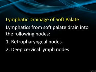
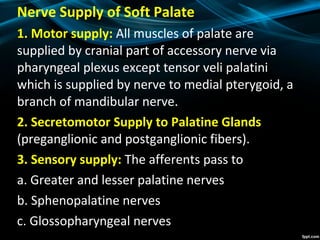
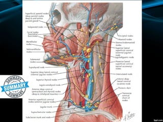
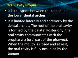
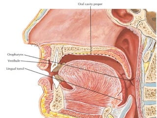
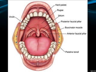
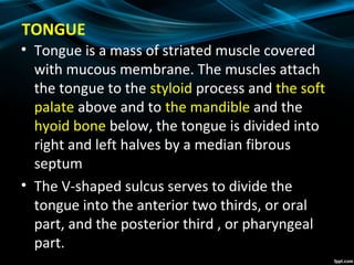
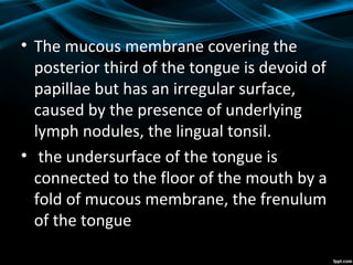
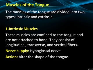
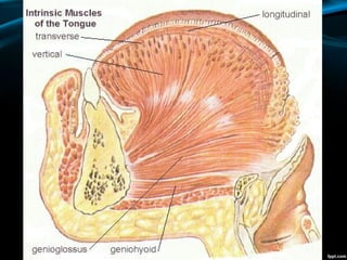
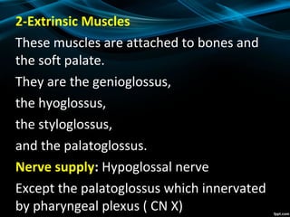
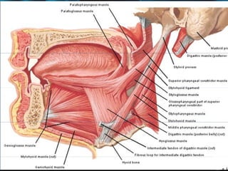
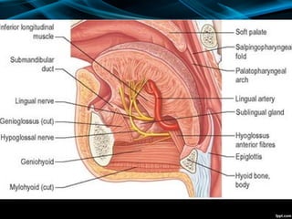
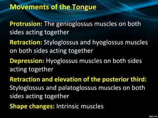
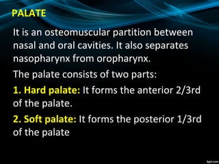
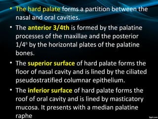
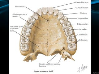
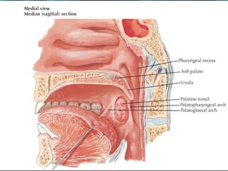
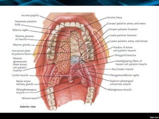
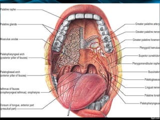
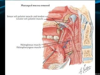
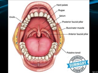
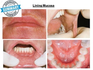
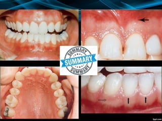
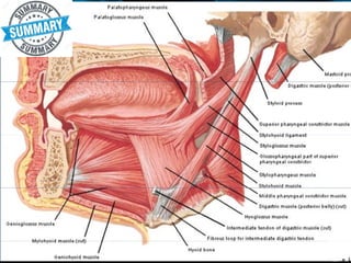
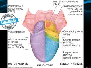
Oral Region
Dr. Haydar Muneer Salih
• Oral cavity is the first part of digestive
tract
1.Ingestion and swallowing
2.Speech
3.Respiration
• It is divided into two parts:
1. Oral cavity proper
2. The vestibule
Oral Cavity Proper
• It is the space between the upper and
the lower dental arches
• It is limited laterally and anteriorly by the
dental arches. The roof of the oral cavity
is formed by the palate. Posteriorly, the
oral cavity communicates with the
oropharynx (oral part of the pharynx).
When the mouth is closed and at rest,
the oral cavity is fully occupied by the
tongue
The oral vestibule
• The oral vestibule is the slit-like space
between the teeth and gingivae (gums)
and the lips and cheeks. The vestibule
communicates with the exterior through
the oral fissure (opening).
• The size of the oral fissure is controlled
by the circumoral muscles
Lips
• Each lip is largely composed of orbicularis oris
muscle. In addition it contains labial (mucous)
glands and blood vessels. It is lined externally
by skin which contains sweat glands and
sebaceous glands with hair follicles (The hairs
are thicker and more numerous in adult
males). They are lined internally with mucus
membrane. The mucocutaneous junction
appears as a reddish pink area visible
externally
• Arterial supply of lips is derived from superior
and inferior labial arteries, branches of facial
artery.
• Lymphatics of lips drain into the following
lymph nodes:
a. Submandibular lymph node
b. Submental lymph nodes
• Skin of lips is innervated by branches of
infraorbital nerve (upper lip) and mandibular
nerve (lower lip). Muscles are supplied by facial
nerve.
Cheek
Cheeks are the fleshy flaps which lie over maxilla
and mandible and form a large part of the face.
Each cheek is continuous in front with the lip.
The junction between the two is marked by the
nasolabial sulcus or the furrow which extends
from the side of the nose to the angle of the
mouth. Like the lips the cheeks are lined
externally by skin and internally by mucous
membrane
From superficial to deep:
1. Skin
2. Superficial fascia containing muscles of facial
expression
3. Buccinator
4. Buccal pad of fat
5. Buccopharyngeal fascia
6. Submucosa, containing buccal (mucous)
glands
7. Mucous membrane
Structure & layers of the cheek:
• Arterial supply of cheeks is derived from
buccal branch of maxillary artery.
• Lymphatics from cheek drain into
submandibular and preauricular lymph
nodes.
• Skin of cheek is innervated by :
zygomaticofacial and infraorbital branches
of maxillary nerve, mucus membrane is
supplied by buccal branch of mandibular
nerve. Muscles are supplied by facial nerve
Oral Mucosa
The mucosal lining of oral cavity continues
with the skin of lips anteriorly and mucosa
of oropharyx posteriorly. It can be divided
into three types according to the
anatomical location and function
1.Lining mucosa
2.Masticatory mucosa
3.Specialized mucosa
1.Lining mucosa:
It consists of non-keratinized stratified
squamous epithelium. It lines the inner
aspect of lips and cheeks, covers soft
palate, ventral surface of tongue, floor of
mouth and lower part of upper and lower
alveolar processes (jaws). It overlies a
loose layer of lamina propria and has
submucosa which contains fat and minor
salivary glands
2.Masticatory mucosa:
• It is the mucosa that lines the upper part
of alveolar process, neck of teeth and the
hard palate. It consists of keratinized
stratified squamous epithelium with
minimal lamina propria that has
connective tissue fibers which adhere
the epithelium to the underlying bone.
There is no submucosa. This modification
• allows the epithelium to bear the stress
of mastication
Gingiva (Gum(
Gingiva is the masticatory mucosa that covers
the alveolar processes of maxilla (upper jaw)
and mandible (lower jaw) and surrounds the
neck of teeth. It can be divided into two parts:
1. Attached gingiva: It is firmly bound to the
periosteum of the alveolar bone and tooth.
2. Free or unattached gingiva: It is the distal 1
mm margin of gingiva that surrounds the neck
of the tooth and is not attached to the bone.
• The gingival tissues derive their blood supply
from branches of maxillary artery (supplies the
buccal and labial surfaces) and lingual artery
(supplies the lingual surfaces).
• Lymphatics from gingivae drain into
submandibular lymph nodes.
• Gingivae of upper jaw are supplied by
branches of maxillary nerve while of lower jaw
are supplied by branches of mandibular nerve
3.Specialized mucosa:
It is the mucosa which covers the dorsal
surface of tongue. It consists of stratified
squamous non-keratinized epithelium
which is directly adherent to the
underlying muscles. There is no
submucosa. It gives rise to a number of
projections called lingual papillae which
are described ahead with tongue.
TONGUE
• Tongue is a mass of striated muscle covered
with mucous membrane. The muscles attach
the tongue to the styloid process and the soft
palate above and to the mandible and the
hyoid bone below, the tongue is divided into
right and left halves by a median fibrous
septum
• The V-shaped sulcus serves to divide the
tongue into the anterior two thirds, or oral
part, and the posterior third , or pharyngeal
part.
• The mucous membrane covering the
posterior third of the tongue is devoid of
papillae but has an irregular surface,
caused by the presence of underlying
lymph nodules, the lingual tonsil.
• the undersurface of the tongue is
connected to the floor of the mouth by a
fold of mucous membrane, the frenulum
of the tongue
Muscles of the Tongue
The muscles of the tongue are divided into two
types: intrinsic and extrinsic.
1-Intrinsic Muscles
These muscles are confined to the tongue and
are not attached to bone. They consist of
longitudinal, transverse, and vertical fibers.
Nerve supply: Hypoglossal nerve
Action: Alter the shape of the tongue
2-Extrinsic Muscles
These muscles are attached to bones and
the soft palate.
They are the genioglossus,
the hyoglossus,
the styloglossus,
and the palatoglossus.
Nerve supply: Hypoglossal nerve
Except the palatoglossus which innervated
by pharyngeal plexus ( CN X)
Sensory Innervation
Anterior two thirds: Lingual nerve branch
of mandibular division of trigeminal nerve
(general sensation) and chorda tympani
branch of the facial nerve (taste)
Posterior third: Glossopharyngeal nerve
(general sensation and taste)
A small posterior region receives sensory
innervation from CN X.
Blood Supply
The lingual artery, the tonsillar branch of the
facial artery, and the ascending pharyngeal
artery supply the tongue.
The veins drain into the internal jugular vein.
Lymph Drainage
Tip: Submental lymph nodes
Sides of the anterior two thirds: Submandibular
and deep cervical lymph nodes
Posterior third: Deep cervical
Movements of the Tongue
Protrusion: The genioglossus muscles on both
sides acting together
Retraction: Styloglossus and hyoglossus muscles
on both sides acting together
Depression: Hyoglossus muscles on both sides
acting together
Retraction and elevation of the posterior third:
Styloglossus and palatoglossus muscles on both
sides acting together
Shape changes: Intrinsic muscles
PALATE
It is an osteomuscular partition between
nasal and oral cavities. It also separates
nasopharynx from oropharynx.
The palate consists of two parts:
1. Hard palate: It forms the anterior 2/3rd
of the palate.
2. Soft palate: It forms the posterior 1/3rd
of the palate
• The hard palate forms a partition between the
nasal and oral cavities.
• The anterior 3/4th is formed by the palatine
processes of the maxillae and the posterior
1/4th
by the horizontal plates of the palatine
bones.
• The superior surface of hard palate forms the
floor of nasal cavity and is lined by the ciliated
pseudostratified columnar epithelium.
• The inferior surface of hard palate forms the
roof of oral cavity and is lined by masticatory
mucosa. It presents with a median palatine
raphe
• Arterial supply of hard palate is dervied
from greater palatine artery, branch of
maxillary artery and ascending palatine
branch of facial artery. The veins drain
into pterygoid plexus of veins.
• Nerve supply of hard palate is derived
from greater palatine and nasopalatine
branches of maxillary nerve through
pterygopalatine ganglion.
Soft Palate
The soft palate is a mobile muscular fold
suspended from the posterior border of
the hard palate like a velum. It is lined by
nonkeratinized stratified squamous
epithelium which encloses muscles,
vessels, nerves, lymphoid tissue and
mucous glands. It appears red in
comparision with the hard palate which is
pink. It separates the nasopharynx from
oropharynx.
• On each side, from the base of uvula, two
curved folds of mucous membrane extend
laterally and downwards along the lateral wall
of oropharynx.
These are:
i. Palatoglossal fold
It is the anterior fold which merges inferiorly
with the sides of the tongue at the junction of
its oral and pharyngeal parts.
ii. Palatopharyngeal fold
It lies posterior to the palatoglossal fold and
merges inferiorly with the lateral wall of the
pharynx.
Muscles of the Soft Palate
The soft palate consist of five pairs of
muscles:
Tensor veli palatini
Levator veli palatini
Palatoglossus
Palatopharyngeus
Musculus uvulae
Functions of the Soft Palate
1. Separates the oropharynx from
nasopharynx during swallowing so that
food does not enter the nose.
2. Isolates the oral cavity from oropharynx
during chewing so that breathing is
unaffected.
3. Helps to modify the quality of voice, by
varying the degree of closure of the
pharyngeal isthmus. 
Arterial Supply of Soft Palate
It is supplied by the following arteries:
1. Ascending palatine artery, branch of facial
artery.
2. Palatine branch of ascending pharyngeal
artery.
3. Greater palatine artery, branch of maxillary
artery.
Venous Drainage of Soft Palate
Veins drain into the pterygoid plexus of veins.
 
Lymphatic Drainage of Soft Palate
Lymphatics from soft palate drain into
the following nodes:
1. Retropharyngeal nodes.
2. Deep cervical lymph nodes
Nerve Supply of Soft Palate
1. Motor supply: All muscles of palate are
supplied by cranial part of accessory nerve via
pharyngeal plexus except tensor veli palatini
which is supplied by nerve to medial pterygoid, a
branch of mandibular nerve.
2. Secretomotor Supply to Palatine Glands
(preganglionic and postganglionic fibers).
3. Sensory supply: The afferents pass to
a. Greater and lesser palatine nerves
b. Sphenopalatine nerves
c. Glossopharyngeal nerves
Oral region
Oral region
Oral region
Oral region
Oral region
Oral region
Oral region
Oral region
Oral region

Oral region

  • 1.
  • 2.
    • Oral cavityis the first part of digestive tract 1.Ingestion and swallowing 2.Speech 3.Respiration • It is divided into two parts: 1. Oral cavity proper 2. The vestibule
  • 3.
    Oral Cavity Proper •It is the space between the upper and the lower dental arches • It is limited laterally and anteriorly by the dental arches. The roof of the oral cavity is formed by the palate. Posteriorly, the oral cavity communicates with the oropharynx (oral part of the pharynx). When the mouth is closed and at rest, the oral cavity is fully occupied by the tongue
  • 5.
    The oral vestibule •The oral vestibule is the slit-like space between the teeth and gingivae (gums) and the lips and cheeks. The vestibule communicates with the exterior through the oral fissure (opening). • The size of the oral fissure is controlled by the circumoral muscles
  • 7.
    Lips • Each lipis largely composed of orbicularis oris muscle. In addition it contains labial (mucous) glands and blood vessels. It is lined externally by skin which contains sweat glands and sebaceous glands with hair follicles (The hairs are thicker and more numerous in adult males). They are lined internally with mucus membrane. The mucocutaneous junction appears as a reddish pink area visible externally
  • 8.
    • Arterial supplyof lips is derived from superior and inferior labial arteries, branches of facial artery. • Lymphatics of lips drain into the following lymph nodes: a. Submandibular lymph node b. Submental lymph nodes • Skin of lips is innervated by branches of infraorbital nerve (upper lip) and mandibular nerve (lower lip). Muscles are supplied by facial nerve.
  • 14.
    Cheek Cheeks are thefleshy flaps which lie over maxilla and mandible and form a large part of the face. Each cheek is continuous in front with the lip. The junction between the two is marked by the nasolabial sulcus or the furrow which extends from the side of the nose to the angle of the mouth. Like the lips the cheeks are lined externally by skin and internally by mucous membrane
  • 15.
    From superficial todeep: 1. Skin 2. Superficial fascia containing muscles of facial expression 3. Buccinator 4. Buccal pad of fat 5. Buccopharyngeal fascia 6. Submucosa, containing buccal (mucous) glands 7. Mucous membrane Structure & layers of the cheek:
  • 17.
    • Arterial supplyof cheeks is derived from buccal branch of maxillary artery. • Lymphatics from cheek drain into submandibular and preauricular lymph nodes. • Skin of cheek is innervated by : zygomaticofacial and infraorbital branches of maxillary nerve, mucus membrane is supplied by buccal branch of mandibular nerve. Muscles are supplied by facial nerve
  • 18.
    Oral Mucosa The mucosallining of oral cavity continues with the skin of lips anteriorly and mucosa of oropharyx posteriorly. It can be divided into three types according to the anatomical location and function 1.Lining mucosa 2.Masticatory mucosa 3.Specialized mucosa
  • 19.
    1.Lining mucosa: It consistsof non-keratinized stratified squamous epithelium. It lines the inner aspect of lips and cheeks, covers soft palate, ventral surface of tongue, floor of mouth and lower part of upper and lower alveolar processes (jaws). It overlies a loose layer of lamina propria and has submucosa which contains fat and minor salivary glands
  • 21.
    2.Masticatory mucosa: • Itis the mucosa that lines the upper part of alveolar process, neck of teeth and the hard palate. It consists of keratinized stratified squamous epithelium with minimal lamina propria that has connective tissue fibers which adhere the epithelium to the underlying bone. There is no submucosa. This modification • allows the epithelium to bear the stress of mastication
  • 23.
    Gingiva (Gum( Gingiva isthe masticatory mucosa that covers the alveolar processes of maxilla (upper jaw) and mandible (lower jaw) and surrounds the neck of teeth. It can be divided into two parts: 1. Attached gingiva: It is firmly bound to the periosteum of the alveolar bone and tooth. 2. Free or unattached gingiva: It is the distal 1 mm margin of gingiva that surrounds the neck of the tooth and is not attached to the bone.
  • 25.
    • The gingivaltissues derive their blood supply from branches of maxillary artery (supplies the buccal and labial surfaces) and lingual artery (supplies the lingual surfaces). • Lymphatics from gingivae drain into submandibular lymph nodes. • Gingivae of upper jaw are supplied by branches of maxillary nerve while of lower jaw are supplied by branches of mandibular nerve
  • 26.
    3.Specialized mucosa: It isthe mucosa which covers the dorsal surface of tongue. It consists of stratified squamous non-keratinized epithelium which is directly adherent to the underlying muscles. There is no submucosa. It gives rise to a number of projections called lingual papillae which are described ahead with tongue.
  • 28.
    TONGUE • Tongue isa mass of striated muscle covered with mucous membrane. The muscles attach the tongue to the styloid process and the soft palate above and to the mandible and the hyoid bone below, the tongue is divided into right and left halves by a median fibrous septum • The V-shaped sulcus serves to divide the tongue into the anterior two thirds, or oral part, and the posterior third , or pharyngeal part.
  • 29.
    • The mucousmembrane covering the posterior third of the tongue is devoid of papillae but has an irregular surface, caused by the presence of underlying lymph nodules, the lingual tonsil. • the undersurface of the tongue is connected to the floor of the mouth by a fold of mucous membrane, the frenulum of the tongue
  • 31.
    Muscles of theTongue The muscles of the tongue are divided into two types: intrinsic and extrinsic. 1-Intrinsic Muscles These muscles are confined to the tongue and are not attached to bone. They consist of longitudinal, transverse, and vertical fibers. Nerve supply: Hypoglossal nerve Action: Alter the shape of the tongue
  • 33.
    2-Extrinsic Muscles These musclesare attached to bones and the soft palate. They are the genioglossus, the hyoglossus, the styloglossus, and the palatoglossus. Nerve supply: Hypoglossal nerve Except the palatoglossus which innervated by pharyngeal plexus ( CN X)
  • 35.
    Sensory Innervation Anterior twothirds: Lingual nerve branch of mandibular division of trigeminal nerve (general sensation) and chorda tympani branch of the facial nerve (taste) Posterior third: Glossopharyngeal nerve (general sensation and taste) A small posterior region receives sensory innervation from CN X.
  • 37.
    Blood Supply The lingualartery, the tonsillar branch of the facial artery, and the ascending pharyngeal artery supply the tongue. The veins drain into the internal jugular vein. Lymph Drainage Tip: Submental lymph nodes Sides of the anterior two thirds: Submandibular and deep cervical lymph nodes Posterior third: Deep cervical
  • 39.
    Movements of theTongue Protrusion: The genioglossus muscles on both sides acting together Retraction: Styloglossus and hyoglossus muscles on both sides acting together Depression: Hyoglossus muscles on both sides acting together Retraction and elevation of the posterior third: Styloglossus and palatoglossus muscles on both sides acting together Shape changes: Intrinsic muscles
  • 41.
    PALATE It is anosteomuscular partition between nasal and oral cavities. It also separates nasopharynx from oropharynx. The palate consists of two parts: 1. Hard palate: It forms the anterior 2/3rd of the palate. 2. Soft palate: It forms the posterior 1/3rd of the palate
  • 43.
    • The hardpalate forms a partition between the nasal and oral cavities. • The anterior 3/4th is formed by the palatine processes of the maxillae and the posterior 1/4th by the horizontal plates of the palatine bones. • The superior surface of hard palate forms the floor of nasal cavity and is lined by the ciliated pseudostratified columnar epithelium. • The inferior surface of hard palate forms the roof of oral cavity and is lined by masticatory mucosa. It presents with a median palatine raphe
  • 46.
    • Arterial supplyof hard palate is dervied from greater palatine artery, branch of maxillary artery and ascending palatine branch of facial artery. The veins drain into pterygoid plexus of veins. • Nerve supply of hard palate is derived from greater palatine and nasopalatine branches of maxillary nerve through pterygopalatine ganglion.
  • 48.
    Soft Palate The softpalate is a mobile muscular fold suspended from the posterior border of the hard palate like a velum. It is lined by nonkeratinized stratified squamous epithelium which encloses muscles, vessels, nerves, lymphoid tissue and mucous glands. It appears red in comparision with the hard palate which is pink. It separates the nasopharynx from oropharynx.
  • 49.
    • On eachside, from the base of uvula, two curved folds of mucous membrane extend laterally and downwards along the lateral wall of oropharynx. These are: i. Palatoglossal fold It is the anterior fold which merges inferiorly with the sides of the tongue at the junction of its oral and pharyngeal parts. ii. Palatopharyngeal fold It lies posterior to the palatoglossal fold and merges inferiorly with the lateral wall of the pharynx.
  • 51.
    Muscles of theSoft Palate The soft palate consist of five pairs of muscles: Tensor veli palatini Levator veli palatini Palatoglossus Palatopharyngeus Musculus uvulae
  • 53.
    Functions of theSoft Palate 1. Separates the oropharynx from nasopharynx during swallowing so that food does not enter the nose. 2. Isolates the oral cavity from oropharynx during chewing so that breathing is unaffected. 3. Helps to modify the quality of voice, by varying the degree of closure of the pharyngeal isthmus. 
  • 54.
    Arterial Supply ofSoft Palate It is supplied by the following arteries: 1. Ascending palatine artery, branch of facial artery. 2. Palatine branch of ascending pharyngeal artery. 3. Greater palatine artery, branch of maxillary artery. Venous Drainage of Soft Palate Veins drain into the pterygoid plexus of veins.  
  • 56.
    Lymphatic Drainage ofSoft Palate Lymphatics from soft palate drain into the following nodes: 1. Retropharyngeal nodes. 2. Deep cervical lymph nodes
  • 57.
    Nerve Supply ofSoft Palate 1. Motor supply: All muscles of palate are supplied by cranial part of accessory nerve via pharyngeal plexus except tensor veli palatini which is supplied by nerve to medial pterygoid, a branch of mandibular nerve. 2. Secretomotor Supply to Palatine Glands (preganglionic and postganglionic fibers). 3. Sensory supply: The afferents pass to a. Greater and lesser palatine nerves b. Sphenopalatine nerves c. Glossopharyngeal nerves