The liver anatomy and physiology
•Download as PPTX, PDF•
4 likes•703 views
1. Define the liver, and describe its location 2. Describe the main features of the liver 3. List and describe the main functions of the liver
Report
Share
Report
Share

Recommended
Liver
The liver is the largest gland in the body, located in the upper right quadrant of the abdomen under the diaphragm and within the rib cage. It has four lobes - two major lobes and two minor lobes - and ducts that carry bile from the liver to the gallbladder and join the pancreatic duct. The liver is made up of lobules containing hepatocytes that radiate outward from a central vein and produce and secrete bile. Blood flows to the liver through the hepatic portal vein and supplies the liver with nutrients and oxygen. The liver performs many important metabolic functions like synthesizing and breaking down substances, and excreting waste from the bloodstream into bile.
Large intestine
The large intestine comes after the small intestine and measures approximately 1.5 meters. It absorbs water and nutrients from waste and protects the body from infections. The large intestine consists of the cecum, colon (ascending, transverse, descending, and sigmoid sections), rectum, and anal canal. It removes remaining water from digested food and stores waste until defecation through contractions that move waste to the rectum for elimination.
Liver anatomy
This is not a substitute for Books. Let it just help you understand some concepts in liver anatomy.
Continuation of this work will depend on your feedback. Stay Blessed.
anatomy of liver
The liver is the largest gland in the human body, weighing approximately 1500 grams. It performs many important functions like absorbing nutrients from the gastrointestinal tract, storing glycogen, and secreting bile. The liver has two lobes - the right lobe, which is the largest, and the left lobe. It is situated in the right upper quadrant of the abdomen and has two surfaces - the diaphragmatic surface and the visceral surface. The liver receives blood from the hepatic portal vein and hepatic artery and is supported by various ligaments.
Anatomy of the small intestine
The small intestine is divided into three parts - the duodenum, jejunum, and ileum. The duodenum receives partially digested food from the stomach and digestive juices from the pancreas and gallbladder. The jejunum, which is around 2.5 meters long, contains villi that increase absorption of nutrients. The ileum, the final 3 meter section, absorbs vitamins and bile acids before connecting to the large intestine.
Secretions of small intestine
The small intestine has four layers and is divided into three regions: the duodenum, jejunum, and ileum. Its interior walls contain circular folds, villi, and microvilli that greatly increase its surface area for nutrient absorption. The small intestine secretes enzymes like enterokinase in the duodenum as well as hormones like gastrin, cholecystokinin, and secretin from endocrine cells in response to food constituents. These secretions aid in digestion and regulate secretions from other organs to maximize nutrient absorption in the small intestine.
Function of stomach
The stomach has five recognizable parts and two curvatures. Sphincters exist at the entry and exit of the stomach to control movement of contents. The stomach stores food, secretes acid and enzymes to digest food into chyme, and empties at a controlled rate into the small intestine. Acid secretion is stimulated by acetylcholine, gastrin, and histamine in three phases: cephalic, gastric, and intestinal. The stomach mucosa protects itself from acid through secretion of mucus and bicarbonate. Peptic ulcers can form if these defenses are overwhelmed.
Small intestine
The small intestine is responsible for most digestion and absorption of nutrients. It is divided into three parts - the duodenum, jejunum, and ileum. The duodenum contains Brunner's glands which secrete mucus to protect the lining and help regulate pH. It connects to the bile ducts and pancreas. The jejunum and ileum further digest food and absorb nutrients through fingerlike villi before waste passes to the large intestine.
Liver anatomy
The liver is divided into eight segments based on its vascular supply. Each segment has its own branch of the portal vein, hepatic artery, and bile duct supplying it. The hepatic veins drain the periphery of each segment. The middle hepatic vein divides the liver into right and left lobes, while the left and right hepatic veins further divide the lobes into segments. Because each segment has its own vascular inflow and outflow, segments can be surgically resected individually without damaging other segments.
Functions of human liver
The human liver has many essential functions:
- It detoxifies chemicals and metabolizes drugs and nutrients. The liver plays a major role in carbohydrate, protein, and lipid metabolism.
- The liver produces bile which aids in fat digestion, as well as proteins involved in blood clotting. It also stores vitamins and minerals.
- Being responsible for these critical functions, the liver is essential for survival, as its functions cannot be compensated for in the long term if liver function is lost.
Stomach physiology
The document summarizes the physiology of the stomach and duodenum. It describes the anatomy, blood supply, nerve supply, and functions of the stomach, including gastric secretion, motility, and emptying. It also discusses gastric glands, cells, phases of gastric secretion, and regulation. For the duodenum, it covers anatomy, blood supply, lymphatic drainage, functions, duodenal glands and secretions. It briefly mentions common disorders of the stomach and duodenum such as gastritis, ulcers, motility disorders, and duodenitis.
Anatomy of stomach
The document summarizes the anatomy and features of the stomach. It describes the stomach's location in the upper left part of the abdomen. It has a J-shape and varies in size, holding 1.5-2 liters in adults. The stomach functions to form a reservoir for food, mix food with gastric juices to form chyme, and control the rate at which chyme enters the small intestine to allow for proper digestion.
ANATOMY OF PANCREAS
The pancreas develops from dorsal and ventral buds originating in the duodenum. During development, the ventral bud rotates posteriorly to fuse with the dorsal bud. The pancreas is located behind the stomach and has both exocrine and endocrine functions. It has a head, neck, body and tail. The main pancreatic duct drains the exocrine pancreas and opens at the major duodenal papilla along with the common bile duct. Developmental anomalies include pancreatic divisum, annular pancreas, ectopic pancreas, agenesis/hypoplasia, and accessory pancreatic lobes.
Mechanism of formation of urine
Mechanism of Formation of Urine involves 4 key processes:
1. Glomerular filtration where plasma is filtered in the nephrons at a normal rate of 125 ml/min. Approximately 178 liters/day are reabsorbed in the renal tubules and only 1-2 liters/day are excreted as urine.
2. Tubular reabsorption where approximately 99% of the filtered load is reabsorbed, mainly through active transport of sodium in the proximal convoluted tubule.
3. Tubular secretion where certain substances like protons and drugs are actively secreted into the tubular fluid.
4. Concentration and acidification of urine where urine becomes concentrated through
Anatomy of the liver and gallbladder
The liver is the largest gland in the body located under the right rib cage. It is divided into four lobes and has two surfaces - a diaphragmatic surface and a visceral surface. The porta hepatis contains the hepatic artery, portal vein and hepatic ducts. Blood flows into the liver through the hepatic artery and portal vein and exits through the hepatic veins. The gallbladder stores and concentrates bile produced by the liver. The biliary system consists of the hepatic ducts, cystic duct, common hepatic duct, gallbladder and common bile duct which empties into the duodenum.
Physiology properties of bile, composition of bile, functions of bile, functi...
This document contains information about the functions of the bile, small intestine, and large intestine. It includes summaries of the properties and composition of bile, as well as its digestive, absorptive, excretory, and other functions. It also describes the functional anatomy of the small intestine, its roles in digestion and absorption of nutrients, and how food exits into the large intestine. Finally, it outlines the absorptive, excretory, secretory, synthetic and other functions of the large intestine, including its role in forming feces and the importance of dietary fiber.
Nephron anatomy
Renal corpuscleGlomerulusBowman’s capsule
Renal tubuleProximal convoluted tubule (PCT)
To Bowman’s capsule:Water Amino acidsGlucoseUreaNa, K, ClHormonesVitamins
Filtration ~ 180 liters filtered out/dayReabsorption ~ 179 liters returned to the blood/day~ 1 liter excreted as urine/day (0.78 mL/min)
SMALL INTESTINE AND LARGE INTESTINE
This document provides an overview of the anatomy and functions of the small and large intestines. It describes the gross anatomy and divisions of the small intestine including the duodenum, jejunum and ileum. It also describes the gross anatomy and divisions of the large intestine including the cecum, colon and rectum. It discusses the structural characteristics of the intestines including the mucosa, submucosa, muscularis and serosa layers. It provides details on the villi, crypts of Lieberkühn and intestinal secretions and their role in absorption.
Gall Bladder & Pancreas.
The document provides information about the gallbladder and pancreas. It describes the location, structure, parts, layers, blood supply, functions, clinical disorders, and removal of the gallbladder. It also details the location, structure, parts, ducts, blood supply, congenital defects, acute pancreatitis, and imaging findings of the pancreas. The document contains detailed anatomical and physiological information about both organs presented through text and diagrams.
1 Stomach
The stomach is a J-shaped muscular sac located in the upper abdomen between the esophagus and small intestine. It has four regions: the cardia, fundus, body, and pyloric part. The stomach is supplied by branches of the celiac artery and drains into the portal vein. Lymph from the stomach drains to nearby lymph nodes. The vagus and splanchnic nerves provide the main innervation to the stomach.
Pancreas
The pancreas is a retroperitoneal gland with both exocrine and endocrine functions. It is 15-20cm in length and divided into the head, neck, body, and tail. The pancreas produces enzymes that are released into the small intestine to aid in digestion and produces hormones like insulin and glucagon that are released into the bloodstream to regulate blood sugar levels. It has both an extensive arterial blood supply and venous drainage that parallels the arteries. The pancreas is innervated by both the sympathetic and parasympathetic nervous systems.
Anatomy of liver
The liver is the largest internal organ located in the right upper quadrant of the abdomen. It has two surfaces - the diaphragmatic surface and visceral surface. The liver is divided into 8 segments based on the Couinaud classification which describes the functional anatomy and vascular supply. This allows for resection of individual segments without damaging other segments. The segments are delineated by the hepatic veins and portal scissurae into right, left, caudate and quadrate lobes.
Stomach
The document summarizes the key anatomical features of the stomach. It describes the organs that surround the stomach, including the liver, pancreas, and intestines. It notes the stomach's size increases from birth through adulthood. The stomach has two openings, two curvatures, and two surfaces. It also contains specialized sphincters like the cardiac and pyloric sphincters that regulate movement of food. The interior of the stomach has rugae and is divided into four regions: the cardia, fundus, body, and pylorus.
Digestive system
The gastrointestinal tract (GI tract) digests food and expels waste. It has 4 layers and is divided into upper and lower tracts. The upper tract includes the mouth, esophagus, and stomach. The stomach acidifies food and the lower tract, including the small and large intestines, further digests and absorbs nutrients before waste is excreted. Accessory organs like the liver, pancreas, and gallbladder produce substances like bile and enzymes to aid digestion. The kidneys filter waste from the blood to produce urine for excretion via the ureters, bladder, and urethra.
Pancreatic juice...
The pancreatic juice is a transparent, isotonic fluid secreted by the pancreas. It has both endocrine and exocrine functions. The exocrine secretions contain digestive enzymes like amylase, lipase, and proteases that aid in digestion. Bicarbonate ions secreted in pancreatic juice neutralize the acidic chime from the stomach and provide an optimal pH for the enzymes. The secretions occur in three phases regulated by both the autonomic nervous system and hormones like secretin and cholecystokinin. Secretin increases the secretion of bicarbonate-rich fluid while cholecystokinin stimulates enzyme secretion after eating. Disorders like pancreatitis and cystic fibrosis can impair pancreatic function and digestion.
Liver anatomy
The document provides an overview of liver anatomy including:
- The liver's position in the right hypochondrium and epigastric region and its weight of 1.5kg on average.
- It has two surfaces: the diaphragmatic surface against the diaphragm and the visceral surface covered in peritoneum except at the gallbladder fossa and porta hepatis.
- The visceral surface is related to other structures like the stomach, duodenum, and right kidney.
- Couinaud described the liver as being divided into 8 segments based on arterial, portal, and biliary drainage.
Anatomy of stomach
The stomach is a J-shaped muscular sac located in the left upper quadrant and umbilical region of the abdomen. It is divided into four regions: the cardia, fundus, body, and pyloric part. The stomach has two openings: the gastroesophageal opening connects to the esophagus and the pyloric opening connects to the small intestine. Blood supply comes from the celiac artery and innervation is provided by the vagus and splanchnic nerves.
Alimentary canal
The document describes the four main layers that make up the walls of the alimentary canal (also known as the gastrointestinal tract): the mucosa, submucosa, muscularis externa, and serosa. The mucosa is the innermost layer that lines the canal and has functions like secreting mucus and enzymes, absorbing nutrients, and protecting against pathogens. It contains epithelial tissue, connective tissue, and smooth muscle. The submucosa lies just outside the mucosa and contains blood vessels, lymphatics, nerves and connective tissue. The muscularis externa is responsible for movements like segmentation and peristalsis through its inner circular and outer longitudinal smooth muscle layers. The outer
liver, pancreas and gall bladder physiology and function and disorders and nu...
The above ppt contains physiological aspects of liver, pancreas, gall bladder and stressed more on disorders and how nutritionally they are managed.
liver and its diseases
The liver is the second largest organ in the human body located in the upper right quadrant of the abdomen. It has many important functions including detoxification, protein synthesis, production of biochemicals for digestion, storage of nutrients like glucose, and hormone production. The liver also plays a key role in carbohydrate and lipid metabolism. It filters blood from the digestive tract and produces bile which aids in digestion. Many disorders can affect the liver such as hepatitis, alcoholic liver disease, and cancer. Maintaining a healthy lifestyle and diet can help support liver function.
More Related Content
What's hot
Liver anatomy
The liver is divided into eight segments based on its vascular supply. Each segment has its own branch of the portal vein, hepatic artery, and bile duct supplying it. The hepatic veins drain the periphery of each segment. The middle hepatic vein divides the liver into right and left lobes, while the left and right hepatic veins further divide the lobes into segments. Because each segment has its own vascular inflow and outflow, segments can be surgically resected individually without damaging other segments.
Functions of human liver
The human liver has many essential functions:
- It detoxifies chemicals and metabolizes drugs and nutrients. The liver plays a major role in carbohydrate, protein, and lipid metabolism.
- The liver produces bile which aids in fat digestion, as well as proteins involved in blood clotting. It also stores vitamins and minerals.
- Being responsible for these critical functions, the liver is essential for survival, as its functions cannot be compensated for in the long term if liver function is lost.
Stomach physiology
The document summarizes the physiology of the stomach and duodenum. It describes the anatomy, blood supply, nerve supply, and functions of the stomach, including gastric secretion, motility, and emptying. It also discusses gastric glands, cells, phases of gastric secretion, and regulation. For the duodenum, it covers anatomy, blood supply, lymphatic drainage, functions, duodenal glands and secretions. It briefly mentions common disorders of the stomach and duodenum such as gastritis, ulcers, motility disorders, and duodenitis.
Anatomy of stomach
The document summarizes the anatomy and features of the stomach. It describes the stomach's location in the upper left part of the abdomen. It has a J-shape and varies in size, holding 1.5-2 liters in adults. The stomach functions to form a reservoir for food, mix food with gastric juices to form chyme, and control the rate at which chyme enters the small intestine to allow for proper digestion.
ANATOMY OF PANCREAS
The pancreas develops from dorsal and ventral buds originating in the duodenum. During development, the ventral bud rotates posteriorly to fuse with the dorsal bud. The pancreas is located behind the stomach and has both exocrine and endocrine functions. It has a head, neck, body and tail. The main pancreatic duct drains the exocrine pancreas and opens at the major duodenal papilla along with the common bile duct. Developmental anomalies include pancreatic divisum, annular pancreas, ectopic pancreas, agenesis/hypoplasia, and accessory pancreatic lobes.
Mechanism of formation of urine
Mechanism of Formation of Urine involves 4 key processes:
1. Glomerular filtration where plasma is filtered in the nephrons at a normal rate of 125 ml/min. Approximately 178 liters/day are reabsorbed in the renal tubules and only 1-2 liters/day are excreted as urine.
2. Tubular reabsorption where approximately 99% of the filtered load is reabsorbed, mainly through active transport of sodium in the proximal convoluted tubule.
3. Tubular secretion where certain substances like protons and drugs are actively secreted into the tubular fluid.
4. Concentration and acidification of urine where urine becomes concentrated through
Anatomy of the liver and gallbladder
The liver is the largest gland in the body located under the right rib cage. It is divided into four lobes and has two surfaces - a diaphragmatic surface and a visceral surface. The porta hepatis contains the hepatic artery, portal vein and hepatic ducts. Blood flows into the liver through the hepatic artery and portal vein and exits through the hepatic veins. The gallbladder stores and concentrates bile produced by the liver. The biliary system consists of the hepatic ducts, cystic duct, common hepatic duct, gallbladder and common bile duct which empties into the duodenum.
Physiology properties of bile, composition of bile, functions of bile, functi...
This document contains information about the functions of the bile, small intestine, and large intestine. It includes summaries of the properties and composition of bile, as well as its digestive, absorptive, excretory, and other functions. It also describes the functional anatomy of the small intestine, its roles in digestion and absorption of nutrients, and how food exits into the large intestine. Finally, it outlines the absorptive, excretory, secretory, synthetic and other functions of the large intestine, including its role in forming feces and the importance of dietary fiber.
Nephron anatomy
Renal corpuscleGlomerulusBowman’s capsule
Renal tubuleProximal convoluted tubule (PCT)
To Bowman’s capsule:Water Amino acidsGlucoseUreaNa, K, ClHormonesVitamins
Filtration ~ 180 liters filtered out/dayReabsorption ~ 179 liters returned to the blood/day~ 1 liter excreted as urine/day (0.78 mL/min)
SMALL INTESTINE AND LARGE INTESTINE
This document provides an overview of the anatomy and functions of the small and large intestines. It describes the gross anatomy and divisions of the small intestine including the duodenum, jejunum and ileum. It also describes the gross anatomy and divisions of the large intestine including the cecum, colon and rectum. It discusses the structural characteristics of the intestines including the mucosa, submucosa, muscularis and serosa layers. It provides details on the villi, crypts of Lieberkühn and intestinal secretions and their role in absorption.
Gall Bladder & Pancreas.
The document provides information about the gallbladder and pancreas. It describes the location, structure, parts, layers, blood supply, functions, clinical disorders, and removal of the gallbladder. It also details the location, structure, parts, ducts, blood supply, congenital defects, acute pancreatitis, and imaging findings of the pancreas. The document contains detailed anatomical and physiological information about both organs presented through text and diagrams.
1 Stomach
The stomach is a J-shaped muscular sac located in the upper abdomen between the esophagus and small intestine. It has four regions: the cardia, fundus, body, and pyloric part. The stomach is supplied by branches of the celiac artery and drains into the portal vein. Lymph from the stomach drains to nearby lymph nodes. The vagus and splanchnic nerves provide the main innervation to the stomach.
Pancreas
The pancreas is a retroperitoneal gland with both exocrine and endocrine functions. It is 15-20cm in length and divided into the head, neck, body, and tail. The pancreas produces enzymes that are released into the small intestine to aid in digestion and produces hormones like insulin and glucagon that are released into the bloodstream to regulate blood sugar levels. It has both an extensive arterial blood supply and venous drainage that parallels the arteries. The pancreas is innervated by both the sympathetic and parasympathetic nervous systems.
Anatomy of liver
The liver is the largest internal organ located in the right upper quadrant of the abdomen. It has two surfaces - the diaphragmatic surface and visceral surface. The liver is divided into 8 segments based on the Couinaud classification which describes the functional anatomy and vascular supply. This allows for resection of individual segments without damaging other segments. The segments are delineated by the hepatic veins and portal scissurae into right, left, caudate and quadrate lobes.
Stomach
The document summarizes the key anatomical features of the stomach. It describes the organs that surround the stomach, including the liver, pancreas, and intestines. It notes the stomach's size increases from birth through adulthood. The stomach has two openings, two curvatures, and two surfaces. It also contains specialized sphincters like the cardiac and pyloric sphincters that regulate movement of food. The interior of the stomach has rugae and is divided into four regions: the cardia, fundus, body, and pylorus.
Digestive system
The gastrointestinal tract (GI tract) digests food and expels waste. It has 4 layers and is divided into upper and lower tracts. The upper tract includes the mouth, esophagus, and stomach. The stomach acidifies food and the lower tract, including the small and large intestines, further digests and absorbs nutrients before waste is excreted. Accessory organs like the liver, pancreas, and gallbladder produce substances like bile and enzymes to aid digestion. The kidneys filter waste from the blood to produce urine for excretion via the ureters, bladder, and urethra.
Pancreatic juice...
The pancreatic juice is a transparent, isotonic fluid secreted by the pancreas. It has both endocrine and exocrine functions. The exocrine secretions contain digestive enzymes like amylase, lipase, and proteases that aid in digestion. Bicarbonate ions secreted in pancreatic juice neutralize the acidic chime from the stomach and provide an optimal pH for the enzymes. The secretions occur in three phases regulated by both the autonomic nervous system and hormones like secretin and cholecystokinin. Secretin increases the secretion of bicarbonate-rich fluid while cholecystokinin stimulates enzyme secretion after eating. Disorders like pancreatitis and cystic fibrosis can impair pancreatic function and digestion.
Liver anatomy
The document provides an overview of liver anatomy including:
- The liver's position in the right hypochondrium and epigastric region and its weight of 1.5kg on average.
- It has two surfaces: the diaphragmatic surface against the diaphragm and the visceral surface covered in peritoneum except at the gallbladder fossa and porta hepatis.
- The visceral surface is related to other structures like the stomach, duodenum, and right kidney.
- Couinaud described the liver as being divided into 8 segments based on arterial, portal, and biliary drainage.
Anatomy of stomach
The stomach is a J-shaped muscular sac located in the left upper quadrant and umbilical region of the abdomen. It is divided into four regions: the cardia, fundus, body, and pyloric part. The stomach has two openings: the gastroesophageal opening connects to the esophagus and the pyloric opening connects to the small intestine. Blood supply comes from the celiac artery and innervation is provided by the vagus and splanchnic nerves.
Alimentary canal
The document describes the four main layers that make up the walls of the alimentary canal (also known as the gastrointestinal tract): the mucosa, submucosa, muscularis externa, and serosa. The mucosa is the innermost layer that lines the canal and has functions like secreting mucus and enzymes, absorbing nutrients, and protecting against pathogens. It contains epithelial tissue, connective tissue, and smooth muscle. The submucosa lies just outside the mucosa and contains blood vessels, lymphatics, nerves and connective tissue. The muscularis externa is responsible for movements like segmentation and peristalsis through its inner circular and outer longitudinal smooth muscle layers. The outer
What's hot (20)
Physiology properties of bile, composition of bile, functions of bile, functi...
Physiology properties of bile, composition of bile, functions of bile, functi...
Similar to The liver anatomy and physiology
liver, pancreas and gall bladder physiology and function and disorders and nu...
The above ppt contains physiological aspects of liver, pancreas, gall bladder and stressed more on disorders and how nutritionally they are managed.
liver and its diseases
The liver is the second largest organ in the human body located in the upper right quadrant of the abdomen. It has many important functions including detoxification, protein synthesis, production of biochemicals for digestion, storage of nutrients like glucose, and hormone production. The liver also plays a key role in carbohydrate and lipid metabolism. It filters blood from the digestive tract and produces bile which aids in digestion. Many disorders can affect the liver such as hepatitis, alcoholic liver disease, and cancer. Maintaining a healthy lifestyle and diet can help support liver function.
Functional anatomy of liver, functional anatomy of biliary system, functions ...
The document discusses the functional anatomy of the liver and biliary system through three presentations - the first discusses the structure and lobes of the liver, the second discusses the biliary system and ducts, and the third discusses the 10 main functions of the liver including metabolic, storage, synthetic, secretory, excretory, and detoxification functions.
Neet physiology of digestion
The document summarizes the human digestive system and the process of digestion. It describes the steps of digestion from ingestion to defecation. The main parts of the digestive system include the mouth, esophagus, stomach, small intestine, large intestine and accessory organs like the liver, pancreas and salivary glands. Digestion involves both mechanical and chemical breakdown of food by enzymes from these organs. Nutrients are then absorbed in the small intestine and transported to the liver and cells before undigested waste is excreted during defecation.
Structure and functions of liver
The liver is the largest gland in the human body, located in the upper right abdominal cavity beneath the diaphragm. It has four lobes and is made up of lobules that contain hepatocytes arranged in plates with blood sinusoids between them. The liver receives blood from the hepatic portal vein and hepatic artery, and filters toxins and produces bile, which is stored in the gallbladder and released into the small intestine after meals to aid in fat digestion. The liver performs many essential metabolic functions including carbohydrate, protein, and fat metabolism, hormone inactivation, and production of bile and proteins.
4 digestion ppt lesson 4
The document discusses the process of assimilation and transport of digested food throughout the body. It defines assimilation as the distribution and use of digested food as an energy source or to build new cells. It describes how sugars, amino acids, and fats are transported via the hepatic portal vein to the liver and then throughout the body. It specifically explains that glucose is used by cells for energy, excess is stored as glycogen in the liver, and insulin stimulates the conversion of glucose to glycogen.
Digestive system in detail
The document provides information about the digestive system. It discusses the organs of the digestive system including the mouth, esophagus, stomach, small intestine, large intestine, rectum and anus. It describes the functions of these organs, such as mechanical and chemical breakdown of food in the mouth, stomach and small intestine. Absorption of nutrients occurs primarily in the small intestine, while the large intestine absorbs water before waste is excreted through the rectum and anus. Glands like the liver, pancreas and salivary glands secrete enzymes and juices to aid in digestion. The six main processes of the digestive system are ingestion, digestion, absorption, assimilation, and excretion.
Human digestive system
The document summarizes the main parts and processes of the human digestive system. It describes the six major processes of digestion - ingestion, propulsion, mechanical and chemical digestion, absorption, and defecation. It then explains the functions and roles of the main digestive organs - mouth, esophagus, stomach, small intestine, and large intestine. Finally, it discusses the accessory organs - liver, pancreas, gallbladder, and salivary glands - and how they aid the digestion process.
liver function tests (LFTs)
This document presents information about liver function tests from a group project. It defines the liver's important functions and explains why liver function tests are important when the liver is damaged or not working properly. The document outlines two main types of liver function tests - special tests and routine tests. It describes several common routine tests, including those that check bilirubin, enzymes, proteins, and other liver markers in the blood to evaluate liver health. Elevated results on certain tests can indicate conditions like liver damage, disease, or blockages.
Humandigestivesystem 090814185124-phpapp02
The document describes the main components and functions of the human digestive system. It discusses the six major processes of digestion: ingestion, propulsion, mechanical and chemical digestion, absorption, and defecation. It names and describes the functions of the main digestive organs - mouth, esophagus, stomach, small intestine, and large intestine. It also outlines the roles of accessory organs like the liver, pancreas, and salivary glands in aiding the digestion process.
Digestive system introduction
This document contains summaries of student presentations on the digestive system from a class called General Physiology. It lists 4 students who each presented on a different topic related to the digestive system on November 1, 2019. The topics included the introduction and functions of the digestive system, the functional anatomy of the digestive system, the functions of primary digestive organs, and the functions of the mouth and properties of saliva. The course outcomes are also listed as presenting the functions of the digestive system and accessory organs.
associated organs in mammalian digestive system
complete assignment on the topic of associated organs in mammalian digestive system
physiology of digestive system.ppt
The document provides an overview of the physiology of the digestive system. It discusses the basic functions of the digestive system which include ingestion, digestion, absorption, and defecation. It describes the organs that make up the gastrointestinal tract (GIT) and their roles, including the mouth, esophagus, stomach, small intestine, large intestine, liver, gallbladder and pancreas. It also discusses the layers of the GIT wall, regulation of digestive functions by nerves and hormones, and the roles of saliva, stomach secretions, bile, and pancreatic juices in digestion.
Functions of Gastrointestinal system
This document provides an overview of the gastrointestinal system, including its main parts and functions. It discusses the digestive tract, which includes the mouth, esophagus, stomach, small intestine and large intestine. It also mentions the accessory organs that help with digestion, such as the teeth, tongue, salivary glands, pancreas, liver and gallbladder. It provides details on the layers of the GI tract wall and nerve supply. It then focuses on specific parts of the digestive system, including the stomach, pancreas, liver and intestines, outlining their structures, secretions and roles in digestion.
Digestive system part 4 (pancreas, liver and gall bladder) english
these slides are prepared to understand digestive system IN EASY WAY Important links- NOTES- https://mynursingstudents.blogspot.com/ youtube channel https://www.youtube.com/c/MYSTUDENTSU... CHANEL PLAYLIST- ANATOMY AND PHYSIOLOGY-https://www.youtube.com/playlist?list=PL93S13oM2gAPM3VTGVUXIeswKJ3XGaD2p COMMUNITY HEALTH NURSING- https://www.youtube.com/playlist?list=PL93S13oM2gAPyslPNdIJoVjiXEDTVEDzs CHILD HEALTH NURSING- https://www.youtube.com/playlist?list=PL93S13oM2gANcslmv0DXg6BWmWN359Gvg FIRST AID- https://www.youtube.com/playlist?list=PL93S13oM2gAMvGqeqH2ZTklzFAZhOrvgP HCM- https://www.youtube.com/playlist?list=PL93S13oM2gAM7mZ1vZhQBHWbdLnLb-cH9 FUNDAMENTALS OF NURSING- https://www.youtube.com/playlist?list=PL93S13oM2gAPFxu78NDLpGPaxEmK1fTao COMMUNICABLE DISEASES- https://www.youtube.com/playlist?list=PL93S13oM2gAOWo4IwNjLU_LCuhRN0ZLeb ENVIRONMENTAL HEALTH- https://www.youtube.com/playlist?list=PL93S13oM2gAPkI6LvfS8Zu1nm6mZi9FK6 MSN- https://www.youtube.com/playlist?list=PL93S13oM2gAOdyoHnDLAoR_o8M6ccqYBm HINDI ONLY- https://www.youtube.com/playlist?list=PL93S13oM2gAN4L-FJ3s_IEXgZCijGUA1A ENGLISH ONLY- https://www.youtube.com/playlist?list=PL93S13oM2gAMYv2a1hFcq4W1nBjTnRkHP facebook profile- https://www.facebook.com/suresh.kr.lrhs/ FACEBOOK PAGE- https://www.facebook.com/My-Student-S... facebook group NURSING NOTES- https://www.facebook.com/groups/24139... FOR MAKING EASY NOTES YOU CAN ALSO VISIT MY BLOG – BLOGGER- https://mynursingstudents.blogspot.com/ Instagram- https://www.instagram.com/mystudentsu... Twitter- https://twitter.com/student_system?s=08
#pancreas, #gallbladder ,#liver ,#BORN,#ASSESSMENT, #APPEARENCE,#PULSE,#GRIMACE,#REFLEX,#RESPIRATION,#RESUSCITATION,#NEWBORN,#BABY,#VIRGINIA, #APGAR, #OXYGEN,#CYANOSIS,#OPTICNERVE, #SARACHNA,#MYSTUDENTSUPPORTSYSTEM, #rashes,#nursingclasses, #communityhealthnursing,#ANM, #GNM, #BSCNURING,#NURSINGSTUDENTS, #WHO,#NURSINGINSTITUTION,#COLLEGEOFNURSING,#nursingofficer,#COMMUNITYHEALTHOFFICER
Lectures 15 Excretion of drug & Enterohepatic Circulation
Lectures 15 Excretion of drug & Enterohepatic CirculationIsra Institute of Rehab Sciences (IIRS), Isra University
This document discusses the main routes of drug excretion from the body. The major routes are renal excretion via the kidneys and biliary excretion via the liver and intestines. Renal excretion involves filtration, secretion, and reabsorption in the nephrons. Biliary excretion involves secretion from the liver into bile and potential reabsorption via enterohepatic circulation. Other minor routes include exhalation from the lungs, excretion in saliva, sweat, milk, and tears. Factors like age, gender, kidney function can impact renal drug clearance and dosing.Physiology of human digestive system
The human digestive system breaks down food through both mechanical and chemical digestion. Food is ingested and broken down mechanically by teeth and enzymes in the mouth, stomach, and small intestine. In the stomach and small intestine, chemicals like acids and enzymes produced by the liver, pancreas, and intestines themselves further break down food into small molecules that can be absorbed into the bloodstream. The digestive system includes the mouth, esophagus, stomach, small and large intestines, and accessory organs like the liver, gallbladder and pancreas that produce digestive juices to break down proteins, lipids, and carbohydrates.
Poultry liver (big organ with big role)
the liver is the central laboratory of a chicken’s body. It is
essential that this organ is kept in an excellent condition in
order to maintain a healthy bird. Understanding the metabolic
function and causes of disruptions in liver functions helps us
to provide the birds with the right feed and health treatment.
Anatomy & Physiology Of The Pancreas (Dm)
The pancreas is an endocrine and exocrine gland located in the abdominal cavity. It has three regions - the head, body, and tail. The pancreas contains exocrine cells that secrete digestive enzymes into the small intestine to break down carbohydrates, lipids, and proteins. It also contains clusters of endocrine cells called islets of Langerhans that secrete hormones like insulin and glucagon to regulate blood sugar levels. Insulin is released from beta cells when blood sugar rises and promotes the uptake of glucose from the bloodstream into cells.
Similar to The liver anatomy and physiology (20)
liver, pancreas and gall bladder physiology and function and disorders and nu...
liver, pancreas and gall bladder physiology and function and disorders and nu...
Functional anatomy of liver, functional anatomy of biliary system, functions ...
Functional anatomy of liver, functional anatomy of biliary system, functions ...
Digestive system part 4 (pancreas, liver and gall bladder) english
Digestive system part 4 (pancreas, liver and gall bladder) english
Lectures 15 Excretion of drug & Enterohepatic Circulation
Lectures 15 Excretion of drug & Enterohepatic Circulation
Recently uploaded
CCSN_June_06 2024_jones. Cancer Rehabpptx
About this webinar: This talk will introduce what cancer rehabilitation is, where it fits into the cancer trajectory, and who can benefit from it. In addition, the current landscape of cancer rehabilitation in Canada will be discussed and the need for advocacy to increase access to this essential component of cancer care.
Hypertension and it's role of physiotherapy in it.
This particular slides consist of- what is hypertension,what are it's causes and it's effect on body, risk factors, symptoms,complications, diagnosis and role of physiotherapy in it.
This slide is very helpful for physiotherapy students and also for other medical and healthcare students.
Here is summary of hypertension -
Hypertension, also known as high blood pressure, is a serious medical condition that occurs when blood pressure in the body's arteries is consistently too high. Blood pressure is the force of blood pushing against the walls of blood vessels as the heart pumps it. Hypertension can increase the risk of heart disease, brain disease, kidney disease, and premature death.
Pneumothorax and role of Physiotherapy in it.
This particular slides consist of- what is Pneumothorax,what are it's causes and it's effect on body, risk factors, symptoms,complications, diagnosis and role of physiotherapy in it.
This slide is very helpful for physiotherapy students and also for other medical and healthcare students.
Here is a summary of Pneumothorax:
Pneumothorax, also known as a collapsed lung, is a condition that occurs when air leaks into the space between the lung and chest wall. This air buildup puts pressure on the lung, preventing it from expanding fully when you breathe. A pneumothorax can cause a complete or partial collapse of the lung.
2024 HIPAA Compliance Training Guide to the Compliance Officers
Join us for a comprehensive 90-minute lesson designed specifically for Compliance Officers and Practice/Business Managers. This 2024 HIPAA Training session will guide you through the critical steps needed to ensure your practice is fully prepared for upcoming audits. Key updates and significant changes under the Omnibus Rule will be covered, along with the latest applicable updates for 2024.
Key Areas Covered:
Texting and Email Communication: Understand the compliance requirements for electronic communication.
Encryption Standards: Learn what is necessary and what is overhyped.
Medical Messaging and Voice Data: Ensure secure handling of sensitive information.
IT Risk Factors: Identify and mitigate risks related to your IT infrastructure.
Why Attend:
Expert Instructor: Brian Tuttle, with over 20 years in Health IT and Compliance Consulting, brings invaluable experience and knowledge, including insights from over 1000 risk assessments and direct dealings with Office of Civil Rights HIPAA auditors.
Actionable Insights: Receive practical advice on preparing for audits and avoiding common mistakes.
Clarity on Compliance: Clear up misconceptions and understand the reality of HIPAA regulations.
Ensure your compliance strategy is up-to-date and effective. Enroll now and be prepared for the 2024 HIPAA audits.
Enroll Now to secure your spot in this crucial training session and ensure your HIPAA compliance is robust and audit-ready.
https://conferencepanel.com/conference/hipaa-training-for-the-compliance-officer-2024-updates
CANSA support - Caring for Cancer Patients' Caregivers
International Cancer Survivors Day is celebrated during June, placing the spotlight not only on cancer survivors, but also their caregivers.
CANSA has compiled a list of tips and guidelines of support:
https://cansa.org.za/who-cares-for-cancer-patients-caregivers/
Luxurious Spa In Ajman Chandrima Massage Center
Chandrima Spa Ajman is one of the leading Massage Center in Ajman, which is open 24 hours exclusively for men. Being one of the most affordable Spa in Ajman, we offer Body to Body massage, Kerala Massage, Malayali Massage, Indian Massage, Pakistani Massage Russian massage, Thai massage, Swedish massage, Hot Stone Massage, Deep Tissue Massage, and many more. Indulge in the ultimate massage experience and book your appointment today. We are confident that you will leave our Massage spa feeling refreshed, rejuvenated, and ready to take on the world.
Visit : https://massagespaajman.com/
Call : 052 987 1315
Top massage center in ajman chandrima Spa
We are one of the top Massage Spa Ajman Our highly skilled, experienced, and certified massage therapists from different corners of the world are committed to serving you with a soothing and relaxing experience. Luxuriate yourself at our spas in Sharjah and Ajman, which are indeed enriched with an ambiance of relaxation and tranquility. We could confidently claim that we are one of the most affordable Spa Ajman and Sharjah as well, where you can book the massage session of your choice for just 99 AED at any time as we are open 24 hours a day, 7 days a week.
Visit : https://massagespaajman.com/
Call : 052 987 1315
LGBTQ+ Adults: Unique Opportunities and Inclusive Approaches to Care
This webinar helps clinicians understand the unique healthcare needs of the LGBTQ+ community, primarily in relation to end-of-life care. Topics include social and cultural background and challenges, healthcare disparities, advanced care planning, and strategies for reaching the community and improving quality of care.
TEST BANK For Accounting Information Systems, 3rd Edition by Vernon Richardso...
TEST BANK For Accounting Information Systems, 3rd Edition by Vernon Richardso...rightmanforbloodline
TEST BANK For Accounting Information Systems, 3rd Edition by Vernon Richardson, Verified Chapters 1 - 18, Complete Newest Version
TEST BANK For Accounting Information Systems, 3rd Edition by Vernon Richardson, Verified Chapters 1 - 18, Complete Newest Version
TEST BANK For Accounting Information Systems, 3rd Edition by Vernon Richardson, Verified Chapters 1 - 18, Complete Newest VersionFACIAL NERVE
The facial nerve, also known as cranial nerve VII, is one of the 12 cranial nerves originating from the brain. It's a mixed nerve, meaning it contains both sensory and motor fibres, and it plays a crucial role in controlling various facial muscles, as well as conveying sensory information from the taste buds on the anterior two-thirds of the tongue.
DRAFT Ventilator Rapid Reference version 2.4.pdf
Ventilator rapid reference for the Zoll Z cvent. Presented for educational purposes only.
MBC Support Group for Black Women – Insights in Genetic Testing.pdf
Christina Spears, breast cancer genetic counselor at the Ohio State University Comprehensive Cancer Center, joined us for the MBC Support Group for Black Women to discuss the importance of genetic testing in communities of color and answer pressing questions.
PET CT beginners Guide covers some of the underrepresented topics in PET CT
This lecture briefly covers some of the underrepresented topics in Molecular imaging with cases , such as:
- Primary pleural tumors and pleural metastases.
- Distinguishing between MPM and Talc Pleurodesis.
- Urological tumors.
- The role of FDG PET in NET.
PrudentRx: A Resource for Patient Education and Engagement
PrudentRx enhances healthcare by educating patients and ensuring effective medication management in a complex, evolving landscape.
Innovative Minds France's Most Impactful Healthcare Leaders.pdf
This edition features a handful of Innovative Minds: France's Most Impactful Healthcare Leaders that are leading us into a better future.
DECODING THE RISKS - ALCOHOL, TOBACCO & DRUGS.pdf
Introduction: Substance use education is crucial due to its prevalence and societal impact.
Alcohol Use: Immediate and long-term risks include impaired judgment, health issues, and social consequences.
Tobacco Use: Immediate effects include increased heart rate, while long-term risks encompass cancer and heart disease.
Drug Use: Risks vary depending on the drug type, including health and psychological implications.
Prevention Strategies: Education, healthy coping mechanisms, community support, and policies are vital in preventing substance use.
Harm Reduction Strategies: Safe use practices, medication-assisted treatment, and naloxone availability aim to reduce harm.
Seeking Help for Addiction: Recognizing signs, available treatments, support systems, and resources are essential for recovery.
Personal Stories: Real stories of recovery emphasize hope and resilience.
Interactive Q&A: Engage the audience and encourage discussion.
Conclusion: Recap key points and emphasize the importance of awareness, prevention, and seeking help.
Resources: Provide contact information and links for further support.
Recently uploaded (20)
Hypertension and it's role of physiotherapy in it.
Hypertension and it's role of physiotherapy in it.
2024 HIPAA Compliance Training Guide to the Compliance Officers
2024 HIPAA Compliance Training Guide to the Compliance Officers
CANSA support - Caring for Cancer Patients' Caregivers
CANSA support - Caring for Cancer Patients' Caregivers
LGBTQ+ Adults: Unique Opportunities and Inclusive Approaches to Care
LGBTQ+ Adults: Unique Opportunities and Inclusive Approaches to Care
TEST BANK For Accounting Information Systems, 3rd Edition by Vernon Richardso...
TEST BANK For Accounting Information Systems, 3rd Edition by Vernon Richardso...
MBC Support Group for Black Women – Insights in Genetic Testing.pdf
MBC Support Group for Black Women – Insights in Genetic Testing.pdf
PET CT beginners Guide covers some of the underrepresented topics in PET CT
PET CT beginners Guide covers some of the underrepresented topics in PET CT
PrudentRx: A Resource for Patient Education and Engagement
PrudentRx: A Resource for Patient Education and Engagement
Innovative Minds France's Most Impactful Healthcare Leaders.pdf
Innovative Minds France's Most Impactful Healthcare Leaders.pdf
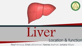
The liver anatomy and physiology
- 1. LiverLocation & function Nouf Almousa, Eman alshammari, Hawraa alsalman, Jumana Alfayez
- 2. Objectives • Define the liver, and describe its location • Describe the main features of the liver • List and describe the main functions of the liver
- 3. Definition & location of liver The liver is a large, meaty organ that sits on the right side of the belly. The liver is reddish-brownin color and feels rubbery to the touch. It’s protected by the rib cage. Nouf Almousa
- 4. The liver is grossly divided into two portions – a right and a left lobe, as viewed from the front from the front surface; but the underside shows it to be divided into four lobes and includes the caudate and quadrate lobes. The gallbladder The gallbladder sits under the liver, along with parts of the pancreas and intestines. The liver and these organs work together to digest, absorb, and process food. Nouf Almousa
- 6. Function 1: Secretion of bile • Bile, greenish yellow secretion that is produced in the liver and passed to concentration, storage, or transport the small intestine (the duodenum). • Its function is to aid in duodenum, bile also serves as route bilirubin, a byproduct of red blood the liver . • Bile is composed of bile acids and phospholipids, cholesterol, pigments, electrolyte chemicals Eman Alshammari
- 7. Function 2 Metabolism of nutrient One of major function of the liver is the metabolic process of major categories of nutrient e.g. (carbohydrate ,lipid ,protein ..) Excess glucose entering the blood after a meal is rapidly taken up by the liver and sequestered as the large polymer, glycogen (a process called glycogenesis) carbohydrate metabolism Deamination and transamination of amino acids, followed by conversion of the non-nitrogenous conversion of the non-nitrogenous part of those molecules to glucose or lipids. Several of the enzymes used in these pathways (for example, alanine and aspartate aminotransferases) are commonly assayed in serum to assess liver damage).protein metabolism Eman Alshammari
- 8. Function 3: Synthesis of plasma proteins • Plasma proteins consist of many known proteins including albumin, fibrinogens and apolipoproteins. Factors involved in hemostasis and fibrinolysis including coagulation factors, anti-trypsin and plasminogen are secreted into the blood as well as carrier proteins such as transferrin and retinol. Examples of plasma proteins include APOB, APOA1, FGG, C2, KNG1, and FGA. Hawraa Alsalman In plasmaIn Liver
- 9. Function 4: Storage of iron • Iron levels within the body need to be tightly regulated. Therefore, excess amounts must be stored in places such as the liver. Most iron stored in the liver as ferritin or hemosiderin, proteins produced by the liver. All cell types within the liver can store iron however the majority is stored within hepatocytes. Hawraa Alsalman
- 10. Function 5: Detoxification of certain hormones and drugs • The liver filters and removes compounds from the body. This includes toxins that our body produces as part of normal metabolism; pathogens like viruses and bacteria, cholesterol, hormones that our body excretes once it's used them eg. estrogen, external toxins - alcohol, drugs, chemicals we encounter through food, breathing, personal care products and our environment. Jumana Alfayez
- 11. Function 6: Removing bacteria by Kupffer cells (Phagocytic cells in Liver) • Kupffer cells (also known as stellate sinusoidal macrophages or Kupffer- macrophages found in the sinusoids of the liver. The main role to clear foreign debris and particles system circulation that pass the Jumana Alfayez
- 13. 1. All these are properties of the liver, except: A. Fleshy B. Lift side of the belly C. Reddish-brown D. Rubbery
- 14. 2. How many lobes are in the liver? A. 2 B. 3 C. 4 D. 5
- 15. 3. Bile transport into small intestine in the? A. The duodenum. B. Jejunum C. Ileum
- 16. 4. Glycogenesis Metabolism of which nutrient? A. Protein B. Carbohydrate C. Lipid
- 17. A. Anti-trypsin ,plasminogen B. Retinol, hemopexin C. Albumin, fibrinogens 5. Plasma proteins consist of many known proteins including ……………… and…………
- 18. A. In ferritin B. In adipose tissue C. In bone marrow 6. Where most of the iron stores in?
- 19. A. Before digestion B. After digestion. C. Before secretion 5.When does detoxification in the liver occur?
- 20. Thank you for listening DONE !