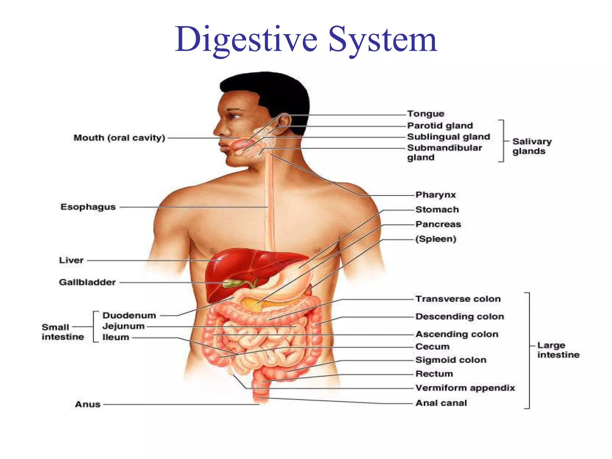
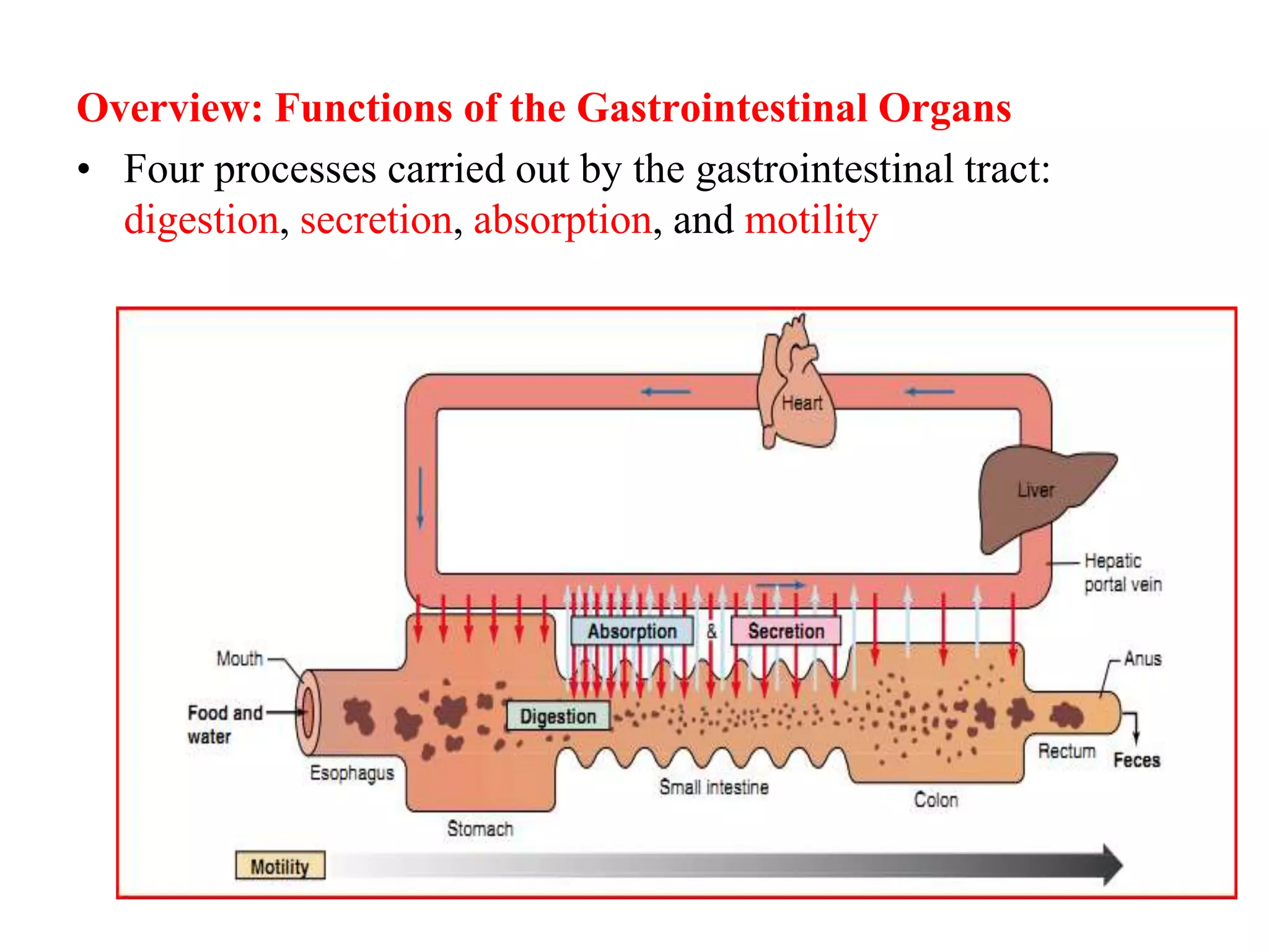
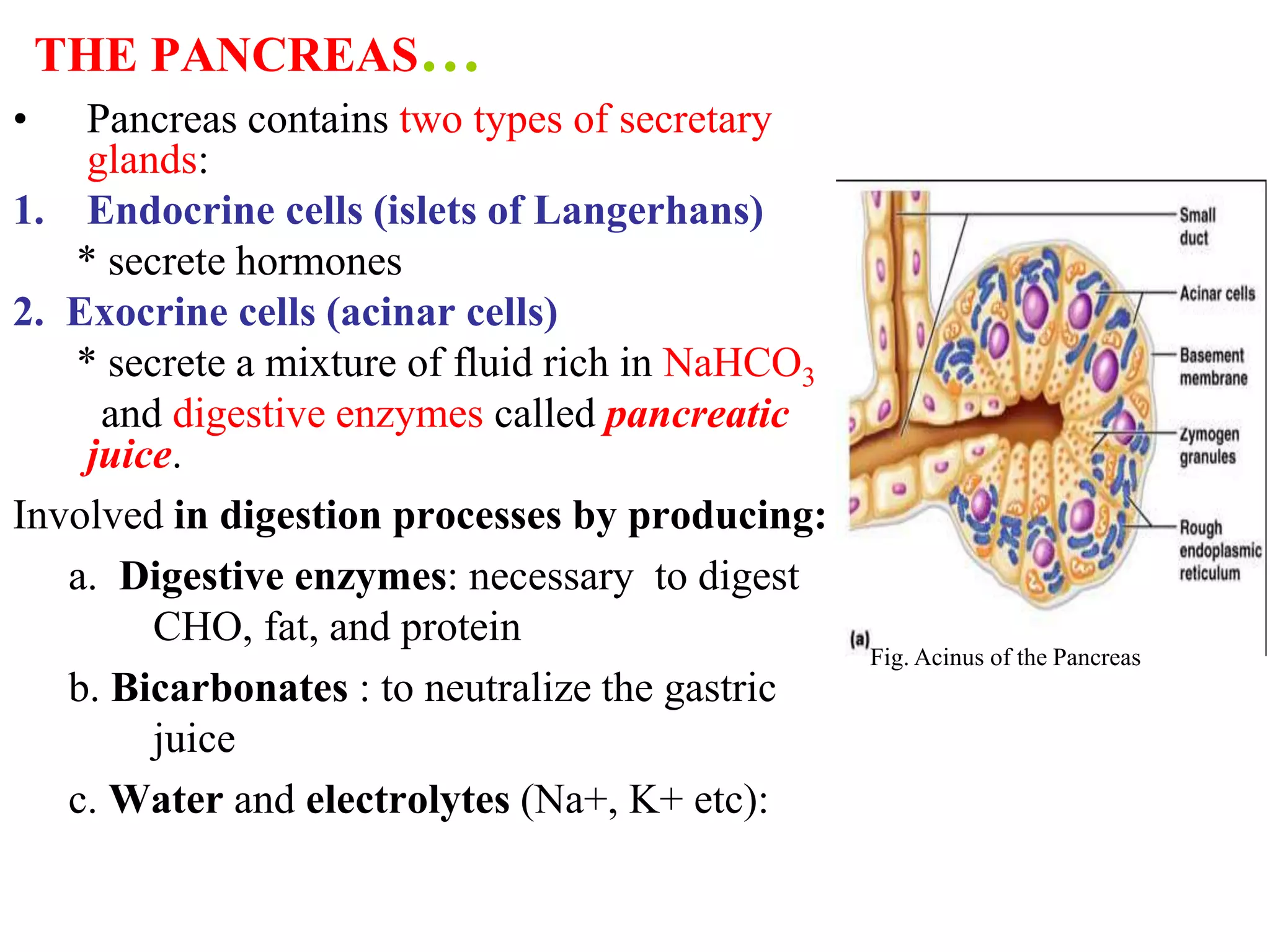
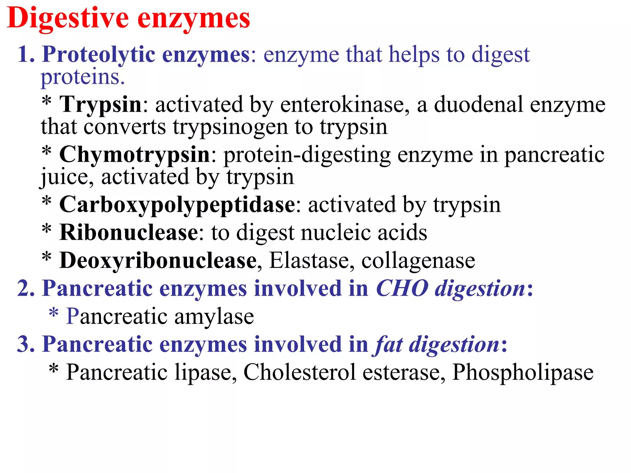
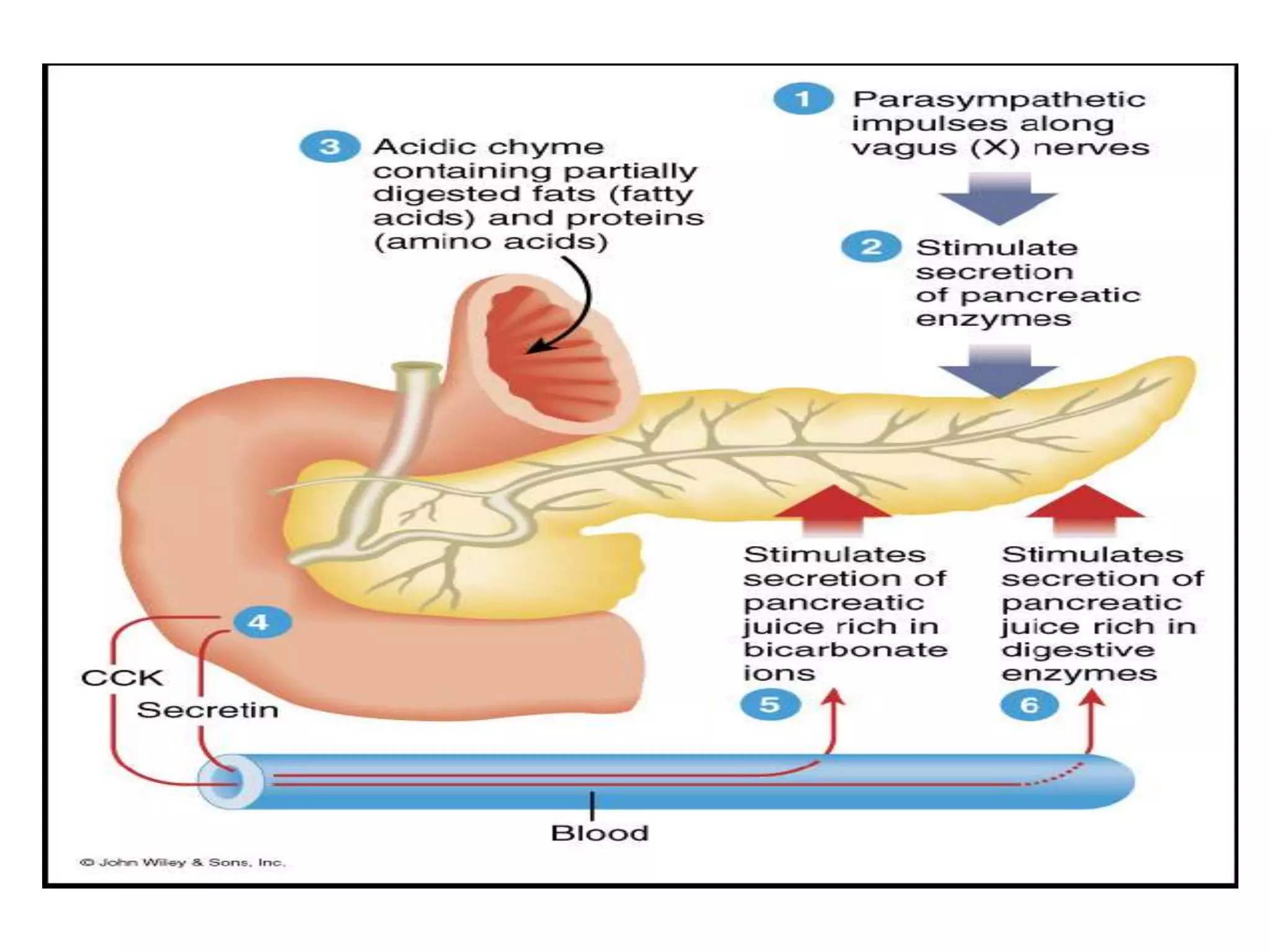
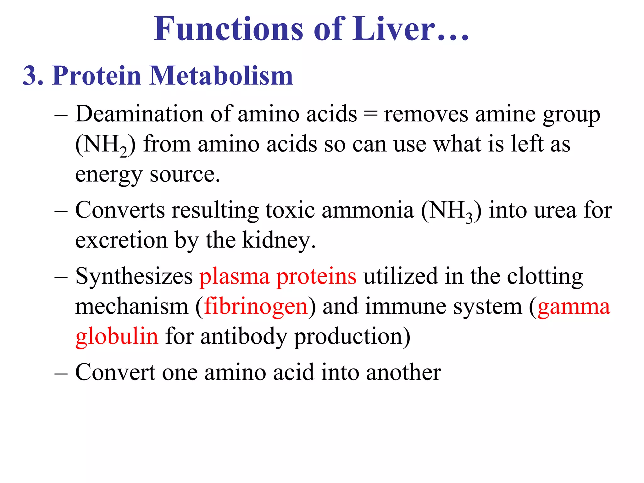
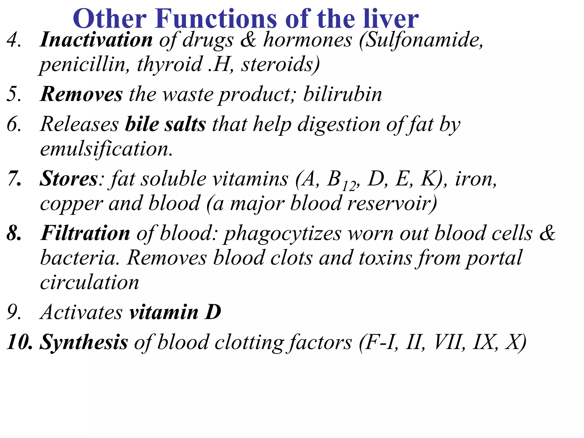
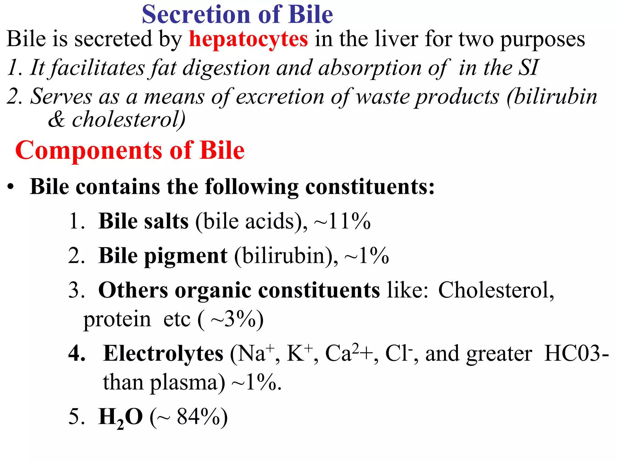
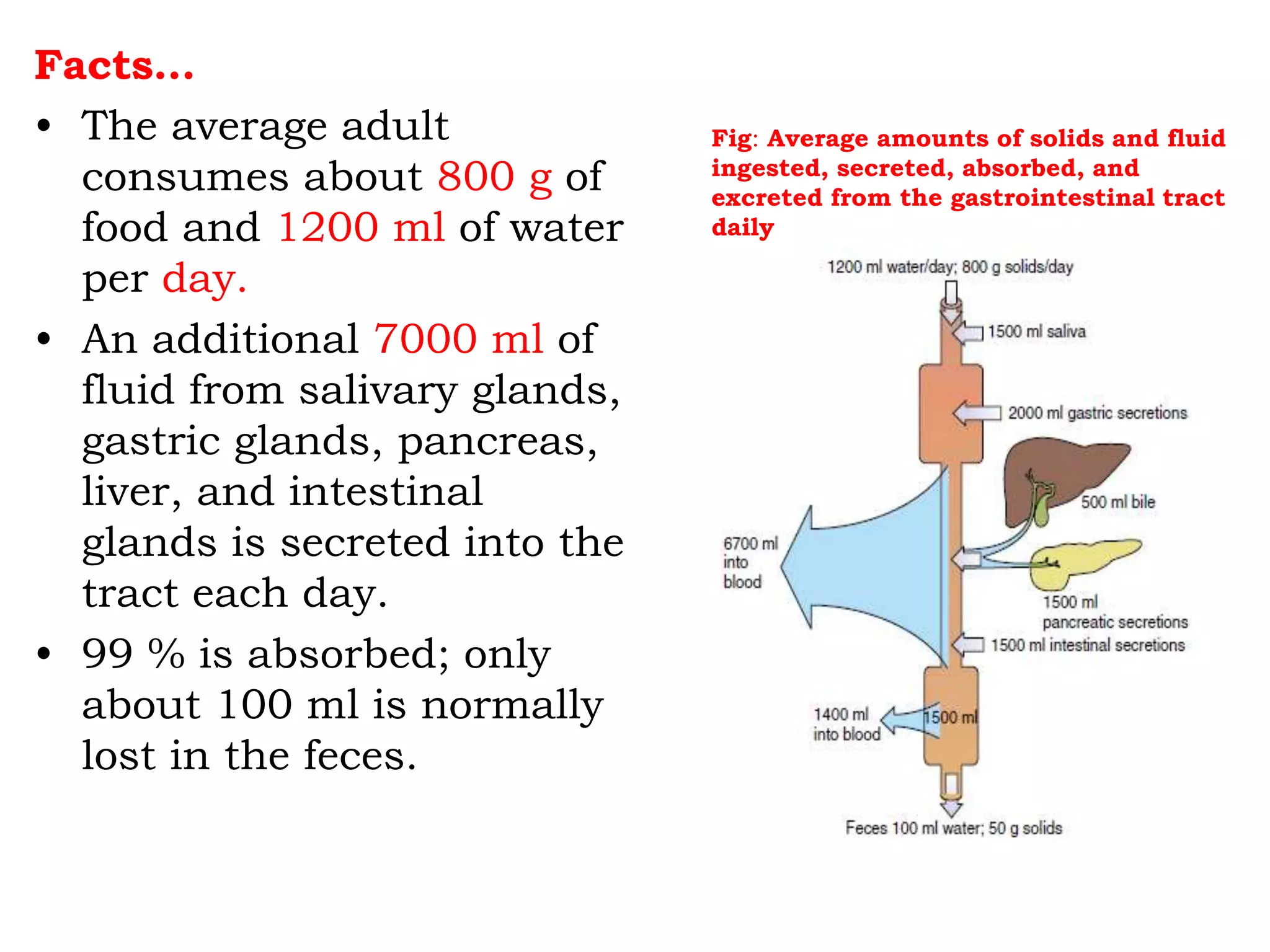
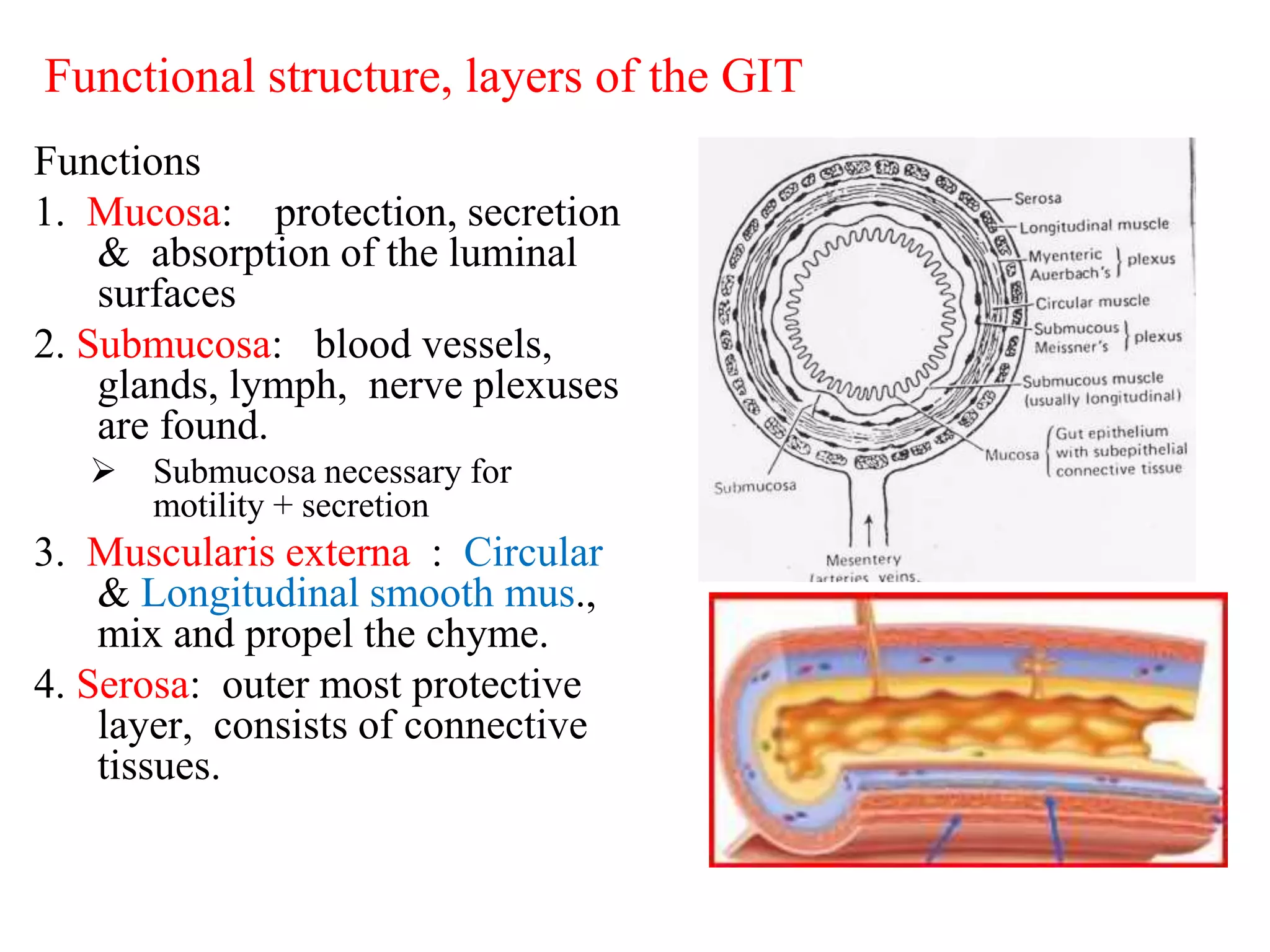
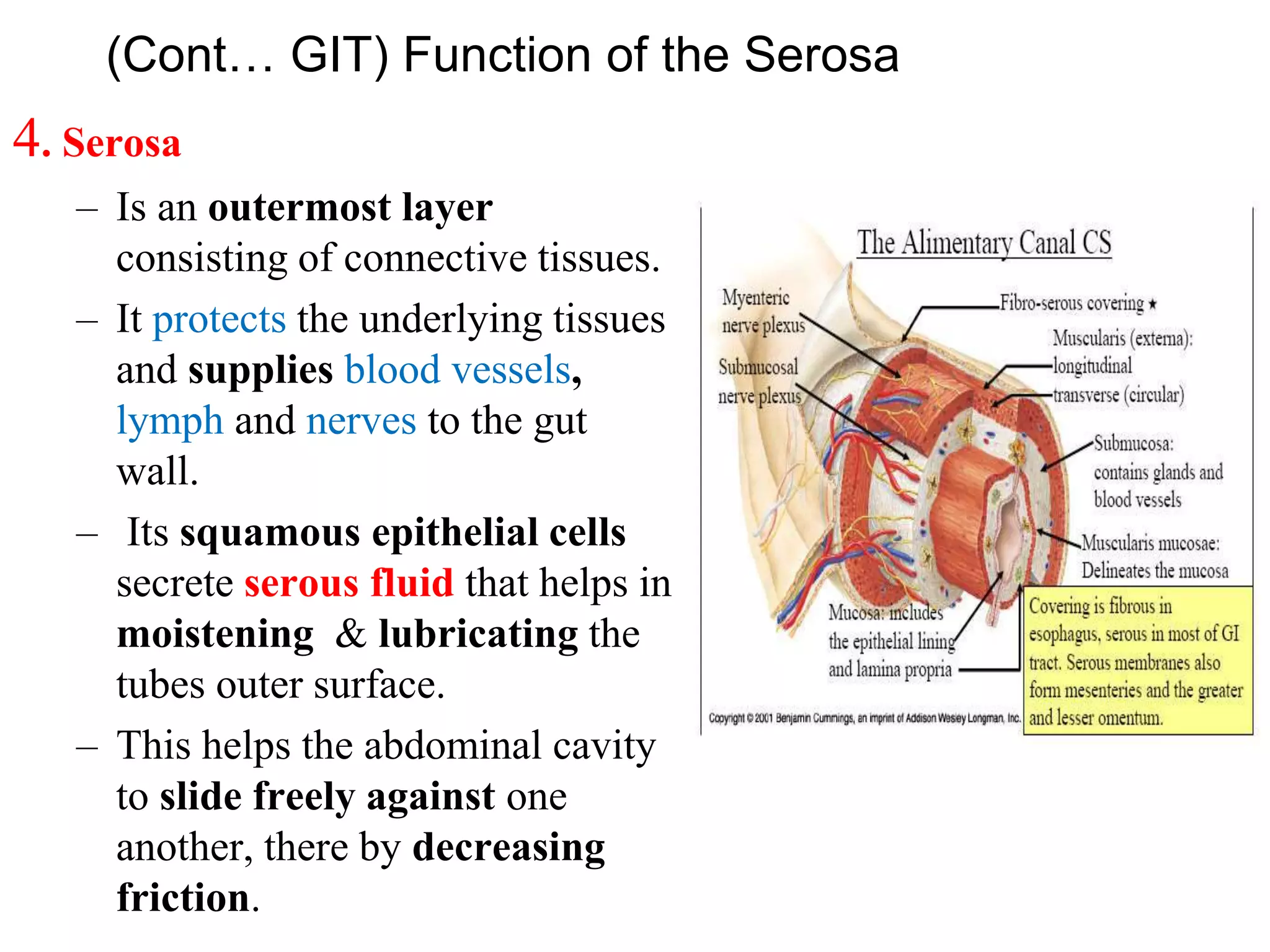
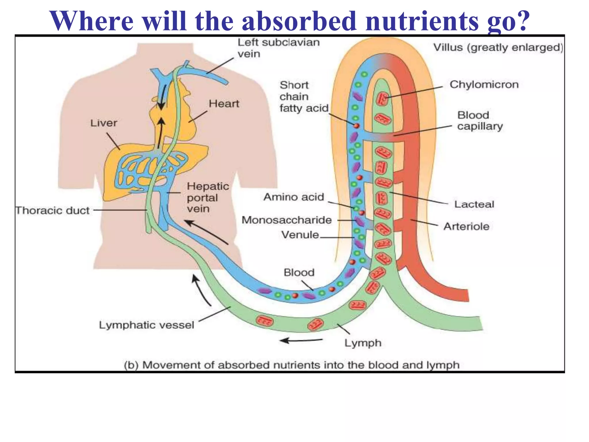
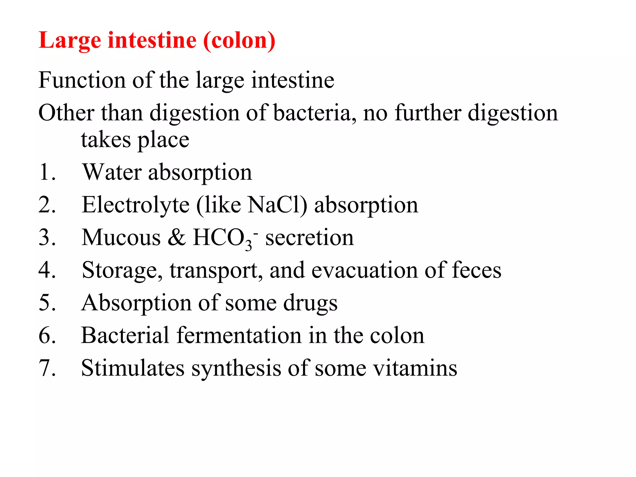
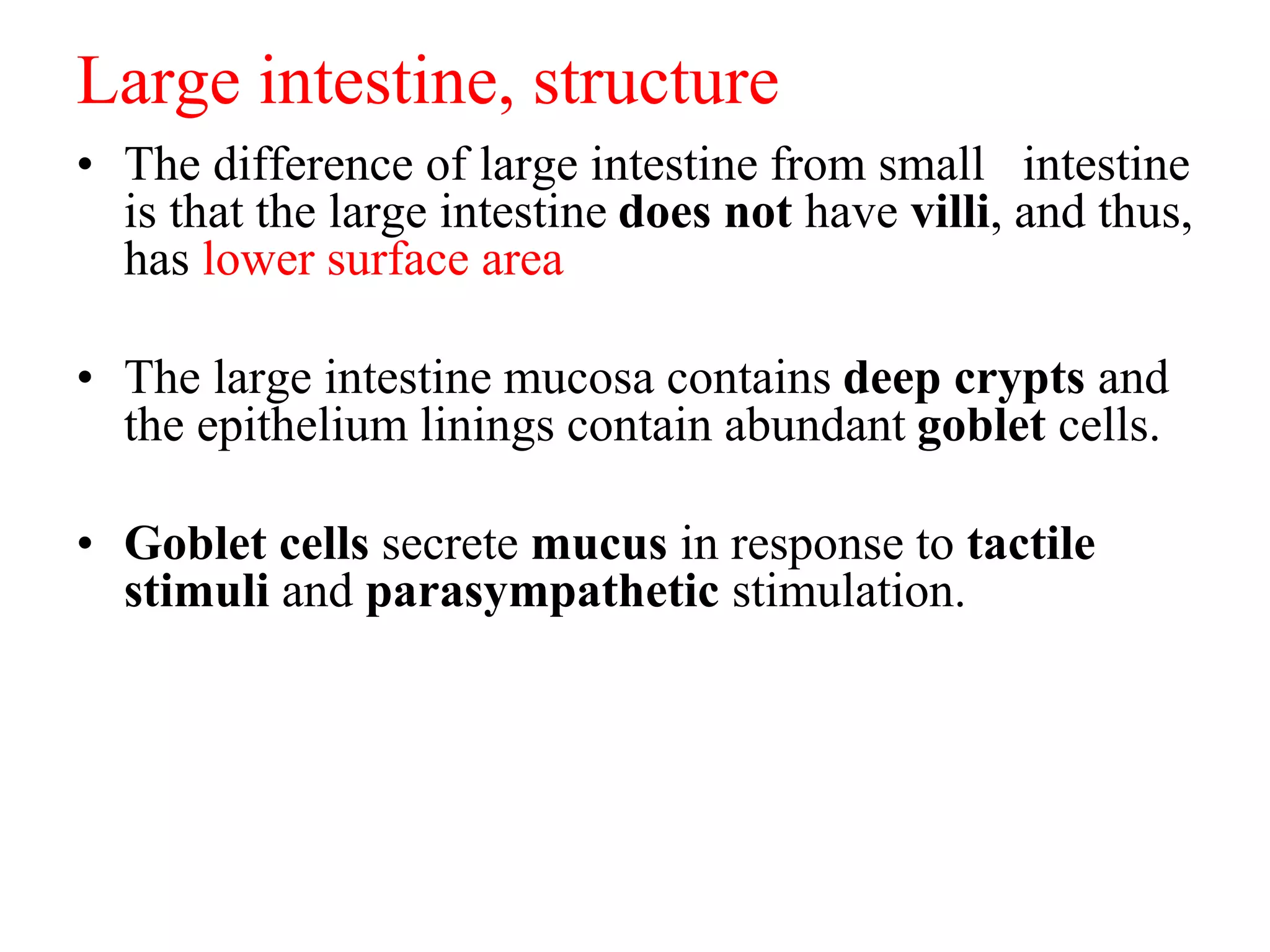
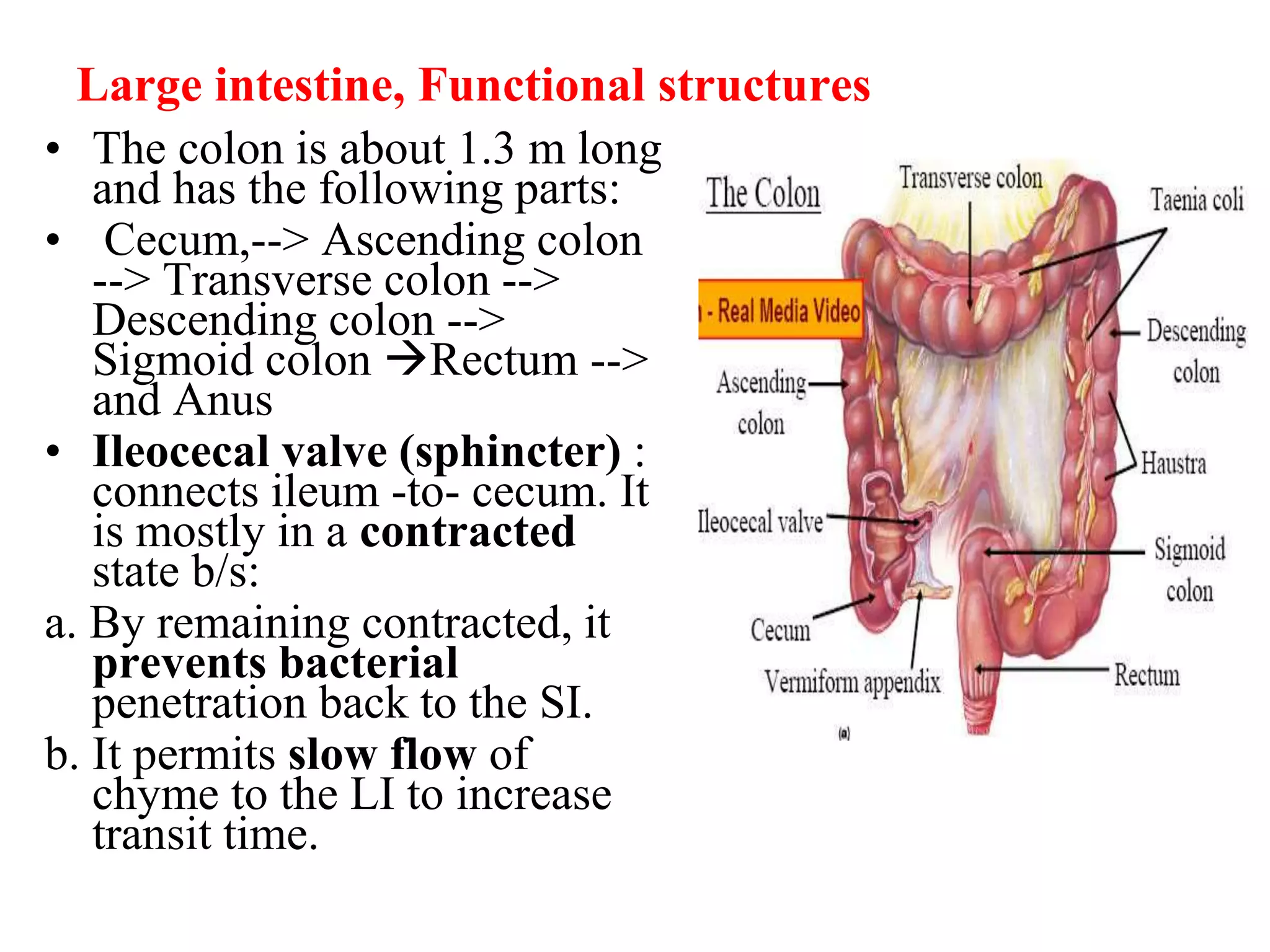
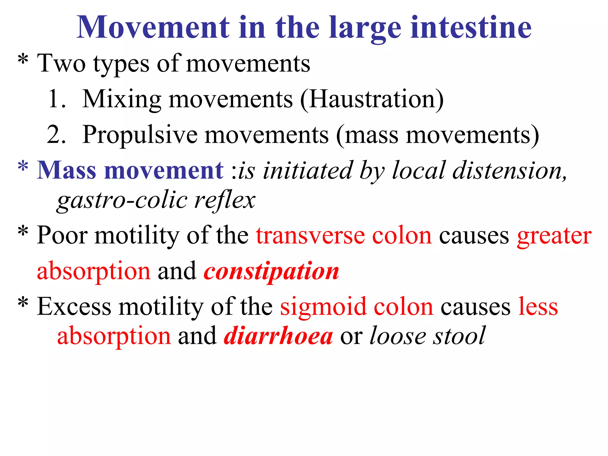
The document provides an overview of the physiology of the digestive system. It discusses the basic functions of the digestive system which include ingestion, digestion, absorption, and defecation. It describes the organs that make up the gastrointestinal tract (GIT) and their roles, including the mouth, esophagus, stomach, small intestine, large intestine, liver, gallbladder and pancreas. It also discusses the layers of the GIT wall, regulation of digestive functions by nerves and hormones, and the roles of saliva, stomach secretions, bile, and pancreatic juices in digestion.
the cardiovascular system and Physiology of heartbhupendra kumar
The document discusses the cardiovascular system and physiology of the heart. It describes the components of the cardiovascular system including the heart, blood vessels, and blood. It explains the basic functions of these parts, including that the heart acts as a pump to circulate blood through two circuits - the pulmonary and systemic circulations. It also provides details on the anatomy and functions of the heart chambers and valves, as well as blood flow, vessels, heart sounds, and blood characteristics.
The pancreatic juice contains water and electrolytes secreted by pancreatic duct cells as well as digestive enzymes secreted by acinar cells. The enzymes include proteases like trypsinogen, lipase for fat digestion, and amylase for carbohydrate digestion. Secretin is released when chyme enters the duodenum and stimulates bicarbonate secretion to neutralize acid. Cholecystokinin stimulates enzyme secretion from acinar cells to digest proteins, fats, and carbohydrates. Together, secretin and cholecystokinin regulate the exocrine pancreatic secretion in response to food in the duodenum.
The document describes the digestive system and digestive process. It details the structures and functions of the gastrointestinal tract including the mouth, esophagus, stomach, small intestine, pancreas, liver, and large intestine. It explains the mechanical and chemical breakdown of nutrients including carbohydrates, lipids, and proteins. Key enzymes and hormones that regulate digestion and nutrient absorption in each organ are also discussed.
1. The heart rate is regulated by the nervous system, specifically the vasomotor center located in the medulla oblongata.
2. The vasomotor center contains vasoconstrictor and vasodilator areas that regulate heart rate by sending sympathetic or parasympathetic signals via the spinal cord. The vasoconstrictor area increases heart rate while the vasodilator area decreases it.
3. Factors like emotions, exercise and respiration can trigger the vasomotor center to adjust heart rate through sympathetic or parasympathetic outflow as part of reflex responses mediated by baroreceptors and chemoreceptors.
The cardiac conduction system sends signals through specialized cardiac muscle cells to coordinate the rhythmic contraction of the heart. It includes the sinoatrial node, atrioventricular node, bundle of His, and Purkinje fibers. The sinoatrial node acts as the pacemaker by spontaneously generating electrical impulses that spread through the internodal pathways and cause the atria to contract. The impulse then travels to and through the atrioventricular node and bundle of His before reaching the Purkinje fibers, which trigger fast, coordinated ventricular contraction.
This document describes the four main heart sounds and how to auscultate them using a stethoscope. It explains that the first heart sound corresponds to closure of the atrioventricular valves and the R wave of an ECG. The second heart sound corresponds to closure of the semilunar valves and the T wave of an ECG. The third heart sound occurs during rapid ventricular filling between the T and P waves. The fourth heart sound corresponds to atrial contraction between the P and Q waves. It identifies the best areas over the heart to auscultate each sound using a stethoscope.
The lymphatic system consists of lymph, lymph nodes, lymph vessels, the spleen, thymus, tonsils, bone marrow and skin-associated lymphoid tissues. The main cells of the lymphatic system are lymphocytes such as B cells, T cells, natural killer cells, and supporting cells that interact with and present antigens to lymphocytes. The lymphatic system protects the body from foreign materials, assists in fluid circulation, and transports dietary fats. The thymus gland aids in the maturation of T cells while the spleen filters blood and mounts an immune response to antigens. Lymph nodes are located around arteries and veins and filter lymph fluid draining tissues.
This document provides an overview of cardiac muscle structure and function. It defines key terms related to the properties of cardiac muscle such as rhythmicity, excitability, conductivity, and contractility. It describes the cardiac syncytium and normal conduction pathway in the heart. It explains excitation-contraction coupling in cardiac muscle and compares it to skeletal muscle. It also compares action potentials in the sinoatrial node and ventricular muscle. Finally, it discusses the significance of the plateau and refractory period in ventricular muscle action potentials.
The document provides an overview of cardiovascular physiology, including:
- The cardiovascular system functions to circulate blood throughout the body, transporting oxygen, nutrients, hormones, and removing waste.
- The heart is the central organ that pumps blood through two main circulations - pulmonary circulation to the lungs and systemic circulation to the rest of the body.
- The functional anatomy of the heart includes four chambers, cardiac muscle tissue, valves that ensure one-way blood flow, and a conducting system that coordinates contractions.
The heart's electrical system, also called the cardiac conduction system, includes three main parts: the sinoatrial (SA) node, the atrioventricular (AV) node, and the His-Purkinje system. The SA node generates electrical signals that travel through the heart and cause the chambers to contract and pump blood through the body in a coordinated, rhythmic manner called sinus rhythm. Electrical signals pass from the atria to the ventricles through the AV node, which causes a delay allowing the atria to contract and empty before the ventricles. The signals then travel through the His-Purkinje system to cause synchronized ventricular contraction and pumping of blood out of the heart.
The document describes the structure and function of the respiratory system. It discusses the upper and lower respiratory tract, including the nose, pharynx, larynx, trachea, bronchi, respiratory bronchioles, alveolar ducts and alveoli. It explains how gas exchange occurs through the alveoli and pulmonary capillaries. The document also covers breathing mechanics, lung anatomy, and how the muscles of respiration work to increase thoracic cavity volume and decrease pressure, allowing air to flow into the lungs.
The heart has an intrinsic system where by the cardiac muscle is automatically stimulated to contract without any external stimulation and this property is called autorythmicity.
The pancreatic juice is a transparent, isotonic fluid secreted by the pancreas. It has both endocrine and exocrine functions. The exocrine secretions contain digestive enzymes like amylase, lipase, and proteases that aid in digestion. Bicarbonate ions secreted in pancreatic juice neutralize the acidic chime from the stomach and provide an optimal pH for the enzymes. The secretions occur in three phases regulated by both the autonomic nervous system and hormones like secretin and cholecystokinin. Secretin increases the secretion of bicarbonate-rich fluid while cholecystokinin stimulates enzyme secretion after eating. Disorders like pancreatitis and cystic fibrosis can impair pancreatic function and digestion.
Properties of gastric juice, composition of gastric juice and functions of ga...Vamsi kumar
This document contains summaries from three student seminar presentations on the topic of gastric juice:
1. Anuj Prabhank Mishra's presentation discussed the properties and composition of gastric juice, including its volume, acidic pH due to hydrochloric acid, and specific gravity.
2. Ashish Mali's presentation covered the composition of gastric juice, which is 99.5% water and 0.5% organic and inorganic solids.
3. Mahir Khan's presentation addressed the functions of gastric juice, including digestive functions of enzymes like pepsin and gastric lipase, its protective role through mucus secretion, and hemopoietic functions of intrinsic factor.
The document summarizes gastric motility and secretion. It discusses the main functions of the stomach including storage, preparing chyme, and absorption. It describes the different cell types in the stomach that secrete gastric juice components like HCl and pepsinogen. It also outlines the roles of gastric motility in serving as a reservoir, breaking down food, and emptying contents at a controlled rate. Key regulators of gastric emptying and secretion include hormones like CCK, gastrin, and secretin.
The heart receives its blood supply from two main coronary arteries - the right and left coronary arteries. The right coronary artery supplies the right atrium and ventricle while the left coronary artery supplies the left atrium and ventricle. The venous drainage of the heart occurs through the coronary sinus, anterior cardiac veins, and minute heart veins which all drain deoxygenated blood back into the right atrium.
Cardiac muscle (The Guyton and Hall Physiology)Maryam Fida
In the heart there is Atrial muscle and Ventricular muscle which are separated from each other by the fibrous AV Rings containing Valves.
ATRIAL MUSCLE: thin walled. There are two sheets, superficial and deep sheet. Superficial sheet is common over both atria. Deep sheet is separate for each atrium. Muscle fibers in the deep sheet are at right angle to the muscle fibers in the superficial sheet.
FUNCTIONS OF THE ATRIUM:
1. Receive venous blood from large veins. So atria act as reservoir.
2. Conduct the blood into the ventricles.
3. Atrial contraction is responsible for last 25 % of ventricular filling.
4. In the right atrium there is SA Node(Pace maker) and AV node.
5. In the wall of the atria, there are low pressure stretch receptors and these are involved in various reflexes like brain bridge reflex and left atrial reflex.
6. Atria also produce a hormone i.e. Atrial Natriuretic Hormone. Whenever NaCl increases in ECF, it causes release of ANH which causes natriuresis.
VENTRICULAR MUSCLE:
Much thicker than atrial muscle. Thickness of right ventricle wall is 3-4 mm and thickness of left ventricle is 8 – 12 mm.
1.Involuntary
2.Has cross striations
3.Each cardiac muscle fiber consists of a number of cardiac cells, united at ends in series. Where as in skeletal muscle each muscle fiber is individual cell.
4.Cardiac muscle cells are branching and interdigitate.
5.Single central nucleus in each cell.
6. Atrial muscle and ventricular muscle act as separate functional syncytium and impulses from atria are conducted to ventricles through the AV Node and AV Bundle.
7. Sarcoplasmic system is present. In skeletal muscle triad is at the junction of A and I bands. In cardiac muscle T Tubules are much large and thus in cardiac muscle if we take a section it may form a diad or a triad. And these diads and triads are present at the level of Z Disks.
8.Between adjacent cardiac cells there are side to side and end to end connections and these are the intercellular junctions. These junctions are Gap Junctions. Or intercalated discs
9.When one part of myocardium is excited the whole muscle is excited.
10.Whole myocardium obeys all or none law as a whole.
11.No spike potential but action potential with plateau.
12.Has got long refractory period.
Absolute refractory period in ventricular muscle is 250 – 300 milli sec.
In atrial muscle Absolute refractory period is 150 milli sec
Because of long refractory period cardiac muscle cannot be tetanized.
Stages & regulation of pancreatic secretionrashidrmc
The document discusses the phases and regulation of pancreatic secretion. It describes three phases: the cephalic phase, gastric phase, and intestinal phase. In the cephalic phase, acetylcholine causes acinar cells to secrete enzymes via parasympathetic stimulation. In the gastric phase, secretion continues in the same way. However, the enzymes from these first two phases generally are not released into the intestine until the intestinal phase. In this third phase, the hormones secretin and cholecystokinin are released in response to acidic chyme and fats in the duodenum. They work to secrete bicarbonate and digestive enzymes through the pancreatic ducts. Together, the endocrine and nervous systems regulate
Intestines(movements and secretions of small and large intestines ) The Guyto...Maryam Fida
Intestines(movements and secretions of small and large intestines)
Distended Portion of small intestine with chyme stretching concentric contractions at intervals lasting a fraction of a minute These contraction causes “Segmentation” of the small intestine ---forms spaced segments new points every time chopping chyme 2-3 times/min mixing with intestinal secretions maximum frequencyof segmentation contraction depends on frequency of BER (Basic electrical rhythm) i.e. In duodenum and proximal jejunum is 12/min and in terminal ileum is 8-9/min.
Atropine blocks the segmentation
law of gut
The peristaltic reflex +anal direction of movement of the peristalsis is called “LAW OF GUT”
This document discusses the conduction system of the heart, including the sinoatrial node, atrioventricular node, bundle of His, and Purkinje fibers. It describes how these structures work together to generate and coordinate the heartbeat. The sinoatrial node initiates impulses at a rate of 70-80 beats per minute. Impulses then spread to the atria and through the atrioventricular node to the ventricles via the bundle of His and Purkinje fibers. The autonomic nervous system can influence heart rate through the release of acetylcholine or norepinephrine on the sinoatrial and atrioventricular nodes.
Cardiac output is defined as the volume of blood pumped by the heart each minute and is regulated intrinsically by factors affecting preload and afterload as well as extrinsically by the autonomic nervous system and hormones. Venous return is a primary extrinsic regulator of cardiac output, increasing stretch of cardiac muscles and stimulating an increase in heart rate. A combination of preload, contractility, afterload and heart rate determine cardiac output under normal resting conditions and during physical activity.
Blood Coagulation and Clotting Mechanism.pptxFarazaJaved
The document summarizes the process of blood coagulation. It discusses that coagulation occurs through a series of reactions activating clotting factors, which convert fibrinogen into fibrin forming a mesh that traps blood cells. The stages are formation of prothrombin activator, conversion of prothrombin to thrombin, and conversion of fibrinogen to fibrin. Anticoagulants like heparin and warfarin prevent clotting by various mechanisms. Tests are used to detect disorders that cause excessive or insufficient clotting.
The cardiovascular system consists of the heart, blood vessels, and blood. The heart pumps blood through the blood vessels, supplying oxygen and nutrients to tissues and removing waste. It has four chambers and is located in the chest behind the sternum. The main components of the circulatory system are the blood, which contains red blood cells, white blood cells, and platelets suspended in plasma; the heart, which pumps blood through the vessels; and a closed system of arteries, veins, and capillaries that carry blood throughout the body.
The document provides information about the cardiovascular system, including the heart and blood vessels. It discusses the anatomy and layers of the heart, including the pericardium, myocardium, and endocardium. It describes the four chambers of the heart - two atria and two ventricles. It outlines the heart's location in the thorax and examines the heart valves and conducting system, including the sinoatrial and atrioventricular nodes. It also reviews the coronary arteries that supply blood to the heart.
The cardiovascular system includes the heart and blood vessels. The heart weighs 200-400 grams and pumps around 7,751 litres of blood daily. It is located behind the sternum and is surrounded by membranes. Blood enters and exits the heart through major vessels while valves regulate flow between chambers. The heart muscle generates electrical impulses and contractions to circulate blood throughout the body. Cardiac output is regulated intrinsically through preload and afterload as well as extrinsically through the nervous and endocrine systems.
The trachea is a cartilaginous tube that extends from the larynx to the lungs. It divides at the carina into the right and left main bronchi. The right bronchus is wider, shorter and more vertical, while the left is smaller but longer. The bronchi continue dividing within the lungs to form the bronchial tree which supplies the lungs. Each lung has a root, hilum, lobes, borders and surfaces. The lungs are supplied by the pulmonary arteries and veins and are innervated by the pulmonary plexus.
The Ultimate Guide to Understanding the Human Digestive SystemDeepanshuYadav2
Human Digestive System: Unraveling the Intricacies of our Inner Factory
Introduction
Welcome to this comprehensive guide on the human digestive system! In this article, we will embark on a fascinating journey through the intricacies of our inner factory, exploring the processes and functions that allow our bodies to break down and absorb nutrients from the food we consume. Join us as we unravel the secrets of digestion, absorption, and elimination, shedding light on the marvelous mechanism that keeps us nourished and energized.
The Human Digestive System: An Overview
The human digestive system is a complex network of organs and processes that work together to facilitate the digestion and absorption of food. From the moment we take a bite to the final elimination of waste, this remarkable system ensures that our bodies receive the vital nutrients needed for growth, repair, and maintenance.
The Mouth: Where It All Begins
The journey of digestion commences in the mouth. As food enters our oral cavity, it undergoes the first stage of mechanical digestion through the process of chewing. The teeth break down the food into smaller pieces, increasing its surface area for efficient chemical digestion. The saliva, secreted by the salivary glands, also plays a crucial role by moistening the food and initiating the breakdown of complex carbohydrates with the enzyme amylase.
The Esophagus: A Pathway to the Stomach
Once food is sufficiently chewed and mixed with saliva, it travels down the esophagus, a muscular tube connecting the mouth to the stomach. Through rhythmic contractions known as peristalsis, the esophagus propels the food downward, allowing it to reach the stomach for further processing.
The Stomach: A Gastric Playground
The stomach serves as a temporary reservoir for food and facilitates both mechanical and chemical digestion. It churns and mixes the food with gastric juices, including hydrochloric acid and enzymes such as pepsin. This powerful combination breaks down proteins and kills harmful bacteria, preparing the food for the next phase of digestion.
The Small Intestine: The Hub of Absorption
The small intestine is where the magic of absorption truly takes place. Divided into three parts—the duodenum, jejunum, and ileum—it receives the partially digested food from the stomach. The walls of the small intestine are lined with finger-like projections called villi, which increase the surface area for nutrient absorption. Here, the nutrients are broken down into their smallest forms and are transported into the bloodstream for distribution to the body's cells.
The Large Intestine: Processing Waste
As the now-depleted food mass enters the large intestine, the focus shifts from digestion to waste processing. The large intestine absorbs water and electrolytes from the remaining undigested material, forming solid waste known as feces. The feces are then stored in the rectum until elimination through the anus occurs.
1. absorption: passage of digested products from the intestinal lumen through mucosal cells and into the bloodstream or lacteals
2. chemical digestion: enzymatic breakdown of food
3. chyme: soupy liquid created when food is mixed with
digestive juices
4. defecation: elimination of undigested substances from the
body in the form of feces
5. ingestion: taking food into the GI tract through the mouth
6. mastication: chewing
7. mechanical digestion: chewing, mixing, and segmentation
that prepares food for chemical digestion
8. peristalsis: muscular contractions and relaxations that propel
food through the GI tract
9. propulsion: voluntary process of swallowing and the
involuntary process of peristalsis that moves food through the
digestive tract
10. segmentation: alternating contractions and relaxations of
non-adjacent segments of the intestine that move food
forward and backward, breaking it apart and mixing it with
digestive juices
This document discusses the physiology of the liver, liver function tests, and pathophysiology of jaundice. It begins by listing the learning objectives which are to understand liver functions, hepatic physiology, bilirubin metabolism, the basis for classifying jaundice, and differences in lab findings for different types of jaundice. It then describes the anatomy and blood supply of the liver, histology of liver lobules, bile secretion, and the many functions of the liver including metabolism, storage, detoxification, and immunity. It also discusses liver function tests and the metabolism of bilirubin before explaining the different types of jaundice and their pathophysiology.
The human digestive system consists of two major component one is the accessory organ like liver pancreas gall bladder salivary gland and other is the Alimentary canal which is started from oral cavity and ends on anal cavity.
in this ppt all parts are described briefly for better understanding.
The digestive system breaks down food and absorbs nutrients and water. It has two major parts - the gastrointestinal tract and accessory organs like the liver and pancreas. Food moves through the mouth, esophagus, stomach, and small and large intestines while digestive enzymes break it down. The small intestine absorbs most nutrients before waste is eliminated in the large intestine and rectum. Accessory organs like the liver, gallbladder and pancreas produce bile and enzymes to further break down food into absorbable components.
The digestive system breaks down food into nutrients that can be absorbed and used by the body. It consists of the gastrointestinal tract and accessory organs. The gastrointestinal tract includes the mouth, esophagus, stomach, small intestine, and large intestine. Accessory organs that contribute to digestion include the teeth, tongue, salivary glands, liver, gallbladder and pancreas. Each organ has a unique role in the multi-step digestive process which includes ingestion, digestion, absorption and elimination.
The document provides an overview of the digestive system, including its main components and functions. It discusses the roles and structures of the mouth, esophagus, stomach, small intestine, large intestine, liver, gallbladder and pancreas. Key points covered include the breakdown of carbohydrates, proteins and fats by digestive enzymes, and the absorption of nutrients into the bloodstream. The digestive tract protects itself through secretions, peristalsis and layers of tissue.
This document provides an overview of the gastrointestinal system, including its main parts and functions. It discusses the digestive tract, which includes the mouth, esophagus, stomach, small intestine and large intestine. It also mentions the accessory organs that help with digestion, such as the teeth, tongue, salivary glands, pancreas, liver and gallbladder. It provides details on the layers of the GI tract wall and nerve supply. It then focuses on specific parts of the digestive system, including the stomach, pancreas, liver and intestines, outlining their structures, secretions and roles in digestion.
Function of liver and effect of bile for digestion of fatRenuYadav3305
1. The liver secretes bile which aids in the digestion and absorption of fats. Bile is composed of bile salts, bile pigments, cholesterol, and other substances.
2. Bile flows from the liver into the gallbladder where it is concentrated and stored and later ejected into the small intestine in response to food.
3. Bile salts are absorbed back in the ileum and recirculate from the liver to the intestine in the enterohepatic circulation, with 95% being reabsorbed. This recycling serves to maintain bile salt concentrations.
The document discusses the key structures and processes of the human digestive system. It begins by outlining the main stages of digestion: ingestion, digestion, and egestion. It then describes the structures involved in ingestion like the mouth, esophagus and stomach. Next, it details the small intestine and how villi and microvilli increase absorption surface area. The document also discusses the roles of the liver, pancreas and large intestine in digestion and nutrient processing.
this lecture gives detailed account of functions of liver as an organ, secretion, regulation and functions of biliary secretion. exocrine and endocrine functions of pancreas. composition of pancreatic secretions
The document summarizes key aspects of human digestion and nutrition. It describes the main stages of digestion that occur in the oral cavity, stomach, small intestine and large intestine. Key points include:
- Digestion involves both physical and chemical breakdown of food facilitated by enzymes and accessory organs like the liver and pancreas.
- The small intestine is the primary site of nutrient absorption into the bloodstream through processes like diffusion, facilitated diffusion and active transport.
- The liver plays an important role in regulating blood composition and nutrient storage/processing through functions like detoxification, bile production, and albumin synthesis.
- A balanced diet with essential nutrients is important for human health, and macronutrients like carbohydrates
This document provides an overview of the gastrointestinal tract and its key parts and functions. It discusses the anatomy and functions of the oral cavity, esophagus, stomach, small intestine, large intestine, salivary glands, liver, and pancreas. It describes the roles of these organs in digestion and absorption, including the secretion and roles of gastric juices, bile, and pancreatic enzymes. It also summarizes diagnostic tools like barium meals and gastroscopy.
The document summarizes the key stages and processes of digestion. It describes the functions of the main parts of the digestive system including the mouth, stomach, small intestine, liver, gallbladder and pancreas. It explains the mechanical and chemical breakdown of food as well as the roles of enzymes and hormones in digesting carbohydrates, proteins and fats. Absorption and motility in the small intestine is also summarized.
The document summarizes the regulation and stages of pancreatic juice secretion. It discusses three phases - cephalic, gastric, and intestinal phases - which are controlled by both nervous and hormonal factors. The cephalic phase involves unconditioned and conditioned reflexes in response to food. The gastric phase is regulated by gastrin hormone. The intestinal phase is controlled by secretin and CCK hormones which stimulate secretion, as well as PP, somatostatin, PYY, and other peptides which inhibit secretion.
The document provides an overview of the digestive system, including:
1. It outlines the functional structures of the gastrointestinal tract and their roles in digestion.
2. It describes the secretions produced in the mouth, stomach, pancreas, liver, and intestines that aid in digestion of carbohydrates, proteins, and fats.
3. It explains how nutrients are absorbed and how metabolism of carbohydrates, proteins, and lipids provides energy for the body.
This document discusses lipid metabolism. It covers the digestion and absorption of lipids in the small intestine through the actions of lingual lipase, gastric lipase, pancreatic lipase, and bile salts. It also discusses the transportation of lipids through the lymphatic system via chylomicrons. Fatty acids are oxidized through beta-oxidation in the liver to produce acetyl-CoA and ketone bodies as an energy source, especially during fasting. Fat-soluble vitamins are absorbed with lipids and transported in the blood and lymph within lipoproteins and chylomicrons.
The small intestine is divided into three regions: the duodenum, jejunum, and ileum. The duodenum secretes bile from the liver, pancreatic juice from the pancreas, and bicarbonate to balance pH. These secretions help to further break down proteins and lipids. Bile emulsifies lipids to aid absorption. Pancreatic juice contains enzymes that metabolize remaining biomolecules. The presence of lipids and proteins triggers the release of cholecystokinin, stimulating the release of bile and pancreatic juice into the small intestine for digestion. Absorption of nutrients occurs in the jejunum and ileum through microvilli in the smooth muscle wall.
Gastrointestinal hormones are secreted in the GI tract to facilitate digestion. The major classifications include gastrin family hormones like gastrin and CCK, secretin family like secretin, and others like ghrelin. Gastrin increases stomach acid and motility. Histamine also increases stomach acid production when stimulated by gastrin. Secretin regulates bicarbonate and pancreas secretions. Hormone release is controlled through neural and endocrine pathways involving feedback loops between the stomach, pancreas, and duodenum.
Similar to physiology of digestive system.ppt (20)
- Video recording of this lecture in English language: https://youtu.be/kqbnxVAZs-0
- Video recording of this lecture in Arabic language: https://youtu.be/SINlygW1Mpc
- Link to download the book free: https://nephrotube.blogspot.com/p/nephrotube-nephrology-books.html
- Link to NephroTube website: www.NephroTube.com
- Link to NephroTube social media accounts: https://nephrotube.blogspot.com/p/join-nephrotube-on-social-media.html
Adhd Medication Shortage Uk - trinexpharmacy.comreignlana06
The UK is currently facing a Adhd Medication Shortage Uk, which has left many patients and their families grappling with uncertainty and frustration. ADHD, or Attention Deficit Hyperactivity Disorder, is a chronic condition that requires consistent medication to manage effectively. This shortage has highlighted the critical role these medications play in the daily lives of those affected by ADHD. Contact : +1 (747) 209 – 3649 E-mail : sales@trinexpharmacy.com
Here is the updated list of Top Best Ayurvedic medicine for Gas and Indigestion and those are Gas-O-Go Syp for Dyspepsia | Lavizyme Syrup for Acidity | Yumzyme Hepatoprotective Capsules etc
Osteoporosis - Definition , Evaluation and Management .pdfJim Jacob Roy
Osteoporosis is an increasing cause of morbidity among the elderly.
In this document , a brief outline of osteoporosis is given , including the risk factors of osteoporosis fractures , the indications for testing bone mineral density and the management of osteoporosis
8 Surprising Reasons To Meditate 40 Minutes A Day That Can Change Your Life.pptxHolistified Wellness
We’re talking about Vedic Meditation, a form of meditation that has been around for at least 5,000 years. Back then, the people who lived in the Indus Valley, now known as India and Pakistan, practised meditation as a fundamental part of daily life. This knowledge that has given us yoga and Ayurveda, was known as Veda, hence the name Vedic. And though there are some written records, the practice has been passed down verbally from generation to generation.
Muktapishti is a traditional Ayurvedic preparation made from Shoditha Mukta (Purified Pearl), is believed to help regulate thyroid function and reduce symptoms of hyperthyroidism due to its cooling and balancing properties. Clinical evidence on its efficacy remains limited, necessitating further research to validate its therapeutic benefits.
2. Outline:
• Digestive System
• Role of GI-system in Homeostasis
• Basic functions of the digestive system
• General characteristics of the GIT
• Functional structure, layers of the GIT
• Accessory organs and their functions
• Regulation of GIT
• Receptors of the GI Tract
• Functional movement of the GIT
• Salivary Glands and Saliva
• Stomach
• Secretary function of GIT
• Small Intestine:
• Large Intestine
4. Digestive System (GIT)
What are the basic
functions of the GIT
(esophagus-to-Anus)?...
It provide the body
with a continuous supply
of:
• Nutrients
• Water and
• Electrolytes
Digestive organs
5. Overview: Functions of the Gastrointestinal Organs
• Four processes carried out by the gastrointestinal tract:
digestion, secretion, absorption, and motility
6. Role of Digestive system in Homeostasis
- Body cells require nutrients to obtain energy and function
properly.
- This supply of energy is accomplished by gastrointestinal system
(GIS) that breaks food by:
a. Mechanical and
b. Chemical processes until the food is changed into its usable form
- Therefore, the GI-system is one of the important homeostatic
organs involved in maintaining the day-to-day balance of food
intake and excretion, thereby sustaining life.
7. Basic functions of the digestive system
The GI system carries out the following basic activities:
1. Ingestion: food intake, which is controlled by the
feeding and satiety center in the HT.
2. Mastication or chewing: mechanical grinding of food
with the aid of the teeth.
3. Swallowing or deglutition: propulsion of food from the
mouth to the stomach.
4. Chemical digestion of food
5. Secretion of enzymes, electrolytes (HCl, NaHCO3),
mucous, and hormones
6. Absorption of nutrients, water & electrolytes into the
blood vessels
7. Defecation: excretion of fecal matter
8. Digestive Process
Figure 23.2
~ Mouth
Food ingestion controlled
by Hypothalamus
~ Esophagus
Simple passage of food
~ Stomach
Temporary storage, Mixing,
digestion, Secretion and
emptying
~ Small intestine
Digestion, secretion
Absorption
~ Large Intestine
Storage, absorption, Passage
of faecal matter
9. GIT and its natural defense
• GIT is hollow at both ends (mouth- to - Anus).
– Therefore, can possibly harbor microorganisms in its luminal
surfaces.
• However, the GI-system can protect itself from such hazards by
some defense mechanisms found in:
– a. Mouth: Saliva contains lysozymes, IgA etc
– b. Stomach: HCl, Pepsin etc. have bactericidal effect
– c. SI (e.g., Payer's patches): Immuno-competent lymph tissues
– d. Macrophages: located in intestinal walls act to defend from
bacterial invasion etc
10.
11. Fig.1: GIT-organs and its accessories
1. Mouth
2. Pharynx
3. Esophagus
4. Stomach
5. Small
intestine
6. Large
intestine
7. Rectum
(Anus)
Main GI-Organs
Accessory Organs
1. Salivary
glands
2. Pancreas
3. Liver &
4. Gallbladder
I. Functional structure of the gut
12. Digestive System…
Organs involved in the process of digestion include:
1. The alimentary canal or gastrointestinal tract (GIT) that used to
digests and absorbs food
– mouth, pharynx, esophagus, stomach, small intestine, and large
intestine
– teeth, tongue, gallbladder, salivary glands, liver, and
pancreas
• Teeth aids mechanical breakdown of food
• Tongue assists chewing and swallowing and speech
• Other accessory organs produce and send secretions that facilitate
chemical breakdown of food
2. Accessory organs
13. Accessory organs
• Lies deep to the greater
curvature of the stomach
• The head is encircled by
the duodenum and the tail
adjacent to the spleen
• Divided into: Head, body
and tail
• Connected to the
duodenum via the main
pancreatic duct and
accessory duct
Anatomy of the Pancreas
14. THE PANCREAS…
• Pancreas contains two types of secretary
glands:
1. Endocrine cells (islets of Langerhans)
* secrete hormones
2. Exocrine cells (acinar cells)
* secrete a mixture of fluid rich in NaHCO3
and digestive enzymes called pancreatic
juice.
Involved in digestion processes by producing:
a. Digestive enzymes: necessary to digest
CHO, fat, and protein
b. Bicarbonates : to neutralize the gastric
juice
c. Water and electrolytes (Na+, K+ etc):
Fig. Acinus of the Pancreas
15. Digestive enzymes
1. Proteolytic enzymes: enzyme that helps to digest
proteins.
* Trypsin: activated by enterokinase, a duodenal enzyme
that converts trypsinogen to trypsin
* Chymotrypsin: protein-digesting enzyme in pancreatic
juice, activated by trypsin
* Carboxypolypeptidase: activated by trypsin
* Ribonuclease: to digest nucleic acids
* Deoxyribonuclease, Elastase, collagenase
2. Pancreatic enzymes involved in CHO digestion:
* Pancreatic amylase
3. Pancreatic enzymes involved in fat digestion:
* Pancreatic lipase, Cholesterol esterase, Phospholipase
16. Hormonal regulation of pancreatic secretion
• Fats and amino acids stimulate the duodenum to
releases CCK and Secretin.
• These hormones flow through the blood and reach
the pancreas.
• In the Pancreas two processes take place
a . CCK stimulates acinar cells to release digestive
enzymes
b. Secretin stimulates duct epithelium to release
H2O & HCO3-
• Both flow through common bile duct to duodenum to do
their specific job.
17.
18. LIVER AND GALLBLADDER
Liver
• The liver is the heaviest
gland in the body and the
second largest organ in the
body after the skin.
• Weighs 1.36kg.
• Located below diaphragm in
the abdomen
• Right lobe larger
• Gallbladder on right lobe
• Size causes right kidney to be
lower than left
19. Liver and Gallbladder…
• The liver is divisible into
left and right lobes,
separated by the
falciform ligament.
• The gallbladder is a sac
located in a depression
on the posterior surface
of the liver.
20. Liver: Associated Structures…
Bile leaves the liver
via:
• Bile ducts, which
fuse into the
common hepatic
duct
• The common hepatic
duct, which fuses
with the cystic duct
• These two ducts
form the common
bile duct
21. Liver: Microscopic Anatomy
• Hexagonal-shaped liver lobules are the structural and
functional units of the liver.
– Composed of hepatocyte (liver cell)
– Portal triads are found at each of the six corners of each
liver lobule
Portal triads consist of:
1. Bile duct
2. Hepatic artery – supplies O2 rich blood to the liver
3. Hepatic portal vein – carries venous blood with nutrients
from digestive viscera
22.
23. Liver: microscopic structures…
• Liver sinusoids – enlarged, leaky capillaries located
between hepatic plates
Cells in the liver
• Kupffer cells – hepatic macrophages found in liver
sinusoids.
• Hepatocytes’ functions include:
– Production of bile
– Processing blood-borne nutrients
– Storage of fat-soluble vitamins
– Detoxification
24. Functions of the liver
1. On Carbohydrate Metabolism:
It is the site of glycogenesis, gluconeogenesis and
glycogenolysis
• Turn proteins into glucose
• Turn triglycerides into glucose
• Turn excess glucose into glycogen & store in the liver
• Turn glycogen back into glucose as needed
2. On Lipid Metabolism
It is the site of β-oxidation, formation of phospholipids,
lipoproteins, synthesis of cholesterol, and conversion of
CHO into fat
25. Functions of Liver…
3. Protein Metabolism
– Deamination of amino acids = removes amine group
(NH2) from amino acids so can use what is left as
energy source.
– Converts resulting toxic ammonia (NH3) into urea for
excretion by the kidney.
– Synthesizes plasma proteins utilized in the clotting
mechanism (fibrinogen) and immune system (gamma
globulin for antibody production)
– Convert one amino acid into another
26. Other Functions of the liver
4. Inactivation of drugs & hormones (Sulfonamide,
penicillin, thyroid .H, steroids)
5. Removes the waste product; bilirubin
6. Releases bile salts that help digestion of fat by
emulsification.
7. Stores: fat soluble vitamins (A, B12, D, E, K), iron,
copper and blood (a major blood reservoir)
8. Filtration of blood: phagocytizes worn out blood cells &
bacteria. Removes blood clots and toxins from portal
circulation
9. Activates vitamin D
10. Synthesis of blood clotting factors (F-I, II, VII, IX, X)
27. Bile Production
• Bile is an alkaline fluid (pH 8)
• secreted in the liver and
functions in emulsification of
fat in the duodenum.
• Emulsification means
changing greater fat globules
into smaller fat-droplets called
micelles
28. Secretion of Bile
Bile is secreted by hepatocytes in the liver for two purposes
1. It facilitates fat digestion and absorption of in the SI
2. Serves as a means of excretion of waste products (bilirubin
& cholesterol)
Components of Bile
• Bile contains the following constituents:
1. Bile salts (bile acids), ~11%
2. Bile pigment (bilirubin), ~1%
3. Others organic constituents like: Cholesterol,
protein etc ( ~3%)
4. Electrolytes (Na+, K+, Ca2+, Cl-, and greater HC03-
than plasma) ~1%.
5. H2O (~ 84%)
29. Regulation of Bile Release
• Acidic, fatty chyme
causes the duodenum to
release:
– Cholecystokinin
(CCK) and
– secretin into the
bloodstream
• CCK and secretin
transported in blood
stimulate the liver to
produce bile
• Vagal stimulation causes
weak contractions of the
gall bladder
30. The Gallbladder
• Thin-walled, green muscular sac on the ventral
surface of the liver
• Stores and concentrates bile by absorbing its water
and ions
• Releases bile via the cystic duct, which flows into the
bile duct
31. Facts…
• The average adult
consumes about 800 g of
food and 1200 ml of water
per day.
• An additional 7000 ml of
fluid from salivary glands,
gastric glands, pancreas,
liver, and intestinal
glands is secreted into the
tract each day.
• 99 % is absorbed; only
about 100 ml is normally
lost in the feces.
Fig: Average amounts of solids and fluid
ingested, secreted, absorbed, and
excreted from the gastrointestinal tract
daily
32. Characteristics, Length of the GIT
- ~ 9 meters long.
- Is open at both ends and highly
susceptible in harboring
microbial agents.
- Voluntary segments of the GIT
are mouth, tongue, upper 1/3 of
the esophagus and the anal
regions.
- The GI-wall possesses several
glands that secrete digestive
juices and enzymes into the
lumen.
33. Functional structure, layers of the GIT
Functions
1. Mucosa: protection, secretion
& absorption of the luminal
surfaces
2. Submucosa: blood vessels,
glands, lymph, nerve plexuses
are found.
Submucosa necessary for
motility + secretion
3. Muscularis externa : Circular
& Longitudinal smooth mus.,
mix and propel the chyme.
4. Serosa: outer most protective
layer, consists of connective
tissues.
34.
35. 1. Mucosa: Functions of the Mucosa
The mucosa is divided into 3-sub-
layers
a. Epithelium:covers the mucosa and
is columnar or stratified squamous
cells.
b. Lamina propria: is under the
epithelium and contains capillaries
for absorption & some lymph
nodules for immunological
defenses.
C. Muscularies mucosa: Located
underneath the lamina.
• It is a thin layer of smooth muscle
and its contraction and relaxation
changes the degree or folding of
the luminal diameter.
36. (Cont… GIT) Function of Submucosa
2. Submucosa
- is located beneath the mucosa
- Contains rich supply of
blood vessels,
nerves,
lymph nodes,
glands, and
nerve plexuses of the enteric
nervous system called
Submucosa (Meissiner’s)
plexus.
37. (Cont…GIT) Function of Muscularis externa
3. Muscularies Externa (has 2-
sublayers) and also contains
myenteric nerve plexus.
a. The inner circular layer their
contractions decrease the diameter
of the lumen.
– At some regions, the circular SM
thicken and forms a sphincter that
prevents backflow of food
contents.
b. The outer longitudinal layer
when it contracts, it decreases the
length of the tract and cause
shortening and lengthening of the
tract.
– The effect of both contractions
helps to mix and propel the chyme
(i.e., towards the anus).
38. (Cont… GIT) Function of the Serosa
4. Serosa
– Is an outermost layer
consisting of connective tissues.
– It protects the underlying tissues
and supplies blood vessels,
lymph and nerves to the gut
wall.
– Its squamous epithelial cells
secrete serous fluid that helps in
moistening & lubricating the
tubes outer surface.
– This helps the abdominal cavity
to slide freely against one
another, there by decreasing
friction.
39. Regulation of GIT
Neural & Hormonal Regulation of the GIT
• Regulation of GI-activities is the role of Neural & Hormonal
mechanisms:
A. Neural regulation of GI-activities: includes that of
a. Extrinsic (Autonomic N fibers)
b. Intrinsic (Enteric fibers)
B. Hormonal regulation: Includes secretion of different hormones
like Gastrin, CCK, Secretin, GIP etc.
C. Paracrine Regulation:
• Paracrine secretions flow into the interstitial fluid and diffuse to
neighboring cells where they exert their effects.
E.g.: - Histamine (stimulates parietal cells to secrete HCl)
- Somatostatin (inhibit secretion of gastrin by G-cells)
40. Neural Innervations of GIT
A. Somatic innervations:
The voluntary muscle fibers at the upper esophagus and lower end
of the GIT are controlled by somatic nerves that are voluntarily in
action.
B. Autonomic innervations (extrinsic regulation):
- have generally a modulatory role (cause strong smooth muscle
contractions by peristalsis)
C. Enteric NS innervations (intrinsic regulation)
41. A. Autonomic Innervations (extrinsic regulation):
• Functions of the parasympathetics & sympathetics nerve fibers.
a. Parasympathetic fibers (cholinergic, Ach):
- is excitatory and causes strong contractions on longitudinal
muscles.
- At sphincters (at circular SM): PS-fibers cause dilation.
b. Sympathetic fibers (adrenergic, NA & adrenaline):
- is generally inhibitory in action and causes dilatation of the
alimentary tract.
- At sphincters, it causes constriction of the circular SM
42. Neural control of the GIT, cont,d
B. Enteric NS: shows local, intrinsic
regulation:
- Can act independently to cause
motility & secretion of the gut
- Are very numerous in No (approx. >
100 mill. neurons) and are sometimes
called little brain.
Types of NTs secreted from these neurons:
Several
e.g., Ach, NE, Dopamine, CCK,
Substance P, Vasoactive intestinal
polypeptide (VIP), Somatostatin, etc.
Components of ENS includes:
1. Myenteric (Auerbach’s) plexus:
- mainly control motility of the GIT
2. Submucosa (Meissner’s) plexus:
- mainly control glandular secretions
43. Receptors of the GI Tract
• Receptors initiate GI-reflexes that:
– Activate or inhibit digestive glands
– Mix lumen contents and move them along
• Mechanoreceptors:
~ respond to distension, spastic contraction
• Chemoreceptors:
~ respond to osmolarity, irritation, pH, presence of fat and
protein food and end products of digested food
• Thermoreceptors: respond to warm food/drinks
Diffusion of gasses and other food contents are affected by temperature. When T- Increases rate of
diffusion also increases
• Pain receptors: respond to tissue injury in the GIT.
44. Nervous Control of the GI Tract:
Intrinsic and extrinsic control
I. Intrinsic control
- Involves the enteric nerve
plexuses present with in the
GIT.
Nerve plexuses near the GI tract
initiate short reflexes:
• Submucosal plexus (plexus of
Meissner)
– controls GI secretary
activities
• Myenteric plexus (plexus of
auerbach)
– controls motility of the gut
45. cont,d
II. Extrinsic control
Involve CNS centers and
extrinsic autonomic nerves
• They control GI motility and
secretion
• They also modify the action of
enteric nerves
~ Sympathetic NS
↓GI function, ↓Motility
↓Secretions
~ Parasympathetic NS ↑GI
function, ↑Motility,↑Secretions
47. GIT reflexes
1. Reflexes that occur entirely within the enteric nervous system.
These include reflexes that control
GI-secretion, peristalsis and mixing-contractions, local inhibitory effects.
(Short reflexes)
48. Reflexes: cont,d
2. Reflexes that arise from the gut
go to the sympathetic ganglia
and then back to the GI-tract.
(intermediate reflexes)
Examples:
a) The gastro-colic reflex: signals
send from the stomach to cause
evacuation of the colon (LI)
b) The entero-gastric reflexes:
signals from the colon & small
intestine to inhibit stomach
motility and secretion.
c) The colono-ileal reflex:
reflexes from the colon to
inhibit emptying of ileal
contents into the colon.
49. Reflexes: cont,d
3. Reflexes from the gut to the spinal card or
brain stem and then back to GIT: (long
reflexes)
• Example: defecation reflex that travel to
– the spinal cord and back again to
produce the powerful colonic, rectal,
abdominal contraction required for
defecation (the defecation reflexes).
50. Hormonal control of GI function
• Entero-endocrine cells produce several GIT- hormones, capable of
regulation motility and secretary activities.
1. Cholecytokinin (CCK, 33aas)
• secreted by the mucosa of the jejunum in response to the presence
of fatty food in the intestine.
• has a very potent effect on gallbladder contractility for expelling
bile into the intestine in order to facilitate fat digestion and
absorption.
• Increase the release of pancreatic lipase from acinar cells.
• CCK inhibits stomach motility in order to give adequate time for fat
digestion.
2. Secretin (27aas)
• Secreted by the mucosa of the duodenum in response to acidic
gastric juice pumped from the stomach.
• It increases NaHCO3 secretion by the pancreas, a buffer that used to
neutralize the acid..
51. Functional movement of the GIT
• Two basic types of movements occur
in the GIT:
1. Propulsive movements:
• which cause food to move forward
along the tract at an appropriate rate
for digestion and absorption.
* Peristalsis: is the basic propulsive
movement of the GIT that appears in
the form of contractile rings around
the gut and propels to the anal ward
direction.
• Peristalsis is an intrinsic property of
the smooth muscles in the GIT that
generate action potential rhythmically
(basic electrical rhythm, BER).
Peristalsis and Segmentation
53. 2. Mixing movements :
• which keep the intestinal contents thoroughly mixed
at all times.
• Mixing contractions are beneficial to mix
the food contents with gastric juice
(chyme).
- Pacemaker cells are normally located at
Fundus region and fire spontaneously
causing the so called basic electrical
rhythm (BER).
- BER are slow waves that result in
tonic/energized and weak contractions of
the stomach walls.
• When slow waves progress to the antrum, the
peristalitic contractions becomes strong and cause
mixing of the stomach contents.
• The contractions also close the pyloric sphincter,
which aids in the repulsion of the chyme back to the
stomach.
• This process helps in mixing the chyme.
54. Clinical
Paralytic ileus:
• Paralysis of intestinal motility that is caused by increased in
sympathetic stimulation and damage to the smooth muscles
Hunger pangs (no food in stomach) can sometimes produce
strong peristalitic contractions.
• Such contractions fuse together and cause pain sensations.
55. Mastication (Chewing)
• Chewing is a process of mechanical breakdown of food.
• Salivary secretion containing amylase involves chemical digestion
and lubrication of chewing.
• Teeth, tongue, jaws and lips are involved in chewing.
• The movt of these organs are controlled by such centers located in
the brainstem, HT, amygdala and cerebral cortex.
• Mastication muscle are supplied mainly by the motor branch of
the trigeminal nerve.
• Chewing center is located in the pons
56. Deglutition (Swallowing)
• It is the propulsion of food from mouth to the esophagus that is
controlled by the swallowing center in the medulla.
• Involves the coordinated activity of the tongue, soft palate,
pharynx, esophagus and many separate muscle groups.
Has 3 stages:
1. Voluntary stage of swallowing: Buccal/oral phase – bolus is
forced into the oropharynx
2. Involuntary stage
A. Pharyngeal stage of swallowing:
– contributes the passage of food through the pharynx to the
esophagus.
– Controlled by the medulla and lower pons.
B. The esophageal stage of swallowing:
– promote the passage of food to the stomach.
57. Secretion of the esophagus
• Mucous glands that surround the esophagus
secrete mucus that provide lubrication and
protection from gastric digestion (HCl) that
may arise from reflux of stomach contents.
58. Lower esophageal (Gastroesophageal) sphincter
• It is a thickened circular smooth muscle at the junction
b/n the esophagus and the stomach.
• Function: prevents the reflux of gastric contents into the
esophagus.
59. Common Disorders of the Esophagus
1. Gastro-esophageal reflux disease
(heart burn).
• It occurs when the cardiac sphincter
remains open (incompetence of the
lower esophageal sphincter) .
2. Achalasia:
• The lower esophageal sphincter
remains contracted (closed).
This effect results in accumulation of food
in the esophagus causing several
disturbances (bad smell) and dilation of
esophageal walls.
(lower esophagus)
60. Digestive Processes
Mechanical Digestion:
~ The physical breakdown of food.
Ex: Grinding of the teeth or churning of the stomach.
Chemical Digestion:
~ The catabolic breakdown of food using enzymes & acids.
Absorption:
~ The passage of digested food from the lumen of the GI
Tract into the blood or lymph.
61. The Mouth
• The mouth (oral cavity) is
responsible for mechanical
digestion of solid food by
mastication.
• Mastication helps mix food
with saliva.
Parts of the oral cavity:
- The cheeks,
- The lips
- The tongue
- The hard and soft palate
- The teeth
- Saliva is released into the oral
cavity
62. Salivary Glands and Saliva
• Saliva is produced in and secreted from salivary glands.
• The basic secretory units of salivary glands are clusters of cells
called an acini.
• These cells secrete a fluid that contains;
– water,
– electrolytes,
– mucus and
– enzymes
• All of which flow out of the acinus into collecting ducts.
63. Types of Salivary Gland cells
• The secretary unit in salivary glands is the acini. There are two
types
• Serous acini: secret watery secretion rich in the enzyme
pthyalin.
• The gland is called serous salivary gland
• Mucous acini: secret thick and sticky secretion rich in mucin,
a glycoprotein, which disolves in water gives mucus.
• The gland is known as mucus salivary gland.
64. Functions of Saliva
a. Digestion: CHO-digestion begins in saliva .
• The enzyme ptyline breaks starch- to-maltose.
• Lingual lipase begins fat digestion in the mouth.
b. Protection: has anti-microbial actions (contains Lysozyme &
thiocyanate that kills microbes).
c. Involved in speech: Clear & fluent articulation is possible in the
presence of saliva.
d. Secretes HCO3
- : good to maintains the pH to neutral range
(6-to-7), the neutral pH is good for ptyline action.
e. Lubrication: Mucin found in saliva facilitates moistening and
swallowing of food.
f. Endocrine function: Sex steroids and some peptides like VIP are
found in saliva to plasma levels.
– So, sex steroids in saliva help to diagnose hypogonadism/ sex
gland secret little or no hormone.
65. Types of Salivary glands and their secretions
a. Parotid 25%: Secrete mainly serous watery fluid rich in ptyline.
b. Submandibular 70%: Produce both serous and mucous fluid.
c. Sublingual ~5%: Secrete mainly thick mucous with little serous
fluid
66. Constituents of saliva
A. H2O (99.5%):
B. Solid ( 0.5%)
– Inorganic (0.2%) Electrolytes : Na+, Cl-, K+,
HCO3- Mg, Iodine, etc.
– organic substances (0.3%) include:
Enzymes: amylase, lingual lipases, lysozymes,
thiocyanate, Glycoproteins, (albumin, globulin),
IgA, mucus, etc)
(Total secretion = about 1-1.5 L/day)
67. Reflex control of salivation:
(Nervous control)
• Sight, smell, and test or thinking of
food Receptors in oral cavity or
smell Sensory fibers from the
tongue to the nuclei in brain stem (MO),
so called Salivatory nuclei
parasympathetic fibers act on salivary
glands to increase copious salivary
secretion and sympathetic fibers to
increase thick and sticky secretion.
• Salivation can also be controlled by
higher centers like hypothalamus
which has nerve connections with
salivatory nuclei in the Medulla
oblongata (MO).
• Higher centers like appetite area in the
hypothalamus are also involved in
reflex control.
68. Phases of Salivary secretions
Three phases of salivary secretions include
1. Cephalic (brain) phase: triggered by thought, smell, or
sight of food (conditioned reflexes)
2. Oral phase: triggered by food that stimulate touch & test
receptors in the mouth (unconditioned reflex)
3. Gastric phase: triggered by substances which stimulate the
gastric mucosa (acids or sour tastes) in the stomach
(unconditioned reflex)
.
69. Stomach
• Chemical breakdown of proteins begins and food is converted to
chyme
• Cardiac region: surrounds the cardiac orifice
• Fundus: dome-shaped region beneath the diaphragm
• Corpus (Body): midportion of the stomach
• Antrum (Pyloric antrum): the dilated part of the stomach before
the pyloric region.
• Pyloric region: made up of the antrum and canal which
terminates at the pylorus
• The pylorus is continuous with the duodenum through the
pyloric sphincter
70.
71. Functions of the stomach
Storage of large quantities of food until it can be pumped into the
duodenum.
Can accommodate large amount of food up to 1.5 liters .
Mixing of food with gastric secretion to form a semi-fluid chyme.
Slow emptying the food from the stomach into the small intestine
at a rate suitable for proper digestion and absorption by the small
intestine.
Secretary function: HCl, mucous, pepsin, gastrin, IF
Sterilization
Digestion
Absorption
Facilitates defecation
72. Functional layers of the stomach
Mucosa: protection, secretion & absorption from the luminal
surface.
Submucosa: contains blood vessels, lymph nodes and some nerve
fibers. Between Submucosa and circular muscles is found a
nerve plexus called meissners plexus which is involved in GIT-
secretions.
Muscularis externa: contains circular & Longitudinal smooth
muscle layers. Between them is found Myeinteric plexus
involved in motility.
Contraction of the circular muscles decreases the diameter of the
lumen and
contraction of the longitudinal, decreases the length of the GIT tract.
The Serosa: outer most protective layer, consists of connective
tissues
73.
74. Microscopic Anatomy of the Stomach
• Muscularis: Allows the
stomach to churn, mix,
and pump food
physically.
• Epithelial lining:
contains Goblet cells
that produce a coat of
alkaline mucus
• The mucous surface
layer traps a bicarbonate-
rich fluid beneath it
• Gastric pits
– contain gastric glands
that secrete gastric
juice, mucus, and
gastrin.
75. Glands of the Stomach Fundus and Body
• Gastric glands of the fundus and body have a variety of secretory
cells
– Mucous neck cells – secrete acid mucus
– Parietal cells – secrete HCl and intrinsic factor
– Chief cells – produce pepsinogen
• Pepsinogen is activated to pepsin by:
– HCl in the stomach
– Pepsin itself via a positive feedback mechanism
– Enteroendocrine cells – secrete gastrin, histamine,
endorphins, serotonin, cholecystokinin (CCK), and
somatostatin
76. Stomach walls, cont,d
- Rugae, increases surface
area as the stomach fills with
chyme.
- Folds have tube like
depressions called gastric
pits that contain different
gastric glands.
- The secretions are released into
the base of the gastric pit.
77. Production of gastric juices in stomach cells
Cells Secretions Location
1. Parietal cells (oxyntic cells) HCl + IF the body and fundus
2. Chief cells Pepsinogen the body and fundus
3. Mucous cells: Mucous the body and fundus
4. G-cells: Gastrin at pyloric antrum
5. D-cells Somatostatin
6. Enterochromaffin-like cells - Histamine throughout the tubular
glands
*Rennin in children (curdles milk)
78. HCl secretion by Parietal cells
1. H+ ions that result from the dissociation of
H2O in the cytoplasm of the parietal cells
are continuously pumped actively through
the membrane of the gland into the gland
lumen (pit).
2. intracellular) CO2 and OH- combine to
produce HCO3-.
3. Cl- ions are transported from the blood into
the parietal cell and finally into the lumen
(pit) of the gland by facilitated diffusion.
4. HCO3
- in exchange to Cl- is transported
in reverse direction (from the cytoplasm
into the blood, charge balance).
5. Finally, H+ and Cl- ions combine in the
lumen of the gland (pit) and produce HCl
• HCl is collected and stored in the pit until
used for different physiological functions.
Secretion of HCl by parietal cells
79. Regulation and Mechanism of HCl Secretion
• The four inputs to parietal cells that regulate acid secretion by
controlling the transfer of the H-K ATPase pumps in
cytoplasmic vesicle membranes to the plasma membrane.
80. Pepsin Secretion
• Several protein-digesting enzymes collectively known as pepsin
• Pepsin is secreted by chief cells in the form of an inactive
precursor called pepsinogen.
• The acidity in the stomach’s lumen converts pepsinogen to
pepsin, the fully active form
• Thus the activation of pepsin is positive-feedback process.
Fig: Conversion of pepsinogen to pepsin in the lumen of the stomach.
81.
82. Emptying of the stomach
• The rate at which the stomach empties depends on the fluidity of the
chyme and its contents.
• Thus, liquids empty faster than solids (fast CHO> protein> fat).
Factors that affect emptying include neural and hormonal
1. Neural factors that affect emptying:
a. Parasympathetic: Causes increase rate of emptying by opening the
pyloric sphincter.
b. Sympathetics: Increases constriction of the pyloric sphincter through
its adrenergic receptors, thus it has the effect of delaying emptying.
c. Enteric nerves (Enterogastric reflex):
When fat or protein chyme reaches the duodenumreceptors detect and send impulses
to enteric nerves of the stomach cause the inhibition of stomach motility and
secretion.
83. (Cont…) Stomach emptying
2. Hormonal factors that slow emptying:
- CCK (stimulated by fat and peptides)
- GIP (gastric inhibitory polypeptide), stimulated by CHO and
fat
- Secretin : stimulated by high acid (HCl)
• The hormones after being secreted pass through the blood
and reach the stomach, where they inhibit stomach motility
and secretion.
• They also cause constriction of the pyloric sphincters thereby
delaying emptying.
84. Physiological advantage of delaying stomach contents
1. It gives ample time for nutrients (e.g., like fat) to
remain longer in the stomach and be digested by
gastric juices.
2. The delay prevents acids (HCl) not to be damped into
the duodenum at higher rates to cause duodenal ulcers.
3. The delay also gives time for pancreatic secretions to
reach duodenum and neutralize the acid.
85. Stomach Lining
• The stomach is exposed to the harshest conditions in the digestive
tract
To keep stomach from digesting itself, the stomach has ;
• A mucosal barrier with:
– A thick coat of bicarbonate-rich mucus on the stomach wall
– Epithelial cells that are joined by tight junctions
– Gastric glands that have cells impermeable to HCl
• Damaged epithelial cells are quickly replaced
86. Absorption from the stomach
An insignificant absorptive functions takes place in the
stomach, these are:
a. Alcohol
b. Certain drugs (aspirin, morphine etc.)
c. Small quantities of H2O.
NB: Organic nutrients (glucose, amino acids, and
FFA) etc are not usually absorbed from the
stomach.
87. Phases of gastric juice secretion
The cephalic (~15%) (neural):
• Afferent impulses from taste, smell, sight, or thought of food are
carried to MO that causes reflex stimulation of the stomach to
secrete gastric juices.
The gastric phase (~70%)
• When food distends the stomach, mechanoreceptors and
chemoreceptor are activated by products of food.
• Sensory information from the receptors pass to the brain stem
(MO)
• Reflex Vagal stimulation on the stomach causes increased gastric
secretions through Ach .
3. The intestinal phase (~5%)
• This stage begins when chyme reaches the duodenum.
• This phase is mostly inhibitory to gastric juice secretion.
• Secretion of hormones like Secretin, CCK, GIP that reflexly inhibit
gastric secretion in the stomach.
88. Small Intestine: Gross Anatomy
• Runs from pyloric sphincter
to the ileocecal valve
• Has three subdivisions:
– duodenum,
– jejunum, and
– ileum
• The bile duct and main
pancreatic duct join the
duodenum
• The jejunum extends from
the duodenum to the ileum
• The ileum joins the large
intestine at the ileocecal valve
89. Small Intestine: gross anatomy
• SI is specialized for completion
of digestion and absorption of
nutrients.
a. Duodenum: ~25 cm, mainly
secretary, mucose, hormones,
enzymes)
b. Jejunum : ~2.5m mainly
absorptive
c. Ileum : ~3.7m mainly
absorptive
90. Small intestine, structure
• Structure of the wall is similar to others and
consists of 4-layers
(mucosa, Submucosa, Muscularis externa and Serosa).
• Enteric and Vagal nerves abundantly innervate the
Small intestine.
• The inner lining is greatly folded to increase the
surface area for absorption, thus
– The microvillus + Villi + greater foldings (plicea
circulares) increase the absorptive surface of Small
intestine by ~ 600-fold).
91. Small Intestine: microscopic anatomy
• Structural modifications of the small intestine wall increase
surface area
• Plicae circulares:
~ deep circular folds of the mucosa and sub mucosa
• Villi
~ finger like extensions of the mucosa
• Microvilli
~ tiny projections of absorptive mucosal cells’ plasma membranes
92.
93. The Villi
• Villi: Are numerous finger like projections
that emerge from the mucous membrane.
• Each Villi consist of a layer of absorptive
epithelial cells that contain minute microvilli
(brush border) at their surfaces.
• The epithelial cells contain digestive enzymes like
sucrase, maltase, lactase, and peptidases in the
membranes of the microvilli.
94. Movement in the small intestine
~ Two types of movements occur in the SI:
1. Mixing movement
2. Propulsive movement
Mixing movements (segmentation contractions)
~ Ring like contractions appear at regular intervals
~ Segmentation contractions exert chopping action
on intestinal chyme and mix it with digestive
juice
95. Propulsive movements
• Are peristaltic waves that propel chyme anal wards
• Initiated by intestinal distension.
• Chyme is propelled in the SI until it reaches the
terminal ileocecal sphincter
96. Ileocecal Sphincter
• Function:
prevents back flow of fecal
matter from the cecum to the
ileum
Factors regulating the sphincter
• Pressure and chemical irritation
of ileum
– relax it and initiates peristalsis.
• Pressure and chemical irritation
of cecum
– inhibit peristalsis of ileum and
closes the sphincter.
97. Digestive enzymes secreted in the SI
1. Peptidase: splits peptides into amino acids
2. Three enzymes hydrolyzing diasaccharides into
monosaccharides: sucrase, maltase and lactase
3. Intestinal lipase: splits neutral fats into glycerol and
fatty acids.
Regulation of SI secretion
• Local factors: tactile, distension, irritation, pH.
• Hormonal: Secretin, CCK, VIP, Glucagon, GIP
• Nervous: vagal stimulation increases intestinal secretion
Sympathetic stimulation decreases intestinal secretion
• Enteric reflexes: stimulation of submucosal plexus
98. Digestion in the Small Intestine
• As chyme enters the duodenum:
– Carbohydrates, proteins and fat digestion have taken
place
– Digestion continues in the small intestine
– Chyme is released slowly into the duodenum
– Because it is hypertonic and has low pH, mixing is
required for proper digestion
• Virtually all nutrient absorption takes place in the small
intestine
99. Digestion in the Small Intestine
1. Digestion of Carbohydrates
Involved Enzymes and their sight of Secretion
• Mouth: salivary amylase
• Esophagus & stomach: nothing happens
• Duodenum: pancreatic amylase
• Brush border enzymes (maltase, sucrase & lactase) act on
disaccharides and produce monosaccharides: fructose, glucose &
galactose
100. Starch
- Salivary amylase (20-40%)
- Pancreatic amylase (50-80%)
Maltose and 3-9
glucose polymers Lactose Sucrose
-Maltase -Lactase -Sucrase
-Dextrase
Glucose + Glucose Glucose and Glucose and
Galactose Fructose
Digestion of Carbohydrates…
101. 2. Proteins Digestion:
Involved Enzymes and their Sight of Secretion
Enzymes used: pepsin in the stomach
• Enzymes acting in the small intestine
* Pancreatic enzymes
– trypsin, chymotrypsin, and carboxypeptidase
* Brush border enzymes
– aminopeptidases, carboxypeptidases, and
dipeptidases
102. Digestion of Proteins, cont,d
Stomach
• HCl denatures or unfolds proteins
• HCl activates pepsinogen into pepsin
• Pepsin turns proteins into peptides
Pancreas
• Digestive enzymes: split peptide bonds between different
amino acids.
Small Intestine
• Brush border enzymes
* enzymes break peptide bonds that attach terminal amino acids to
carboxyl ends of peptides (carboxypeptidases)
* enzymes break peptide bonds that attach terminal amino acids to
amino ends of peptides (aminopeptidases)
* enzymes split dipeptides to amino acids (dipeptidase)
103. 3. Chemical Digestion of Fats
• Happens mainly in the small intestine
Two phases:
1. Emulsification
– By bile salts
– Emulsion droplets
2. Chemical digestion
• Pancreatic lipase
–Free fatty acids
–Monoglycerides
104. Dietary source of fat
• Neutral fats (triglycerides), Cholesterol and cholesterol esters
Phospholipids
Fat Emulsified fat FFA + Glycerides
-Lingual lipase
-Gastric lipase
-Pancreas lipase
Cholesterol Bile salt FFA + Glycerides
Cholesterol esters Cholesterol Esterase
Phospholipids-A2 Phospholipase FFA + Phopholipids
Digestion of Lipids
Bile salt
105. Absorption in the Small Intestine
• Absorption
~ is the passage of the end
products of digestion
from the GI tract into
blood or lymph
Occurs by
• diffusion,
• facilitated diffusion,
• osmosis, and
• active transport.
106. Small intestine, Absorption
• Absorption & transport
– Carbohydrates are degraded into their simpler
forms; glucose, galactose, and fructose are
absorbed by secondary active transport (diffusion
together with Na+ ion).
– Fats are degraded into FFA + glycerol and taken
by lacteals that enter the thoracic duct and finally
join the circulation.
– Protein are absorbed as amino acids by active
transport.
– Electrolytes Na+, Ca2+, iron etc are absorbed by
active transport.
108. Water Absorption
• 95% of water is absorbed in the small intestines
by osmosis
• Water moves in both directions across intestinal
mucosa
• Net osmosis occurs whenever a concentration
gradient is established by active transport of
solutes into the mucosal cells
• Water uptake is coupled with solute uptake, and
as water moves into mucosal cells, substances
follow along their concentration gradients.
110. Large Intestine
• Is subdivided into
– the cecum,
• appendix,
– colon,
– rectum, and
• anal canal
• The cecum:
– Contains appendix
111. Colon
• Has distinct regions:
– ascending colon,
– transverse colon,
– descending colon, and
– sigmoid colon
• The sigmoid colon joins the rectum
• The anal canal, the last segment of the large intestine, opens to
the exterior at the anus
112. Valves and Sphincters of the Rectum and Anus
• Valves of the rectum stop feces from being passed with gas
• The anus has two sphincters:
– Internal anal sphincter composed of smooth muscle
– External anal sphincter composed of skeletal muscle
• These sphincters are closed except during defecation
113. Large intestine (colon)
Function of the large intestine
Other than digestion of bacteria, no further digestion
takes place
1. Water absorption
2. Electrolyte (like NaCl) absorption
3. Mucous & HCO3
- secretion
4. Storage, transport, and evacuation of feces
5. Absorption of some drugs
6. Bacterial fermentation in the colon
7. Stimulates synthesis of some vitamins
114. • The large intestine takes about 16 hours to finish up the
remaining processes of the digestive system.
• Food is no longer broken down at this stage of digestion.
• The colon absorbs vitamins which are created by the
colonic bacteria
• It also compacts feces, and stores fecal matter in the
rectum until it can be discharged via the anus in
defecation.
115. Bacteria
• The bacteria of the large intestine consist of:
– Bacteria surviving the small intestine that enter the cecum
– Those entering via the anus
• The large intestine houses over 700 species of bacteria that
perform a variety of functions.
• The large intestine absorbs some of the products formed by
the bacteria inhabiting this region.
116. Bacteria in the large intestine:
– Ferment indigestible carbohydrates, undigested
polysaccharides (fiber) are metabolized to short-
chain fatty acids by bacteria in the large intestine
and absorbed by passive diffusion.
– Release irritating acids and gases (flatus). The
bicarbonate that the large intestine secretes helps to
neutralize the increased acidity
– They produce large amounts of vitamins,
especially vitamin K and biotin (a vitamin B), for
absorption into the blood.
117. Large intestine, structure
• The difference of large intestine from small intestine
is that the large intestine does not have villi, and thus,
has lower surface area
• The large intestine mucosa contains deep crypts and
the epithelium linings contain abundant goblet cells.
• Goblet cells secrete mucus in response to tactile
stimuli and parasympathetic stimulation.
118. Large intestine, Functional structures
• The colon is about 1.3 m long
and has the following parts:
• Cecum,--> Ascending colon
--> Transverse colon -->
Descending colon -->
Sigmoid colon Rectum -->
and Anus
• Ileocecal valve (sphincter) :
connects ileum -to- cecum. It
is mostly in a contracted
state b/s:
a. By remaining contracted, it
prevents bacterial
penetration back to the SI.
b. It permits slow flow of
chyme to the LI to increase
transit time.
119. Movement in the large intestine
* Two types of movements
1. Mixing movements (Haustration)
2. Propulsive movements (mass movements)
* Mass movement :is initiated by local distension,
gastro-colic reflex
* Poor motility of the transverse colon causes greater
absorption and constipation
* Excess motility of the sigmoid colon causes less
absorption and diarrhoea or loose stool
120. Defecation reflex
• Rectal Stimulation
distension of myenteric plexus
Initiates peristalsis in descending, sigmoid colon and rectum
Inhibition of internal anal sphincter (IAS) by
• Parasympathetic stimulation
• myenteric plexus
Peristaltic wave forces feces to the anus
Voluntary Relaxation of external anal sphincter
►Defecation
121.
122. Secretion of the large intestine
Glands
• Cryps of Lieberkuhn Secret H2O, electrolytes
• Goblet cells and mucous
Regulated by local (tactile) factors