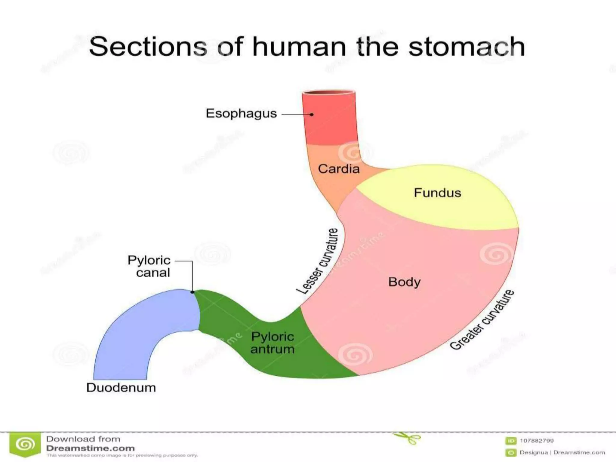
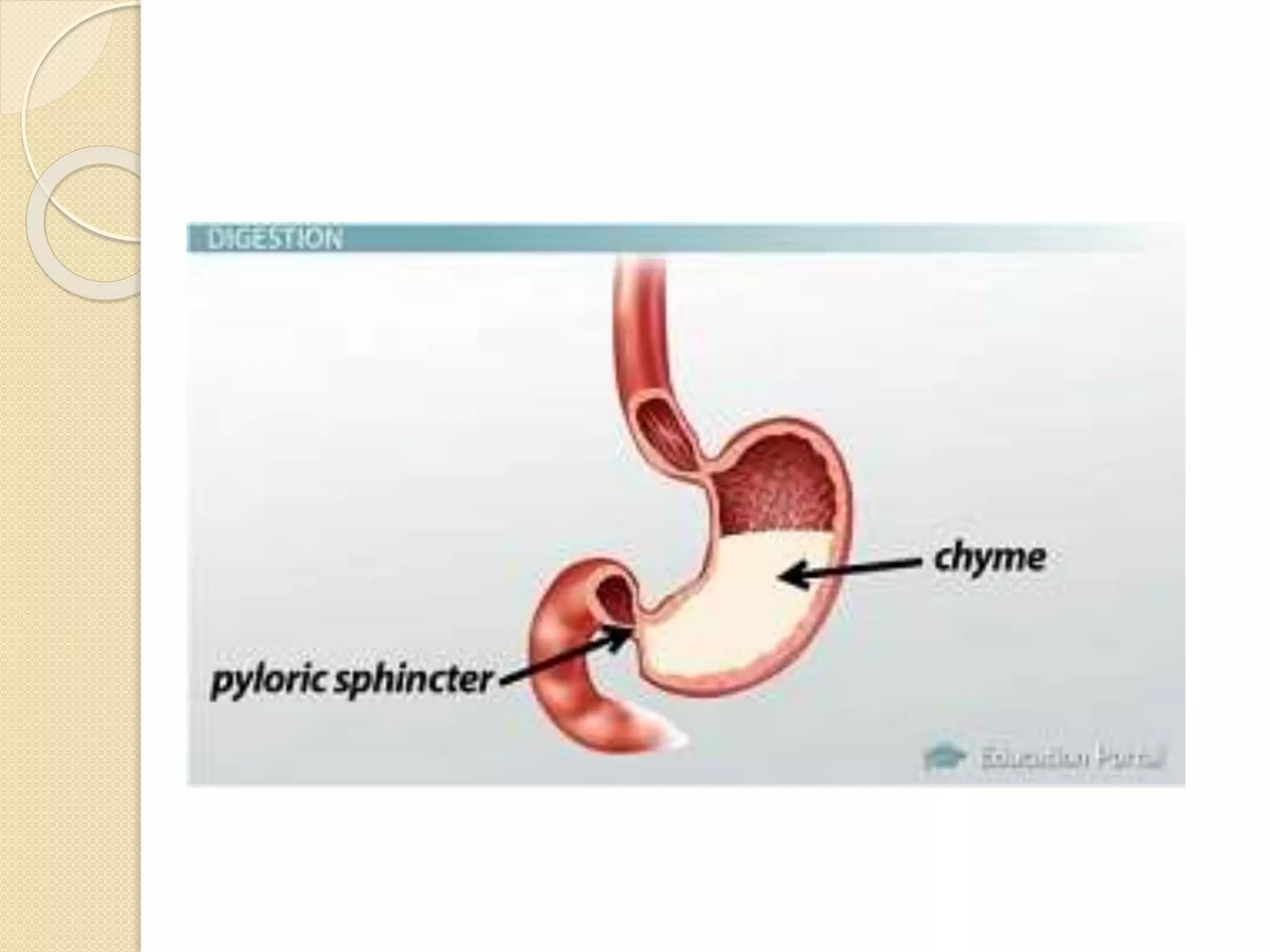
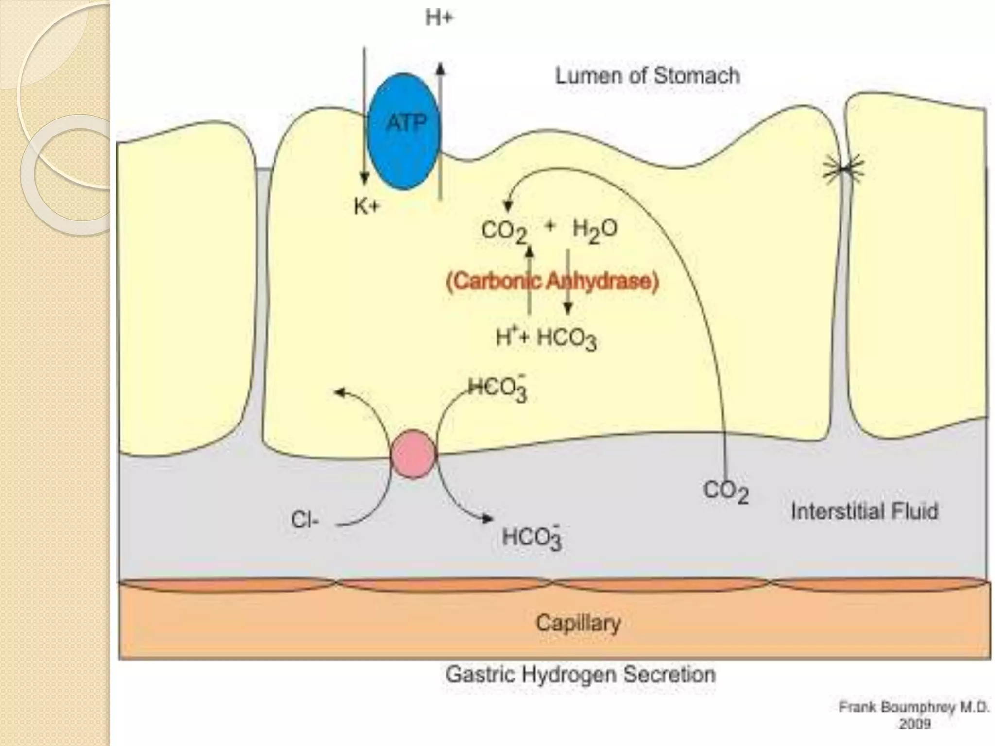
The stomach has five recognizable parts and two curvatures. Sphincters exist at the entry and exit of the stomach to control movement of contents. The stomach stores food, secretes acid and enzymes to digest food into chyme, and empties at a controlled rate into the small intestine. Acid secretion is stimulated by acetylcholine, gastrin, and histamine in three phases: cephalic, gastric, and intestinal. The stomach mucosa protects itself from acid through secretion of mucus and bicarbonate. Peptic ulcers can form if these defenses are overwhelmed.
3. ο . The stomach has five recognizable
parts and two curvatures; parts β the
cardia (around the oesophageal
opening), the fundus (above the level of
the oesophageal opening), the body
(central portion), the antrum - also
referred to as the pyloric antrum β (lower
part) and the pylorus (the most distal
part)
ο curvatures β the lesser curvature
(forming the upper concave border) and
the greater curvature (forming the lower
and longer convex border).
4.
5. ο Sphincters exist at the entry and exit
parts of the stomach. Although there is
no distinct specialization of stomach
muscle fibres at the cardia, several
mechanisms have been implicated in
preventing reflux of stomach contents
into the oesophagus; these mechanisms
will be outlined. At the pyloric end, the
circular muscle coat is thickened to
produce the pyloric sphincter which
controls the flow of stomach contents
into the duodenum.
6.
7. Functional and applied anatomy
of the stomach
ο The adult stomach can hold 2 to 3
litres of food.
ο The size and position of the stomach
varies, depending upon body shape,
degree of distension and posture.
8. Physiology of the Stomach
ο Food is ingested faster than it can be
digested. The stomach stores food whilst
subjecting it to preliminary physical and
chemical disruption to form chyme, which
is then delivered at a controlled rate to the
intestines.
9.
10. ο This is achieved by a complex of
secretion and specialised motility. The
stomach secretes acid and enzymes,
mostly proteolytic. This generates a
luminal environment very hostile to
biological material, so the stomach
mucosa must be protected by further
secretion of mucus and alkali.
11. Stomach acid
ο The luminal pH is normally below 2.0,
with a concentration of up to
100mmol.l-1 of hydrochloric acid. This
is secreted by the parietal (oxyntic)
cells located in the gastric pits.
12. ο Most body fluids are slightly alkaline,
so if H+ ions are to be secreted they
must first be created in quantity. This
happens at numerous mitochondria in
parietal cells by, in effect, splitting
water. This inevitably generates OH-,
which then combines with CO2 from
metabolism to form HCO3-, which is
exported to the blood.
13. ο Very importantly therefore, every mol
of H+ secreted into the stomach
results in 1 mol of HCO3-, entering the
blood, the so-called βalkaline tideβ.
This is subsequently re-secreted into
the GI tract by the pancreas and liver
to neutralise the acid as it leaves the
stomach.
14.
15. ο The H+ ions produced by the
mitochondria are concentrated by
proton pumps in the walls of canaliculi
which invaginate the luminal surface
of parietal cells and carry the
concentrated acid into the stomach
contents through the gastric pits.
16. ο At the same time the chief cells, also
in the gastric pits secrete enzymes,
principally pepsin, in the form of an
inactive precursor pepsinogen, which
is cleaved by acid in the stomach into
its active form
17. ο Acid and enzyme secretion is
controlled by a complex of neural and
endocrine systems. Parietal cells are
stimulated by Acetylcholine, Gastrin
and Histamine, which act through
separate receptors to promote acid
secretion.
18. ο Acetylcholine is released from post
ganglionic, parasympathetic nerves,
stimulated by gastric distension as
food arrives and acts upon muscarinic
receptors.
19. ο Gastrin is released from endocrine
cells in the stomach. It is a
polypeptide from the Gastrin/CCK
family, produced by G cells. These
are stimulated by amino acids and
peptides in the stomach content, but
inhibited by low pH.
20. ο Histamine is released from mast cells,
and diffuses locally to parietal cells
where it acts via H2 receptors which
are almost exclusive to the stomach.
Histamine release is stimulated by
both Gastrin and Acetylcholine, so it
amplifies their action.
21. Phases of gastric secretion
ο There are three phases of gastric
secretion.
ο 1. Cephalic phase
ο 2. Gastric phase
ο 3. Intestinal phase
22.
23. ο The sight and smell of food, and the
act of swallowing, activates the
parasympathetic system, which
stimulates the release of
Acetylcholine. This is known as the
Cephalic Phase
24. ο Once food reaches the stomach its initial
effect is to distend the stomach, further
stimulating Acetylcholine release, and to
raise the pH of the stomach contents by
buffering the relatively small amount of
acid present between meals. The rise in
pH disinhibits gastrin secretion. Acid and
enzymes then act on proteins to produce
peptides which further stimulate gastrin
release as the pH falls and that
disinhibition is removed. This is the
gastric phase.
25. ο Once chyme leaves the stomach in
significant quantities it stimulates the
release of chemicals from the
intestines (Cholecystokinin and
Gastric Inhibitory Polypeptide) which
reduce acid secretion. This is the
Intestinal Phase.
26. Stomach Defences
ο The surface cells of the stomach
mucosa secrete a thick layer of
alkaline mucus, which offers some
mechanical protection, and traps H+
ions diffusing into it from the stomach
lumen, by reacting with HCO3- ions
also produced from the surface cells.
This prevents the pH of the surface of
the mucosa cells from falling too low.
The production of defences is
stimulated by prostaglandins.
27.
28. Peptic Ulceration
ο If the defences of the stomach are
damaged or overwhelmed the acids
attacks cells and produces gastritis
then ulceration. Defences are
damaged by infection with H Pylori by
excess alcohol ingestion, and by non-
steroidal anti inflammatory drugs
which inhibit prostaglandin production.
29. Reducing Acid Secretion
ο Acid secretion may be reduced by
drugs antagonising the action of
histamine at H2 receptors (eg
cimetidine), or by drugs which inhibit
proton pumps.
30. Gastric Motility
ο When food is swallowed a vagal reflex
produces receptive relaxation, which
causes the resting tension in the walls
of the stomach to reduce. Food is
therefore accommodated without a
rise in intragastric pressure, reducing
the risk of reflux of acid into the
oesophagus.
31. ο The full stomach begins regular
peristaltic contractions, triggered by a
pacemaker, in the cardiac region about
three times a minute. These sweep over
the stomach from cardia to pylorus,
accelerating as they move. This
combined with the funnel shape of the
stomach both mixes the contents and
decants liquid chyme into the pyloric
region. A small squirt of chyme leaves
the pylorus with each peristaltic wave
before the pylorus shuts.
32. ο Gastric emptying rate is controlled by
feedback from the duodenum via
chemical signals, so that the rate of
emptying of the stomach is
appropriate for further digestion and
absorption. Fats greatly slow gastric
emptying as they take more time to
digest and absorb.
33. ο When empty, the gastric muscosa is
thrown into longitudinal folds (called
rugae); a gastric canal forms
temporarily between the gastric folds
along the lesser curvature to allow
saliva and other fluids and small
amounts of masticated food to pass
along to the pyloric part.
34. ο The gastric mucosa has three
histologically different zones. The cardia
contains mostly mucus- secreting
glands. The fundus and the body
contain gastric glands that consist of
mucus-secreting neck cells, acid-
secreting (parietal or oxyntic) cells, chief
peptic cells (that secrete pepsinogen, a
precursor of the enzyme pepsin) and
other local hormone-producing (APUD)
cells. The pyloric region contains glands
whose cells secrete mucus and hormone
gastrin (produced by G-cells).
35. ο Gastric ulcers occur commonly in the
antrum and along the lesser curvature
of the stomach. Perforation of ulcers
leads to the spillage of gastric
contents into the peritoneal cavity; the
spillage may affect abdominal
structures such as the pancreas and
associated blood vessels lying in close
proximity to the stomach.
36. ο Knowledge of the anatomy of the
stomach and its immediate relations is
important in predicting structures at
risk to damage due to spillage of
gastric contents onto them.
37. ο Reflux of gastric contents into the
oesophagus is a common condition. If
it is frequent and/or clearance of reflux
material is deficient, βheartburnβ
and/or inflammation with ulceration
may result.
38. ο Chronic reflux may result in
metaplastic changes in the mucosa
(Barrettβs oesophagus) with an
increased risk of developing
oesophageal carcinoma. Alcohol and
smoking are other factors that may be
involved in oesophageal cancer which
occurs commonly in the lower part of
the tube. The cancer may obstruct the
lumen, leading to dysphagia (painful
and difficulty in swallowing).
39. ο Inflammation (gastritis) of the stomach
may be acute (caused by aspirin and
non-steroidal anti-inflammatory drugs or
by alcohol) causing exfoliation of the
surface epithelial cells and decreasing
the secretion of protective mucus.
Gastritis may also be chronic (caused by
infection with the bacterium Helicobacter
pylori; inflammatory changes in the
mucosa result in atrophy and epithelial
metaplasia (which may develop into
carcinoma).