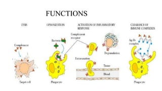
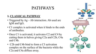
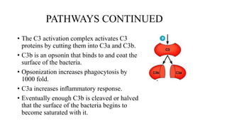
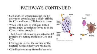
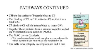
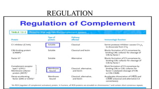
2. GROUP MEMBERS
• Vanessa Ohui Dadeboe UE20015417
• Priscilla Bandah UE20014517
Koomson Abigail UE20016917
3. OBJECTIVES
• History
• Overview of the complement system.
• Functions
• Three main pathways
• Regulation
• Role in disease
• References
4. HISTORY
• Discovered in the 1890’s by Jules Bordet, a young
Belgian scientist in Paris.
• She said the serum has two components. The heat
stable component responsible for specific immunity
and the heat sensitive component responsible for non-
specific immunity.
• Paul Ehrlich named the heat-sensitive component
complement.
5. OVERVIEW
• The complement system is a part of the innate immune system
that enhances the ability of antibodies and phagocytic cells to
clear microbes and damaged cells from an organism.
• It can be activated by antibodies generated by the adaptive
immune system
• It is a defensive system consisting of over 30 proteins and protein
fragments produced by the liver and found in circulating blood
serum.
• It functions in a cascade system.
• Complement proteins are often designated by “C”. From C1 –
C9.
6. OVERVIEW CONTINUED
• Proteolysis results in large fragments “b” and smaller
fragments “a”. Eg. C3a and C5b
• The only exception is C2, where C2a is the large fragment
and C2b is the smaller fragment.
8. PATHWAYS
1. CLASSICAL PATHWAY
• Triggered by Ag – Ab interaction. Ab used are
IgM and IgG.
• C1 complex is activated when it binds to the ends
of antibodies.
• Once C1 is activated, it activates C2 and C4 by
cutting them in halves giving C2a and C2b, C4a
and C4b.
• C2b and C4b bind to form a C3 activation
complex on the surface of the bacteria while the
C2a and C4a diffuse away.
9. PATHWAYS CONTINUED
• The C3 activation complex activates C3
proteins by cutting them into C3a and C3b.
• C3b is an opsonin that binds to and coat the
surface of the bacteria.
• Opsonization increases phagocytosis by
1000 fold.
• C3a increases inflammatory response.
• Eventually enough C3b is cleaved or halved
that the surface of the bacteria begins to
become saturated with it.
10. PATHWAYS CONTINUED
• C2b and C4b which make up the C3
activation complex has a slight affinity
for C3b and hence C3b binds to them.
• When C3b binds to C2b and C4b it
forms a new complex referred to as the
C5 activation complex.
• The C5 activation complex activates C5
proteins by cutting them into C5a and
C5b
• C5b begins to coat the surface of the
bacteria because many are produced.
• C5a disperses away from the bacteria.
11. PATHWAYS CONTINUED
• C5b on the surface of bacteria binds to C6
• The binding of C6 to C5b activates C6 so that it can
bind to C7
• C7 binds to C8 which in turn binds to many C9’s
• Together these proteins form a circular complex called
the Membrane attack complex (MAC).
• The MAC causes Cytolysis.
• The circular membrane attack complex acts as a channel in
which cytoplasm can rush out of and water rushes in.
• The cells inner integrity is compromised and it dies
12. PATHWAYS CONTINUED
2. ALTERNATIVE PATHWAY
• It is slower than the classical pathway.
• Activated by C3b binding to microbial cell surfaces.
• C3 contains an unstable thioester bond which slows spontaneous
hydrolysis to C3b and C3a.
• C3b is able to bind to foreign surface antigens.
• The C3b on the surface of a foreign cell binds to another plasma
protein called Factor B.
13. PATHWAYS CONTINUED
• The binding of C3b to factor B allows a protein
enzyme called Factor D to cleave Factor B to
Ba and Bb.
• Factor Bb remains bound to C3b while Ba and
Factor D disperse away.
• Properdin, also called factor P, binds to the
C3bBb complex to stabilize it.
• C3bBbP make up the C3 activation complex for
the alternative pathway
14. PATHWAYS CONTINUED
• The C3 activation complex causes the
production of more C3b.
• This allows the initial steps of this pathway to
be repeated and amplified.
• When an additional C3b binds to the C3
activation complex it converts it into a C5
activation complex.
• The C5 activation complex cleaves C5 into C5a
and C5b.
• C5b begins the production of the MAC.
15. PATHWAYS CONTINUED
3. LECTIN PATHWAY
• Lectins are carbohydrate binding proteins.
• It is similar to the classical pathway but with the opsonin, mannose-
binding lectin (MBL) and ficolins instead of C1q.
• Activated by the binding of MBL to mannose residues which activates
the MBL associated serine proteases, MASP-1 and MASP-2 which is
similar to C1r and C1s respectively.
• This complex activates C2 and C4 by cutting them into C2a and C2b,
C4a and C4b.
• C4b binds to C2b to form the C3-convertase as in the classical
pathway.
17. ROLE IN DISEASES
COMPLEMENT DEFICIENCY
• Example, deficiency in the MAC components of complements is
associated with Neisseria meningitidis and N. gonorrhoeae which are
bacterial infections.
COMPLEMENT REGULATORS DEFICIENCY
• Example, mutations in the complement regulators like factor H and
membrane cofactor protein have been associated with atypical
hemolytic uremic syndrome.
Other examples are Paroxysmal nocturnal hemoglobinuria, asthma,
Alzheimer’s disease, multiple sclerosis and rejection of transplanted
organs.
18. REFERENCES
• Roitt I, Brostoff J, Male D (2001). Immunology (6th ed), 480p. St. Louis:
Mosby, ISBN 0-7234-3189-2
• Parham P (2005). The immune system. New York: Garland, ISBN 0-
8153-4093-1
• DeFranco AL, Locksley RM, Robertson M (2007). Immunity: The
immune response in infectious and inflammatory disease. London;
Sunderland, MA: New Science Press; Sinauer Associates, ISBN 987-0-
9539181-0-2.
• Stoemer, Kristina A, Morrison, Thomas E. (15 March 2011).
Complement and Viral pathogenesis, Virology. 411 (2): 362-373
Central.