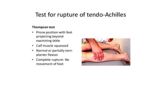
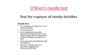
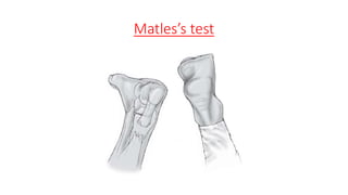
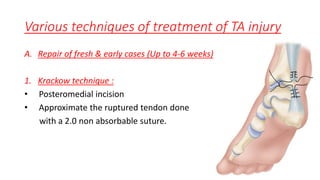
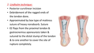
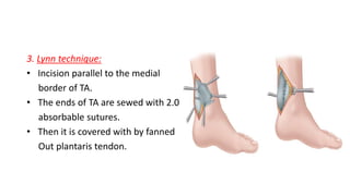
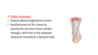
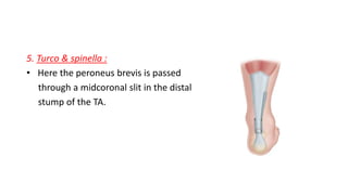
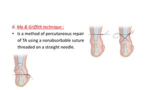
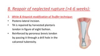
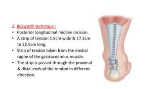
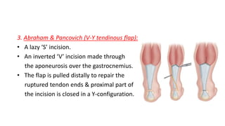
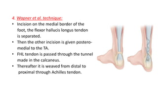
TendoAchilles rupture
Dr. Md. Ashiqur Rahman
Resident
Orthopedics
DMCH
Questions
1. What is the problem?
2. Duration?
3. Any H/O trauma?
4. What were you doing during trauma?
5. Any H/O injection in the ankle?
6. Do you take any medicine regularly?
Exposure : Both lower limb upto ankle
On examination:
Look:
1. Any scar mark, skin change, skin change, blackening behind the
ankle.
2. Visible gap in TA.
3. Gait abnormalities.
4. Heel & toe walking.
Feel:
1. There is tenderness at the rupture site.
2. A gap felt 5cm above the insertion. This gap is more prominent with
dorsiflexion of ankle joint.
3. Thompson’s squeeze test/Simmond’s test: Positive
4. Matle’s test : Positive
5. O’brien’s test : Positive
O’Brien’s needle test
Matles’s test
Move:
1. Ankle movement : Plantar flexion is weak.
Tendoachilles injury can be classified can be classified into 02
categories:
1. Tendoachilles rupture: Due to degenerative cause : Steroid injection,
spontaneous.
2. Tendoachilles tear: Due to trauma : Direct blow. Cut injury,
commode injury.
Mechanism
A. Indirect :
• Pushing off with the weight bearing forefoot while extending the
knee.
• Sudden unexpected dorsiflexion of the ankle.
• Violent dorsiflexion of the plantar flexed foot, as in a fall from a
height.
B. Direct:
• Direct blow to the contracted tendon or from a laceration.
Site:
• Rupture of TA usually takes place about 2-6cm/5cm from the
insertion (Watershed line).
Clinical features
• Most patients are aged over 40 years.
• Feeling is has been stuck just above the heel.
• Unable to tip-toe & push-off.
• A gap can be seen and felt 2 to 5cm above the insertion of the
tendon.
• Simmond’d test/Thompson squeeze test, Mateles test, O’brien’s
needle test : Positive.
• Investigations: X-ray on ankle joint to exclude any fractures
USG
MRI
Precipitating factors
• Aging process
• Tendinitis
• Tendinosis
• Peritendinitis
• Local steroid injection
• DM
• Repetitive microtrauma
Principle of TA injury treatment
• Cut injury, traumatic injury & degenerative ruptures presented early
can be repaired end to end.
• All neglected ruptures need reconstruction by various techniques.
Various techniques of treatment of TA injury
A. Repair of fresh & early cases (Up to 4-6 weeks)
1. Krackow technique :
• Posteromedial incision
• Approximate the ruptured tendon done
with a 2.0 non absorbable suture.
2. Lindholm technique:
• Posterior curvilinear incision
• Debridement of the ragged ends of
the tendon done.
• Approximated by box type of mattress
suture of heavy nonabsorb. Suture
• 02 flaps from the proximal tendon &
gastrocnemius aponeurosis taken &
sutured to the distal stump of the tendon
& to one another to cover the site of
rupture completely.
3. Lynn technique:
• Incision parallel to the medial
border of TA.
• The ends of TA are sewed with 2.0
absorbable sutures.
• Then it is covered with by fanned
Out plantaris tendon.
4. Teuffer technique :
• Postero-lateral longitudinal incision
• Reinforcement of TA is done by
passing the peroneus brevis tendon
through a drill hole in the calcaneal
tuberosity to produce a dynamic loop.
5. Turco & spinella :
• Here the peroneus brevis is passed
through a midcoronal slit in the distal
stump of the TA.
6. Ma & Griffith technique :
• Is a method of percutaneous repair
of TA using a nonabsorbable suture
threaded on a straight needle.
B. Reapair of neglected rupture (>4-6 weeks):
1. White & Kraynick modification of Teuffer technique:
• Postero-lateral incision.
• TA is repaired by harvested plantaris
tendon in figure of eight fashion.
• Reinforced by peroneus brevis tendon
by passing it through a drill hole in the
calcaneal tuberosity.
2. Bosworth technique :
• Posterior longitudinal midline incision.
• A strip of tendon 1.5cm wide & 17.5cm
to 22.5cm long.
• Strip of tendon taken from the medial
raphe of the gastrocnemius muscle.
• The strip is passed through the proximal
& distal ends of the tendon in different
direction.
3. Abraham & Pancovich (V-Y tendinous flap):
• A lazy ‘S’ incision.
• An inverted ‘V’ incision made through
the aponeurosis over the gastrocnemius.
• The flap is pulled distally to repair the
ruptured tendon ends & proximal part of
the incision is closed in a Y-configuration.
4. Wapner et al. technique:
• Incision on the medial border of the
foot, the flexor hallucis longus tendon
is separated.
• Then the other incision is given postero-
medial to the TA.
• FHL tendon is passed through the tunnel
made in the calcaneus.
• Thereafter it is weaved from distal to
proximal through Achilles tendon.
Priciple of vulpius procedure:
• Equinous deformity due to gastrocnemius contracture is corrected by
this procedure.
• Aponeurotic tendon of the gastrocnemius is divided and its distal part
is allowed to retract distally but is not sutured to the soleus.
Post-operative management of ruptured TA
• Long leg full plaster with window or anterior slab with knee in 45°-60°
flexed & ankle in equinous position.
• Stitches are removed after 14 days.
• LLFP is converted to SLFP freeing the knee after 4-6 weeks.
• Plaster discarded after 8-10 weeks & walking with high heel shoe is
allowed.
• Heel heigh is reduced gradually & made planti-grade by next one
month.
• Protection of the tendon from stress is needed for at least 6 months.

TendoAchilles Rupture

  • 1.
    TendoAchilles rupture Dr. Md.Ashiqur Rahman Resident Orthopedics DMCH
  • 2.
    Questions 1. What isthe problem? 2. Duration? 3. Any H/O trauma? 4. What were you doing during trauma? 5. Any H/O injection in the ankle? 6. Do you take any medicine regularly?
  • 3.
    Exposure : Bothlower limb upto ankle On examination: Look: 1. Any scar mark, skin change, skin change, blackening behind the ankle. 2. Visible gap in TA. 3. Gait abnormalities. 4. Heel & toe walking.
  • 4.
    Feel: 1. There istenderness at the rupture site. 2. A gap felt 5cm above the insertion. This gap is more prominent with dorsiflexion of ankle joint. 3. Thompson’s squeeze test/Simmond’s test: Positive 4. Matle’s test : Positive 5. O’brien’s test : Positive
  • 6.
  • 7.
  • 8.
    Move: 1. Ankle movement: Plantar flexion is weak. Tendoachilles injury can be classified can be classified into 02 categories: 1. Tendoachilles rupture: Due to degenerative cause : Steroid injection, spontaneous. 2. Tendoachilles tear: Due to trauma : Direct blow. Cut injury, commode injury.
  • 9.
    Mechanism A. Indirect : •Pushing off with the weight bearing forefoot while extending the knee. • Sudden unexpected dorsiflexion of the ankle. • Violent dorsiflexion of the plantar flexed foot, as in a fall from a height. B. Direct: • Direct blow to the contracted tendon or from a laceration.
  • 10.
    Site: • Rupture ofTA usually takes place about 2-6cm/5cm from the insertion (Watershed line).
  • 11.
    Clinical features • Mostpatients are aged over 40 years. • Feeling is has been stuck just above the heel. • Unable to tip-toe & push-off. • A gap can be seen and felt 2 to 5cm above the insertion of the tendon. • Simmond’d test/Thompson squeeze test, Mateles test, O’brien’s needle test : Positive. • Investigations: X-ray on ankle joint to exclude any fractures USG MRI
  • 12.
    Precipitating factors • Agingprocess • Tendinitis • Tendinosis • Peritendinitis • Local steroid injection • DM • Repetitive microtrauma
  • 13.
    Principle of TAinjury treatment • Cut injury, traumatic injury & degenerative ruptures presented early can be repaired end to end. • All neglected ruptures need reconstruction by various techniques.
  • 14.
    Various techniques oftreatment of TA injury A. Repair of fresh & early cases (Up to 4-6 weeks) 1. Krackow technique : • Posteromedial incision • Approximate the ruptured tendon done with a 2.0 non absorbable suture.
  • 15.
    2. Lindholm technique: •Posterior curvilinear incision • Debridement of the ragged ends of the tendon done. • Approximated by box type of mattress suture of heavy nonabsorb. Suture • 02 flaps from the proximal tendon & gastrocnemius aponeurosis taken & sutured to the distal stump of the tendon & to one another to cover the site of rupture completely.
  • 16.
    3. Lynn technique: •Incision parallel to the medial border of TA. • The ends of TA are sewed with 2.0 absorbable sutures. • Then it is covered with by fanned Out plantaris tendon.
  • 17.
    4. Teuffer technique: • Postero-lateral longitudinal incision • Reinforcement of TA is done by passing the peroneus brevis tendon through a drill hole in the calcaneal tuberosity to produce a dynamic loop.
  • 18.
    5. Turco &spinella : • Here the peroneus brevis is passed through a midcoronal slit in the distal stump of the TA.
  • 19.
    6. Ma &Griffith technique : • Is a method of percutaneous repair of TA using a nonabsorbable suture threaded on a straight needle.
  • 20.
    B. Reapair ofneglected rupture (>4-6 weeks): 1. White & Kraynick modification of Teuffer technique: • Postero-lateral incision. • TA is repaired by harvested plantaris tendon in figure of eight fashion. • Reinforced by peroneus brevis tendon by passing it through a drill hole in the calcaneal tuberosity.
  • 21.
    2. Bosworth technique: • Posterior longitudinal midline incision. • A strip of tendon 1.5cm wide & 17.5cm to 22.5cm long. • Strip of tendon taken from the medial raphe of the gastrocnemius muscle. • The strip is passed through the proximal & distal ends of the tendon in different direction.
  • 22.
    3. Abraham &Pancovich (V-Y tendinous flap): • A lazy ‘S’ incision. • An inverted ‘V’ incision made through the aponeurosis over the gastrocnemius. • The flap is pulled distally to repair the ruptured tendon ends & proximal part of the incision is closed in a Y-configuration.
  • 23.
    4. Wapner etal. technique: • Incision on the medial border of the foot, the flexor hallucis longus tendon is separated. • Then the other incision is given postero- medial to the TA. • FHL tendon is passed through the tunnel made in the calcaneus. • Thereafter it is weaved from distal to proximal through Achilles tendon.
  • 24.
    Priciple of vulpiusprocedure: • Equinous deformity due to gastrocnemius contracture is corrected by this procedure. • Aponeurotic tendon of the gastrocnemius is divided and its distal part is allowed to retract distally but is not sutured to the soleus.
  • 25.
    Post-operative management ofruptured TA • Long leg full plaster with window or anterior slab with knee in 45°-60° flexed & ankle in equinous position. • Stitches are removed after 14 days. • LLFP is converted to SLFP freeing the knee after 4-6 weeks. • Plaster discarded after 8-10 weeks & walking with high heel shoe is allowed. • Heel heigh is reduced gradually & made planti-grade by next one month. • Protection of the tendon from stress is needed for at least 6 months.