The document discusses spinal canal stenosis, including:
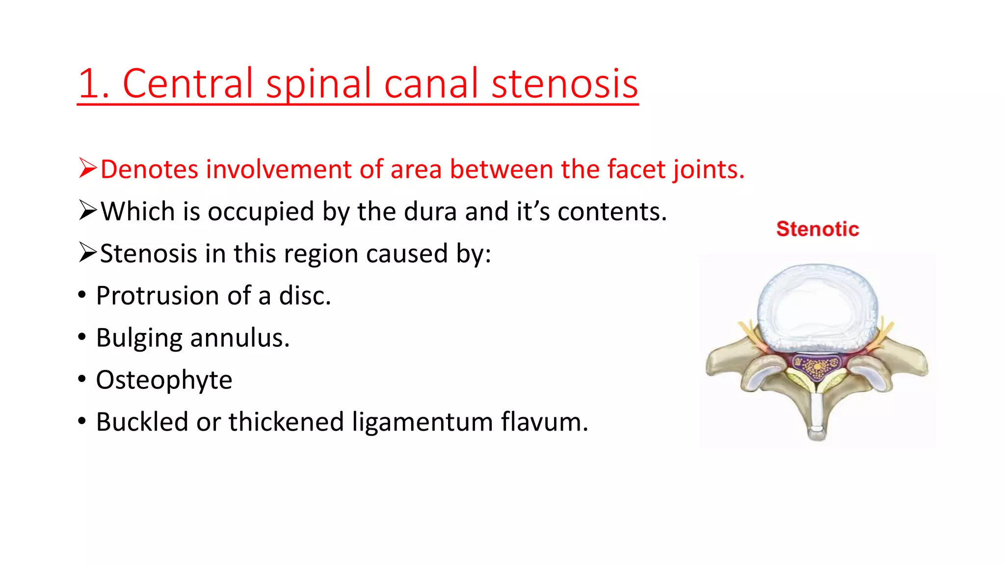
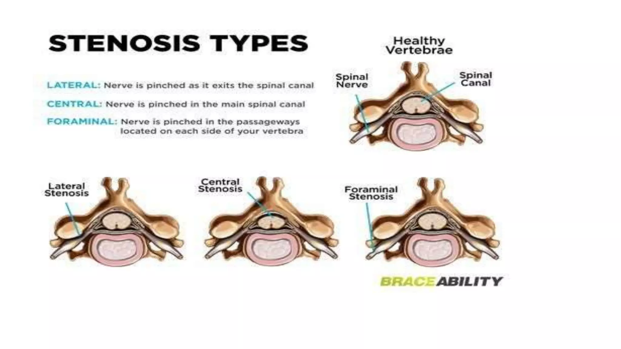
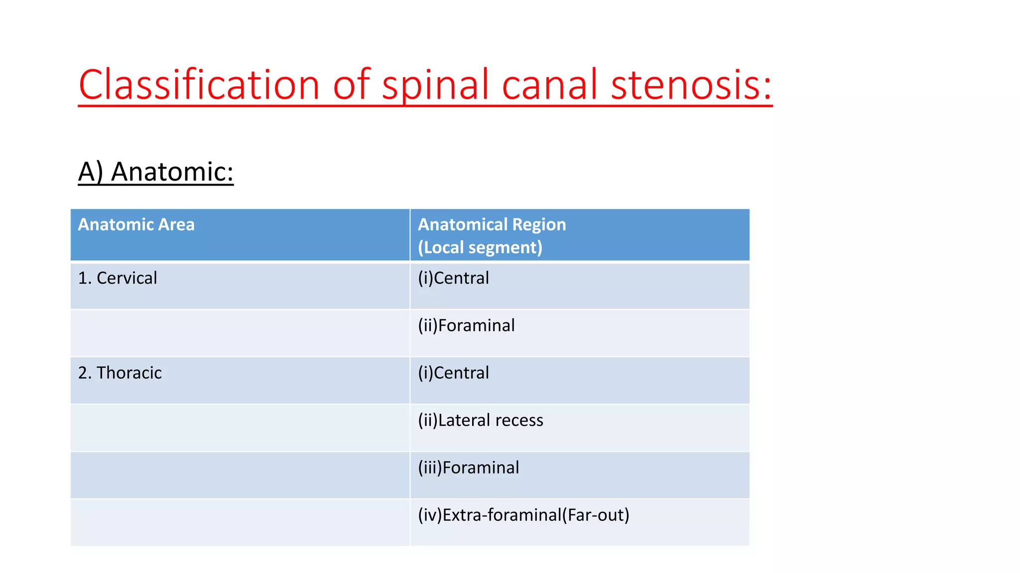
1. It describes spinal canal stenosis as the narrowing of the spinal canal and compression of the spinal cord and nerve roots, most commonly occurring in the lumbar vertebrae.
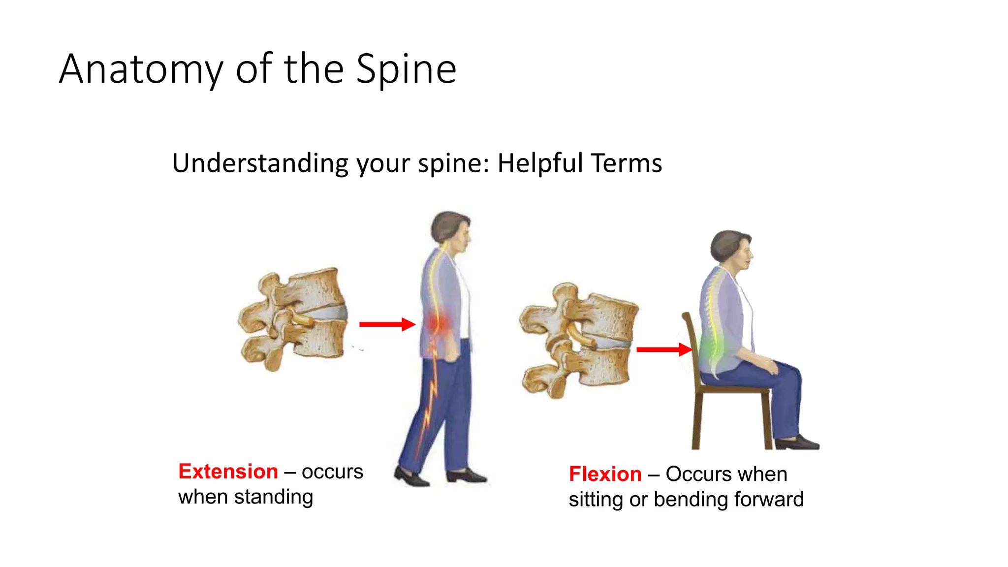
2. Symptoms include back pain radiating into the legs, numbness, and weakness that is relieved by bending forward and made worse by standing upright or walking.
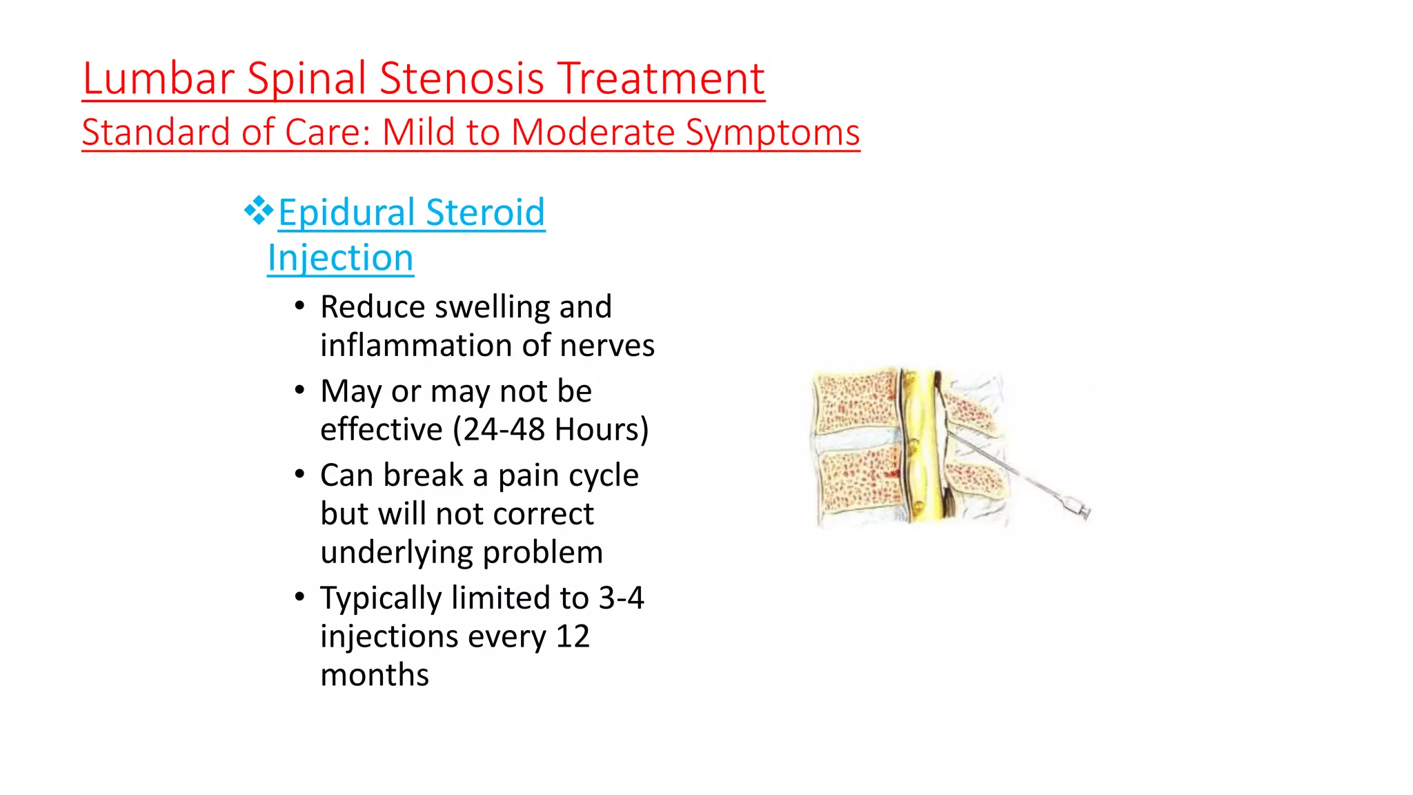
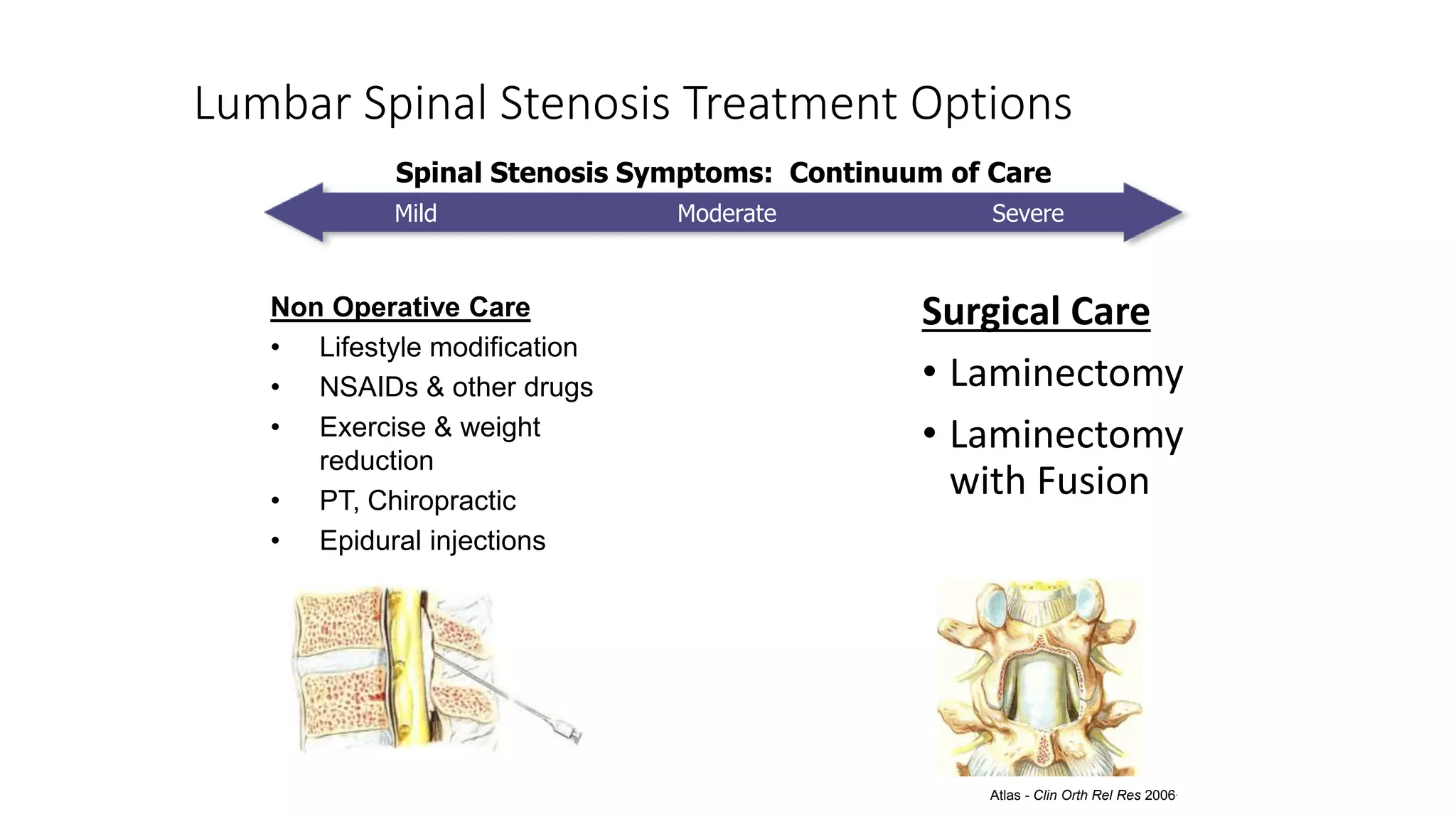
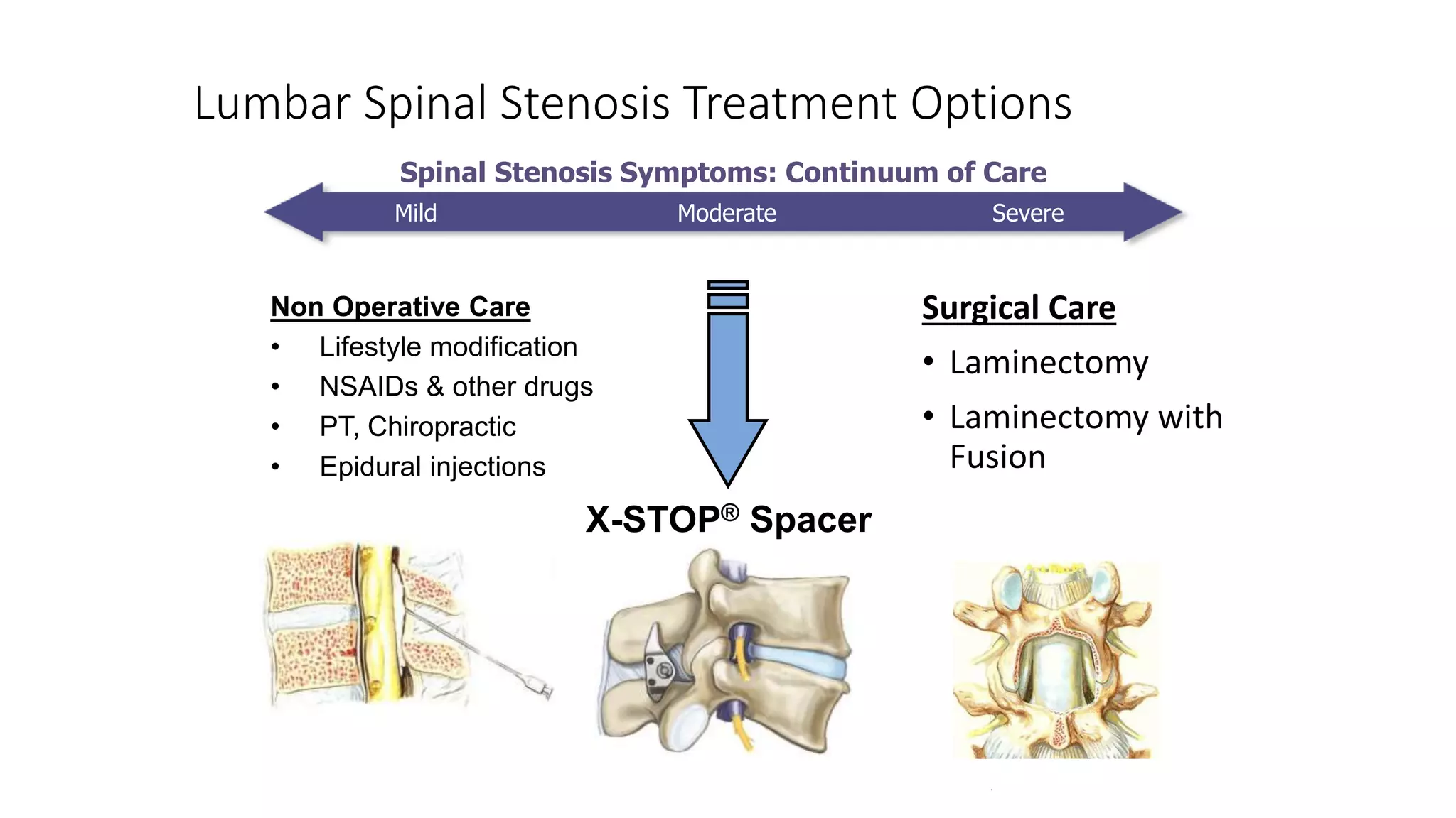
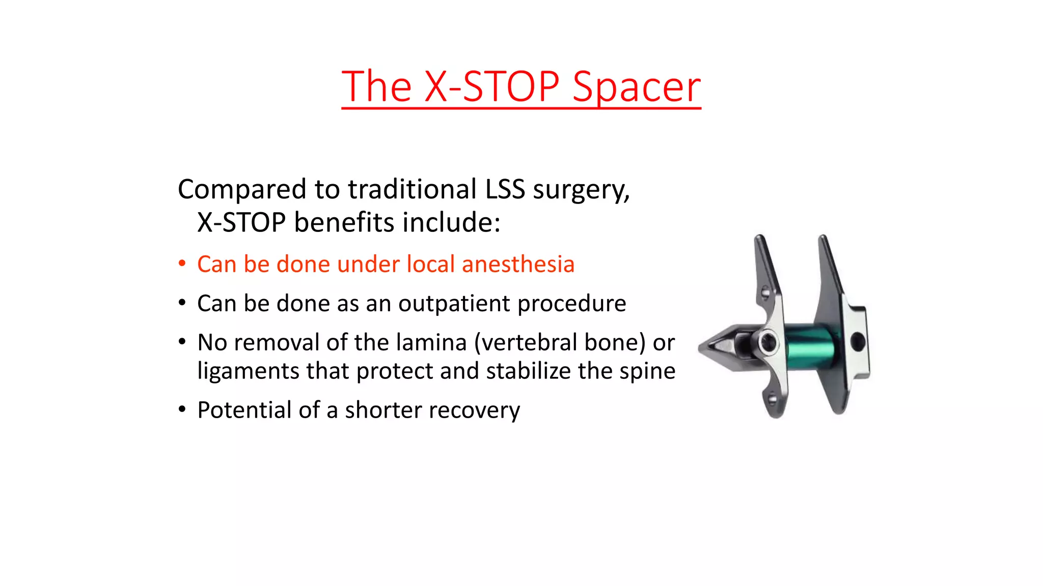
3. Treatment options range from non-surgical approaches like medication, physical therapy, and epidural injections for mild-to-moderate cases to surgical decompression like laminectomy or the X-STOP implant for more severe cases.
1. Basics Of Spinal canal
stenosis
Prepared by
Dr. Md. Ashiqur Rahman
Resident,DMCH
2. Introduction:
It is the narrowing of the spinal canal and the consequent
compression of cord and nerve roots.
It may affect cervical, thoracic or lumbar spine.
Common in lumbar vertebrae.
3. Diameter of spinal canal & foramen:
AP 15mm and transverse diameter 20mm.
AP<11mm and transverse diameter <16mm is abnormal.
4. Vascular symptoms typically are felt in the upper calf, are relieved
after a short rest (5mins.) while still standing, do not require sitting or
bending and worsen despite walking uphill or riding stationary
bicycle.
Neurogenic claudication improve improves with trunk flexion,
stooping or lying but may require 20 minutes to improve.
5. Extension – occurs
when standing
Flexion – Occurs when
sitting or bending forward
Anatomy of the Spine
Understanding your spine: Helpful Terms
6.
7.
8. Lateral canal
Lateral to the dura is the lateral
canal, which contains nerve roots.
1. Lateral recess also known as
‘lee’s entrance zone’.
2. Foraminal region also known as
‘lee’s midzone’.
3. Extra-foraminal region also
known as ‘Exit zone’
9. 1. Central spinal canal stenosis
Denotes involvement of area between the facet joints.
Which is occupied by the dura and it’s contents.
Stenosis in this region caused by:
• Protrusion of a disc.
• Bulging annulus.
• Osteophyte
• Buckled or thickened ligamentum flavum.
10. 2.Lateral recess stenosis
(This is where the nerve roots exit the dura & courses distally & laterally under the superior
articular facet)
Boundary:
Anteriorly: Disc and the posterior ligamentous complex.
Posteriorly : The superior articular facet.
Laterally : Medial border of Pedicle.
Medially : The central canal.
Stenosis in this region caused by :
Facet arthritis
Vertebral body spur
Annulus pathology
11. 3.Foraminal stenosis
Boundary : (Which lies ventral to the pars)
• Anteriorly : Posterior vertebral body & disc.
• Posteriorly : Pars interarticularis
• Medially : Lateral recess
• Laterally : Lateral border of the pedicle.
Stenosis in this region:
• Pars fracture
• Lateral disc herniation
• Thickening of ligamentum flavum
• A spur from underlying surface of pars
12. 4. Extra-foraminal(Far-out)
Boundary :
• The exit zone is identified as the
area lateral to the facet joint.
Stenosis in this region :
• A ‘far lateral’ disc.
• Spondylolisthesis
• Associated subluxation
• Facet arthritis
13.
14.
15. Stenotic
Lumbar Vertebra
• Vertebrae provide body support
• Discs act as “shock absorbers”
• Vertebra protects spinal cord and nerves
• Nerves have space and are not pinched
• As we age, ligaments and bone can
thicken
• Narrowing is called “stenosis”
• Narrowing squeezes nerves in spinal
canal and nerve roots exiting spine to
legs
• Result - pain & numbness in back and
legs
Nerve Root
Spinal
Canal
Bone (Facet
Joint)
Healthy
Intervertebral
Disc
Thickened
Ligament
Flavum
Pinched
Nerve Root
Narrowed
Spinal Canal
16.
17. Spinal Pain
• Axial Pain – Back Pain
• From bones, joints, muscles, discs
• Neurogenic Pain – Leg Pain +/- Tingles
• From nerve irritation
18. Classification of spinal canal stenosis:
A) Anatomic:
Anatomic Area Anatomical Region
(Local segment)
1. Cervical (i)Central
(ii)Foraminal
2. Thoracic (i)Central
(ii)Lateral recess
(iii)Foraminal
(iv)Extra-foraminal(Far-out)
19. B) Pathologic:
1. Congenital:
Achondroplastic (Dwarfism)
Congenital forms of spondylolisthesis
Scoliosis
Kyphosis
2. Idiopathic
21. Difference between neurogenic claudication & vascular claudication
Evaluation Vascular neurogenic
Walking distance Fixed Variable
Palliative factors Standing Sitting/Bending
Provocative
factors
Walking Walking/Standing
Walking uphill painful Painless
Bicycle test Positive (painful) Negative
Pulses Absent Present
Weakness Rarely Occasionally
Back pain Occasionally Commonly
Back motion Normal Limited
Pain character Cramping-distal to proximal Numbness, aching-proximal to distal
Atrophy Uncommon Occasional
22. • Sitting or
bending forward
relieves
symptoms
• Standing provokes
symptoms
• Pain/weakness in
the legs
• Patients lean
forward while
walking to relieve
symptoms
Symptoms of Lumbar Spinal Stenosis
(Elevator Syndrome)
23. Symptoms of Lumbar Spinal Stenosis
Classic Presentation:
• Dull or aching back pain spreading to
your legs
• Numbness and “pins and needles” in
your legs, calves or buttocks
• Weakness, or a loss of balance
• A decreased endurance for physical
activities
24. Clinical features:
Male>50 years
Low backache
Cauda equina claudication (Most common symptom)
Stoop test : Positive---walking---increased pain---stooped---decreased
pain(due to canal length increased by 2.2mm).
Bicycle test : Positive
Walking test: Positive.
25. Investigations:
X-ray L/S spine – AP, Lateral & Oblique view.
o Decrease inter-pedicle distance
o Hypertrophy & stenosis of the facet joint
o Decreased intra-laminar distance
o Short, stout spinous process
o Associated features : Presence of listhesis, prolapsed disc,
osteophytes.
26. Myelography :
oWaist like narrowing of the dural sac.
oIdentation of dural sac due to disc prolapse.
MRI
29. Lumbar Spinal Stenosis Treatment
Standard of Care: Mild to Moderate Symptoms
Non-operative care:
• Avoid activities that bring on pain (24 Hour Thermostat)
• Impact aerobics
• Frequent bending, twisting, lifting
• Non-Steroidal Anti-Inflammatory Drugs (NSAIDs), Herbals
• Physical Therapy, Chiropractic, Exercise & Weight Reduction
• To help stabilize the spine
• Lessen the burden on the spine
• Reduce irritation of pain sensitive structures
30. Lumbar Spinal Stenosis Treatment
Standard of Care: Mild to Moderate Symptoms
Epidural Steroid
Injection
• Reduce swelling and
inflammation of nerves
• May or may not be
effective (24-48 Hours)
• Can break a pain cycle
but will not correct
underlying problem
• Typically limited to 3-4
injections every 12
months
31. Principles of spinal stenosis surgery
Decompression by laminectomy or a fenestration procedure is the
treatment of choice for lumbar spinal stenosis.
Laminectomy may be required is older patients and severe multilevel
stenosis.
Whereas fenestration procedure, consisting of bilateral laminotomies
and partial facetectomies, that preserve midline structures, are an
alternative in younger patients with intact disc.
32. Principles of spinal stenosis surgery
If radical decompression of only one root is necessary, additional
stabilization by fusion with or without instrumentation is usually
unnecessary.
The removal of more than one complete facet joint may require
instrumented fusion.
37. Lumbar Spinal Stenosis Treatment
Standard of Care: More Severe Symptoms
Laminectomy
• Un-roofing the spine, Opening
the “pipe”
• Removal of parts of the
vertebra, including:
• Lamina (bone)
• Attached ligaments
• Facets (bone)
• Goal: relieve pressure on
nerves by increasing size of
spinal canal and nerve exit
openings
• Most common surgery for
stenosis, may require a fusion
• General anesthesia
• In-patient procedure
• 6-12 week recovery
.
38.
39. Lumbar Spinal Stenosis Treatment Options
Surgical Care
• Laminectomy
• Laminectomy
with Fusion
Non Operative Care
• Lifestyle modification
• NSAIDs & other drugs
• Exercise & weight
reduction
• PT, Chiropractic
• Epidural injections
Spinal Stenosis Symptoms: Continuum of Care
Mild SevereModerate
Atlas - Clin Orth Rel Res 2006.
40. Lumbar Spinal Stenosis Treatment Options
Surgical Care
• Laminectomy
• Laminectomy with
Fusion
Non Operative Care
• Lifestyle modification
• NSAIDs & other drugs
• PT, Chiropractic
• Epidural injections
X-STOP® Spacer
Spinal Stenosis Symptoms: Continuum of Care
Mild SevereModerate
.
41. The X-STOP®
Spacer
• Spacer only limits extension
• Wings prevent side-to-side and
upward migration
• Preserves your supraspinous
ligament, which prevents
backward migration
• Preserves anatomy
• Treats LSS symptoms, not
“anatomy”
Supraspinous
ligament
Spinous
process
42. X-STOP®
Superior to Non-operative Care
Patients with Clinically Significant Improvement
(Indicated Population, 24-month follow-up)
6%
54%56%
73%
66%64%
6%
24%
17%17%
0%
25%
50%
75%
100%
Symptom
Severity
Physical
Function
Patient
Satisfaction
ZCQ
Success
Overall
Treatment
Success
X-STOP (n = 73)
Control (n = 66)
Differences between X-STOP and Control groups statistically significant (p < 0.001) at all follow-up intervals.
(all 3 criteria)
SOURCE: X-STOP® IPD® System Summary of Safety and Effectiveness (SSE); Includes all study sites.
43. The X-STOP Spacer
Compared to traditional LSS surgery,
X-STOP benefits include:
• Can be done under local anesthesia
• Can be done as an outpatient procedure
• No removal of the lamina (vertebral bone) or
ligaments that protect and stabilize the spine
• Potential of a shorter recovery
44. Are you a candidate?
The X-STOP Spacer is indicated for:
• People aged 50 or older
• Pain or weakness in the legs
• Confirmed diagnosis of lumbar spinal stenosis
• Moderately impaired physical function
• Experience symptom relief in flexion (sitting)
• Completed 6 months of non-operative treatment
• Operative treatment indicated at one or two lumbar
levels (but no more than 2 levels)
45. X-STOP
®
IPD
®
System
Instructions For Use (IFU)
Contraindications
The X STOP is contraindicated in patients with:
• an allergy to titanium or titanium alloy;
• spinal anatomy or disease that would prevent implantation of the device or cause the device to be unstable
in situ, such as:
• significant instability of the lumbar spine, e.g., isthmic spondylolisthesis or degenerative spondylolisthesis greater
than grade 1.0 (on a scale of 1 to 4);
• an ankylosed segment at the affected level(s);
• acute fracture of the spinous process or pars interarticularis
• significant scoliosis (Cobb angle greater than 25 degrees);
• cauda equina syndrome defined as neural compression causing neurogenic bowel or bladder dysfunction;
• diagnosis of severe osteoporosis, defined as bone mineral density (from DEXA scan or some comparable
study) in the spine or hip that is more than 2.5 SD below the mean of adult normals in the presence of one
or more fragility fractures;
• active systemic infection or infection localized to the site of implantation.