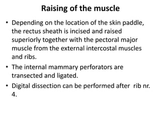
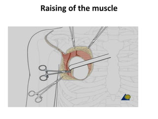
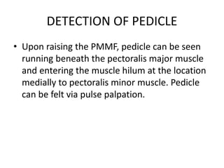
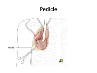
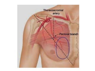
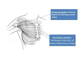
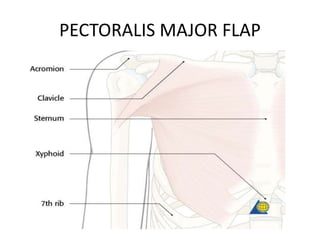
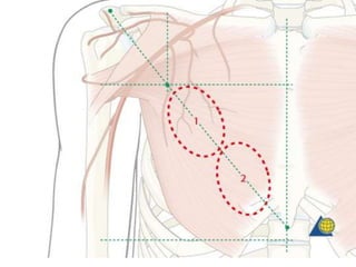
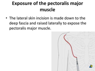
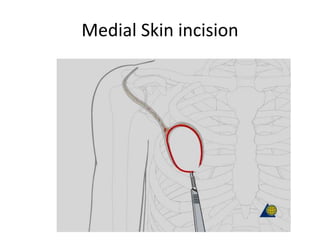
PECTORALIS MAJOR FLAP
Dr Jameel Kifayatullah
Senior dental surgeon
Khyber college of dentistry,Peshawar
PECTORALIS MAJOR FLAP
• Anatomy
• Blood supply from the thoraco acromial artery
and the secondary segmental perforators
arising from the internal mammory artery.
• The lateral thoracic artery does not usually
contribute significantly to the vasculature of
the pectoralis muscle.
Landmarks
• Flap design
• The following landmarks are identified and
marked:
• Acromion
• Clavicle
• Sternum
• Xyphoid
• 7th rib
PECTORALIS MAJOR FLAP
Size limitations
• A line from the acromium to the tip of the xyphoid is drawn.
Then a second line is made perpendicular to the clavicle
starting between mid-clavicle and the distal third of the
clavicle. The crossing point of the two lines is used as the
superior limit of the skin paddle.
• The limits of the skin paddle are:
• The 7th rib inferiorly.
• Laterally the extent of the pectoralis major muscle.
• Medially the border is at the midline of the sternum
• The skin paddle position over the muscle will depend on the
location of the neck/oral defect. The skin paddle illustrated in
this procedure is suitable for a neck defect. For oral defects
the skin paddle would be positioned more inferomedially.
• The pectoralis major muscle is harvested in full thickness. The
width and length of the muscle harvested will depend on the
size of the defect.
Size limitations
Skin paddle outline
• The skin flap is designed as a curved ellipse to facilitate
wound closure.
• Here the skin paddle is outlined fairly proximally (1)
limiting the arc of rotation of the flap, making it
suitable for neck defects.
• Should the surgeon need to reconstruct an intraoral
defect, or cheek defect, placing the skin paddle more
distally (2) allows for a greater arc of rotation and
hence more reach of the skin paddle.
Exposure of the pectoralis major
muscle
• The lateral skin incision is made down to the
deep fascia and raised laterally to expose the
pectoralis major muscle.
Medial skin incision
• The medial skin incision is made
down to the deep fascia and raised
medially to expose the pectoralis major
muscle.
• Part of the rectus sheath can be seen running
longitudinally at inferior edge.
Medial Skin incision
Raising of the muscle
• Depending on the location of the skin paddle,
the rectus sheath is incised and raised
superiorly together with the pectoral major
muscle from the external intercostal muscles
and ribs.
• The internal mammary perforators are
transected and ligated.
• Digital dissection can be performed after rib nr.
4.
Raising of the muscle
DETECTION OF PEDICLE
• Upon raising the PMMF, pedicle can be seen
running beneath the pectoralis major muscle
and entering the muscle hilum at the location
medially to pectoralis minor muscle. Pedicle
can be felt via pulse palpation.
Pedicle
Dissection of the pedicle
• The thoraco acromial pedicle can be identified
medially to the pectoralis minor muscle.
• The lateral thoracic artery is ligated as the insertion
of the pectoralis major muscle to the humeral head
is transected laterally. However, in some cases
where the lateral thoracic is the dominant vessel,
then the lateral thoracic artery should be preserved
as it is the main blood supply to the muscle.
DISSECTION OF PEDICLE
THORACOAROMIAL ARTERY
BRANCHES
THORACOAROMIAL ARTERY
BRANCHES
• The three main branches of the thoraco
acromial pedicle (clavicle, deltoid, acromial)
can be ligated. However, this is usually not
necessary unless it significantly limits the arc
of the rotation of the pedicle.
SUBCUTANEOUS TUNNELING
• A subcutaneous tunnel is developed between
the donor and the recipient sites, superficial
to the clavicle. The pedicle can then be
passed.
Now, the flap is ready to be passed through
the tunnel and transposed into defect region.
Closure
• The chest wall is undermined and the incision
is closed in layers.
• A suction drain is used to drain the surgical
site and prevent the formation of a hematoma
or late seroma.
Indications
• Tongue and floor of the mouth repair
• Pharyngoesophageal transit reconstruction
• Repair of cervical loss of skin
• Repair of facial loss of skin
• Pharynx reconstruction
• Repair of pharyngocutaneous fistula
• Salvage reconstruction after rotational flap necrosis
Contraindications
• Excessive trauma sustained by the subclavian
artery during the placement of a central venous
catheter, whereby the integrity of the
thoracoacromial artery has been compromised.
Preoperative angiography is indicated when an
injury is suspected to this primary pedicle that
might result in inadequate vascularity to the
pectoralis major muscle if myocutaneous flap
development were performed
Midfacial, upper third facial, and maxillary soft
tissue defects where the arc of rotation and
length of the pectoralis major myocutaneous
flap are insufficient
Contraindications
Excessively large skin paddle required to
perform the reconstruction. Women are able
to undergo pectoralis myocutaneous flap
development with larger skin paddles than
major men due to the redundancy of the
female breast. Skin grafting the chest wall is
not advisable in the event of inability to
achieve primary closure of the chest wall due
to the likely development of postoperative
costochondriti
Complications
• Neck wound infection
• Loss of skin/partial dehiscence
• Total necrosis
• Partial necrosis
• fistula
• Donor site complications
• Stenosis
Advantages
• Excellent flap viability
• The bulkiness of the flap allows good carotid protection
• Ease of elevation
• Minimal donor site morbidity
• This flap offers one-stage reconstruction.
• Patient's position need not be changed
intraoperatively.
• This flap provides a large cutaneous island that can be
used for defects involving 2 epithelial surfaces.
• The muscular part covers neck structures protecting
the carotid artery, especially in patients who have
undergone radiation therapy.
DISADVANTAGES OF PECTORLIS
MAJOR FLAP
• The flap can conceal recurrences, making follow-
up in the neck area more complicated.
• In women, the flap might include breast tissue,
which may lead to breast asymmetry.
• In males, hirsute chest skin is placed intraorally.
• This flap causes loss of pectoralis muscle function
in arm adduction and/or rotation.
• In patients who are overweight, the flap is bulky,
which leads to postoperative contour deformiti
DISADVANTAGES OF PECTORLIS
MAJOR FLAP
• Gravitational pull on pedicle and contraction
• Reduced neck mobility
• Limited arc of rotation
Flap monitoring
• No tracheotomy ties, gown ties, or pressure on
pedicle in neck or over clavicle
• "No Pressure" sign taped on patient's chest near
pedicle or written with marking pen on skin
• The flap is monitored by observation of color and
needle-prick bleeding. Poor flap appearance may
be improved with the use of dextran, steroids,
and/or hyperbaric oxygen. Pressure points and
excessive torque should be assessed and
improved if possible with changes in the head
and arm positions or neck suture removal.
• Flap failure is most likely to occur at the superior
aspect of the flap/mucosa closure.
Pectoralis major flap

Pectoralis major flap

  • 1.
    PECTORALIS MAJOR FLAP DrJameel Kifayatullah Senior dental surgeon Khyber college of dentistry,Peshawar
  • 3.
    PECTORALIS MAJOR FLAP •Anatomy • Blood supply from the thoraco acromial artery and the secondary segmental perforators arising from the internal mammory artery. • The lateral thoracic artery does not usually contribute significantly to the vasculature of the pectoralis muscle.
  • 6.
    Landmarks • Flap design •The following landmarks are identified and marked: • Acromion • Clavicle • Sternum • Xyphoid • 7th rib
  • 7.
  • 8.
    Size limitations • Aline from the acromium to the tip of the xyphoid is drawn. Then a second line is made perpendicular to the clavicle starting between mid-clavicle and the distal third of the clavicle. The crossing point of the two lines is used as the superior limit of the skin paddle. • The limits of the skin paddle are: • The 7th rib inferiorly. • Laterally the extent of the pectoralis major muscle. • Medially the border is at the midline of the sternum • The skin paddle position over the muscle will depend on the location of the neck/oral defect. The skin paddle illustrated in this procedure is suitable for a neck defect. For oral defects the skin paddle would be positioned more inferomedially. • The pectoralis major muscle is harvested in full thickness. The width and length of the muscle harvested will depend on the size of the defect.
  • 9.
  • 10.
    Skin paddle outline •The skin flap is designed as a curved ellipse to facilitate wound closure. • Here the skin paddle is outlined fairly proximally (1) limiting the arc of rotation of the flap, making it suitable for neck defects. • Should the surgeon need to reconstruct an intraoral defect, or cheek defect, placing the skin paddle more distally (2) allows for a greater arc of rotation and hence more reach of the skin paddle.
  • 12.
    Exposure of thepectoralis major muscle • The lateral skin incision is made down to the deep fascia and raised laterally to expose the pectoralis major muscle.
  • 13.
    Medial skin incision •The medial skin incision is made down to the deep fascia and raised medially to expose the pectoralis major muscle. • Part of the rectus sheath can be seen running longitudinally at inferior edge.
  • 14.
  • 15.
    Raising of themuscle • Depending on the location of the skin paddle, the rectus sheath is incised and raised superiorly together with the pectoral major muscle from the external intercostal muscles and ribs. • The internal mammary perforators are transected and ligated. • Digital dissection can be performed after rib nr. 4.
  • 16.
  • 17.
    DETECTION OF PEDICLE •Upon raising the PMMF, pedicle can be seen running beneath the pectoralis major muscle and entering the muscle hilum at the location medially to pectoralis minor muscle. Pedicle can be felt via pulse palpation.
  • 18.
  • 19.
    Dissection of thepedicle • The thoraco acromial pedicle can be identified medially to the pectoralis minor muscle. • The lateral thoracic artery is ligated as the insertion of the pectoralis major muscle to the humeral head is transected laterally. However, in some cases where the lateral thoracic is the dominant vessel, then the lateral thoracic artery should be preserved as it is the main blood supply to the muscle.
  • 20.
  • 21.
  • 22.
    THORACOAROMIAL ARTERY BRANCHES • Thethree main branches of the thoraco acromial pedicle (clavicle, deltoid, acromial) can be ligated. However, this is usually not necessary unless it significantly limits the arc of the rotation of the pedicle.
  • 23.
    SUBCUTANEOUS TUNNELING • Asubcutaneous tunnel is developed between the donor and the recipient sites, superficial to the clavicle. The pedicle can then be passed. Now, the flap is ready to be passed through the tunnel and transposed into defect region.
  • 24.
    Closure • The chestwall is undermined and the incision is closed in layers. • A suction drain is used to drain the surgical site and prevent the formation of a hematoma or late seroma.
  • 26.
    Indications • Tongue andfloor of the mouth repair • Pharyngoesophageal transit reconstruction • Repair of cervical loss of skin • Repair of facial loss of skin • Pharynx reconstruction • Repair of pharyngocutaneous fistula • Salvage reconstruction after rotational flap necrosis
  • 27.
    Contraindications • Excessive traumasustained by the subclavian artery during the placement of a central venous catheter, whereby the integrity of the thoracoacromial artery has been compromised. Preoperative angiography is indicated when an injury is suspected to this primary pedicle that might result in inadequate vascularity to the pectoralis major muscle if myocutaneous flap development were performed Midfacial, upper third facial, and maxillary soft tissue defects where the arc of rotation and length of the pectoralis major myocutaneous flap are insufficient
  • 28.
    Contraindications Excessively large skinpaddle required to perform the reconstruction. Women are able to undergo pectoralis myocutaneous flap development with larger skin paddles than major men due to the redundancy of the female breast. Skin grafting the chest wall is not advisable in the event of inability to achieve primary closure of the chest wall due to the likely development of postoperative costochondriti
  • 32.
    Complications • Neck woundinfection • Loss of skin/partial dehiscence • Total necrosis • Partial necrosis • fistula • Donor site complications • Stenosis
  • 33.
    Advantages • Excellent flapviability • The bulkiness of the flap allows good carotid protection • Ease of elevation • Minimal donor site morbidity • This flap offers one-stage reconstruction. • Patient's position need not be changed intraoperatively. • This flap provides a large cutaneous island that can be used for defects involving 2 epithelial surfaces. • The muscular part covers neck structures protecting the carotid artery, especially in patients who have undergone radiation therapy.
  • 34.
    DISADVANTAGES OF PECTORLIS MAJORFLAP • The flap can conceal recurrences, making follow- up in the neck area more complicated. • In women, the flap might include breast tissue, which may lead to breast asymmetry. • In males, hirsute chest skin is placed intraorally. • This flap causes loss of pectoralis muscle function in arm adduction and/or rotation. • In patients who are overweight, the flap is bulky, which leads to postoperative contour deformiti
  • 35.
    DISADVANTAGES OF PECTORLIS MAJORFLAP • Gravitational pull on pedicle and contraction • Reduced neck mobility • Limited arc of rotation
  • 36.
    Flap monitoring • Notracheotomy ties, gown ties, or pressure on pedicle in neck or over clavicle • "No Pressure" sign taped on patient's chest near pedicle or written with marking pen on skin • The flap is monitored by observation of color and needle-prick bleeding. Poor flap appearance may be improved with the use of dextran, steroids, and/or hyperbaric oxygen. Pressure points and excessive torque should be assessed and improved if possible with changes in the head and arm positions or neck suture removal. • Flap failure is most likely to occur at the superior aspect of the flap/mucosa closure.