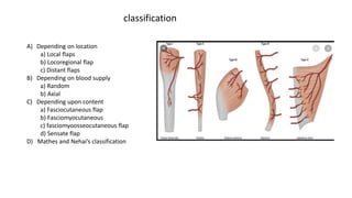
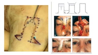
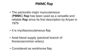
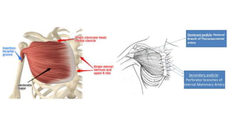
SOFT TISSUE FLAPS
Reconstructive
ladder :- A heirachy of
options available for
closing a wound
Flap physiology
A) Depending on location
a) Local flaps
b) Locoregional flap
c) Distant flaps
B) Depending on blood supply
a) Random
b) Axial
C) Depending upon content
a) Fasciocutaneous flap
b) Fasciomyocutaneous
c) fasciomyoosseocutaneous flap
d) Sensate flap
D) Mathes and Nehai’s classification
classification
Advancement flap
Rotational flap
A
B
C
To increase mobility
1) Back cut
2) Burrow’s triangle
Transposition Flap
A
B C
D
E
Bilobed flap
Rhomboid Flap
Locoregional flaps
1)Nasolabial flap
2)PMMC
3)Forehead flap
4)Deltopectoris flap
5)Latissmus dorsi flap
Commonly used locoregional flaps for head and neck reconstruction
• Random flap
• Apex of the flap
should 3-4 mm
below medial
canthus of eye
• The medial border
of the flap lies on
nasolabial groove
• Lateral border of
the flap is marked
depending upon
laxity of the
available skin after
performing pinch
test
• Flap is raised in
supramascular
plane
• Tunnel is created
• Inset of the flap Is
done through
created tunnel and
donor area is
closed primarily
PMMC flap
• The pectoralis major myocutaneous
(PMMC) flap has been used as a versatile and
reliable flap since its first description by Ariyan in
1979
• It is myofasciocutaneous flap
• Axial blood supply (pectoral branch of
thoracoacromion artery)
• Considered as workhorse flap
• Skin paddle is marked after
measuring the length from the
defect
• 3 points are marked – sternum,
xiphisternum, acromion
• Sternum –acromion line is
joined and xiphisternum
acromion line is joined
• Line drawn from center of
sternum – acromion line on
xiphisternum acromion line
• Pedicle is marked on this new
line
• Incision is marked from axilla to
skin paddle
1.Incision is taken from axilla and superior and
inferior skin flaps are raised around the skin
paddle
2.Tagging sutures on skin paddle are taken
3.Flap is then raised above pectoris minor muscle
4.Pectoris major pedicle is then identified
5.Tunnel is created above clavicle
6.PMMC flap is then passed through tunnel
gently and inset of the flap is done in oncology
defect
7.Donor area is closed primarily
Flap Harvesting
-The deltopectoral flap was popularized by Bakamijian in the
mid-1960 and remained popular for some time as a workhorse for
reconstruction of oropharyngeal and hvpopharvngeal defects
-The medially based deltopectoral flap was considered in the past
to be the workhorse for reconstruction of oropharyngeal and
pharyngoesophageal defects.
-The flap derives its primary blood
supply from the perforating branches of the internal mammary
artery through the second, third and fourth intercostal spaces.
-The proximal part of the flap therefore has an axial blood supply.
However, the blood supply to the distal one-third of the
deltopectoral flap is of random pattern through the subdermal
plexus.
-The skin overlying the deltoid region derives its blood
supply from the anterior perforating branch of the acrorniothoracic
artery. This artery, however, is divided when the deltopectoral flap
is elevated and therefore the distal third of the flap derives its
blood supply through random pattern blood vessels of the
subcutaneous plexus.
Deltopectoral flap
• Deltopectoral flap marking which includes 1st,
2nd & 3rd IMA perforator
• The incision line is extending deltopectoral
grove
• Subfascial flap is raised & inset of flap is done in
oncology defect
• Donor area is closed with primary or STSG
• 2nd surgery is performed after 3 weeks for
pedicle detachment
Forehead flap
• Also called as temporl flap
• In 800BC sushruta, described a nasal reconstruction approach based on
pedicled forehead flap
• Later described by McGregor in 1963
• Its an axil pattern myocutaneous flap provide large area of skin &
subcutaneous tissue
• Based on the site 1) median forehead flap (based on supratrochlear + dorsal
nasal)
• 2)paramedian forehead flap ( based on supratrochlear +
supraorbital)
• 3) laterally based forehead flap (based on superfacial
temporal + posterior auricular)
Modification of laterally based forehead flap
• In this case, laterally based forehead flap was used for reconstruction of lower lip defect
• Donor area was closed with STSG
• Vertical extension of the incision of the base of the flap was done to increase the flap reach
• 2nd surgery was performed after 3 weeks for detachment of the peduncle of the flap
• This flap can be used for intraoral defects by creating tunnel.
Antero-lateral thigh flap
• First described by Song et al
• It’s a fasciocutaneous or
myofasciocutaneous free flap
• It has axial blood supply from
descending or oblique branch of
lateral circumflex femoral artery
• Musculocutaneous perforators are
more than septocutaneous
perforators
ALT marking
• After marking superior incision is taken
• Identification of RF muscle
• Lift the muscle to identify descending branch of latral
circumflex femolar artery and vena comitans
• Identification of perforators
• Deroofing of perforators and trace till main pedicle
• Identify and separate femoral nerve branches from
pedicle
• Complete inferior incision and raise flap
• Check for bright red color bleeding from edges
• Detach pedicle as superior as possible
• Primary closure of donor area
Harvesting technique
Advantages:-
1) Can be used for large defects
2) Simple anatomy and easy to
harvest
3) Most of the time donor area can
be closed primarily
4) No donor side morbidity
Disadvantages :-
1) Injury to cutaneous branch of
femoral nerve can cause numbness
in lateral thigh region
2) Defattening is required in Patient
with very thick thigh
Radial forearm flap
• Yang Goufan described this flap
for the first time
• It is mainly used as
fasciocutaneous flap but also can
be used as osseocutaneous flap
• It is axial pattern flap based on
radial artery and its vena
comitans
• Mainly used for reconstruction of
tongue defects
• Preoperatively Allen’s test is done
• Marking for flap is done
• Tourniquet is then activated
• Vertical incision in suprafascial layer is taken to
identify cephalic vein & dissection of cephalic
vein is done
• Identification and separation of ante-brachial
cutaneous nerve
• Superior incision of the skin paddle is then taken
and superfacial branch of radial nerve is
identified and separated from the skin paddle
• Complete the skin paddle incision and the radial
artery and its vena comitans are identified and
double ligated with linen thread and liga clips
• Again check for blood circulation of the hand
• Slowly dissect the radial artery along with skin
paddle between brachioradialis and flexor carpi
radialis
• Pedicle can be harvested upto 20 cm long
• Deactivate the tourniquet
• Radial artery and its venae comitans along with
cephalic vein are ligated and detached
• Donor area can be closed with STSG
Advantages :-
• Easy to harvest, also called as beginners flap
• Long pedicle is available, upto 20 cm
• Diameter of radial artery is 2-2.5 mm
• Cephalic vein can be used as back up vein in case of congestion
• Pollicis longus tendon can be harvested at the same time for
total lower lip recon
Disadvantages :-
• Donor site morbidity
• Injury to superfacial radial nerve can cause loss of sensation in
1st anatomical snuff box region
• Whenever it is raised as osseocutaneous flap, pathological
fracture of radius can occur

Soft tissue flaps

  • 1.
  • 2.
    Reconstructive ladder :- Aheirachy of options available for closing a wound
  • 6.
  • 10.
    A) Depending onlocation a) Local flaps b) Locoregional flap c) Distant flaps B) Depending on blood supply a) Random b) Axial C) Depending upon content a) Fasciocutaneous flap b) Fasciomyocutaneous c) fasciomyoosseocutaneous flap d) Sensate flap D) Mathes and Nehai’s classification classification
  • 11.
  • 13.
  • 14.
    To increase mobility 1)Back cut 2) Burrow’s triangle
  • 15.
  • 16.
  • 17.
  • 21.
    Locoregional flaps 1)Nasolabial flap 2)PMMC 3)Foreheadflap 4)Deltopectoris flap 5)Latissmus dorsi flap Commonly used locoregional flaps for head and neck reconstruction
  • 22.
  • 23.
    • Apex ofthe flap should 3-4 mm below medial canthus of eye • The medial border of the flap lies on nasolabial groove • Lateral border of the flap is marked depending upon laxity of the available skin after performing pinch test • Flap is raised in supramascular plane • Tunnel is created • Inset of the flap Is done through created tunnel and donor area is closed primarily
  • 29.
    PMMC flap • Thepectoralis major myocutaneous (PMMC) flap has been used as a versatile and reliable flap since its first description by Ariyan in 1979 • It is myofasciocutaneous flap • Axial blood supply (pectoral branch of thoracoacromion artery) • Considered as workhorse flap
  • 31.
    • Skin paddleis marked after measuring the length from the defect • 3 points are marked – sternum, xiphisternum, acromion • Sternum –acromion line is joined and xiphisternum acromion line is joined • Line drawn from center of sternum – acromion line on xiphisternum acromion line • Pedicle is marked on this new line • Incision is marked from axilla to skin paddle
  • 32.
    1.Incision is takenfrom axilla and superior and inferior skin flaps are raised around the skin paddle 2.Tagging sutures on skin paddle are taken 3.Flap is then raised above pectoris minor muscle 4.Pectoris major pedicle is then identified 5.Tunnel is created above clavicle 6.PMMC flap is then passed through tunnel gently and inset of the flap is done in oncology defect 7.Donor area is closed primarily Flap Harvesting
  • 34.
    -The deltopectoral flapwas popularized by Bakamijian in the mid-1960 and remained popular for some time as a workhorse for reconstruction of oropharyngeal and hvpopharvngeal defects -The medially based deltopectoral flap was considered in the past to be the workhorse for reconstruction of oropharyngeal and pharyngoesophageal defects. -The flap derives its primary blood supply from the perforating branches of the internal mammary artery through the second, third and fourth intercostal spaces. -The proximal part of the flap therefore has an axial blood supply. However, the blood supply to the distal one-third of the deltopectoral flap is of random pattern through the subdermal plexus. -The skin overlying the deltoid region derives its blood supply from the anterior perforating branch of the acrorniothoracic artery. This artery, however, is divided when the deltopectoral flap is elevated and therefore the distal third of the flap derives its blood supply through random pattern blood vessels of the subcutaneous plexus. Deltopectoral flap
  • 35.
    • Deltopectoral flapmarking which includes 1st, 2nd & 3rd IMA perforator • The incision line is extending deltopectoral grove • Subfascial flap is raised & inset of flap is done in oncology defect • Donor area is closed with primary or STSG • 2nd surgery is performed after 3 weeks for pedicle detachment
  • 36.
    Forehead flap • Alsocalled as temporl flap • In 800BC sushruta, described a nasal reconstruction approach based on pedicled forehead flap • Later described by McGregor in 1963 • Its an axil pattern myocutaneous flap provide large area of skin & subcutaneous tissue • Based on the site 1) median forehead flap (based on supratrochlear + dorsal nasal) • 2)paramedian forehead flap ( based on supratrochlear + supraorbital) • 3) laterally based forehead flap (based on superfacial temporal + posterior auricular)
  • 38.
    Modification of laterallybased forehead flap
  • 39.
    • In thiscase, laterally based forehead flap was used for reconstruction of lower lip defect • Donor area was closed with STSG • Vertical extension of the incision of the base of the flap was done to increase the flap reach • 2nd surgery was performed after 3 weeks for detachment of the peduncle of the flap • This flap can be used for intraoral defects by creating tunnel.
  • 40.
    Antero-lateral thigh flap •First described by Song et al • It’s a fasciocutaneous or myofasciocutaneous free flap • It has axial blood supply from descending or oblique branch of lateral circumflex femoral artery • Musculocutaneous perforators are more than septocutaneous perforators
  • 41.
  • 42.
    • After markingsuperior incision is taken • Identification of RF muscle • Lift the muscle to identify descending branch of latral circumflex femolar artery and vena comitans • Identification of perforators • Deroofing of perforators and trace till main pedicle • Identify and separate femoral nerve branches from pedicle • Complete inferior incision and raise flap • Check for bright red color bleeding from edges • Detach pedicle as superior as possible • Primary closure of donor area Harvesting technique Advantages:- 1) Can be used for large defects 2) Simple anatomy and easy to harvest 3) Most of the time donor area can be closed primarily 4) No donor side morbidity Disadvantages :- 1) Injury to cutaneous branch of femoral nerve can cause numbness in lateral thigh region 2) Defattening is required in Patient with very thick thigh
  • 43.
    Radial forearm flap •Yang Goufan described this flap for the first time • It is mainly used as fasciocutaneous flap but also can be used as osseocutaneous flap • It is axial pattern flap based on radial artery and its vena comitans • Mainly used for reconstruction of tongue defects
  • 44.
    • Preoperatively Allen’stest is done • Marking for flap is done • Tourniquet is then activated • Vertical incision in suprafascial layer is taken to identify cephalic vein & dissection of cephalic vein is done • Identification and separation of ante-brachial cutaneous nerve • Superior incision of the skin paddle is then taken and superfacial branch of radial nerve is identified and separated from the skin paddle • Complete the skin paddle incision and the radial artery and its vena comitans are identified and double ligated with linen thread and liga clips • Again check for blood circulation of the hand • Slowly dissect the radial artery along with skin paddle between brachioradialis and flexor carpi radialis • Pedicle can be harvested upto 20 cm long • Deactivate the tourniquet • Radial artery and its venae comitans along with cephalic vein are ligated and detached • Donor area can be closed with STSG
  • 45.
    Advantages :- • Easyto harvest, also called as beginners flap • Long pedicle is available, upto 20 cm • Diameter of radial artery is 2-2.5 mm • Cephalic vein can be used as back up vein in case of congestion • Pollicis longus tendon can be harvested at the same time for total lower lip recon Disadvantages :- • Donor site morbidity • Injury to superfacial radial nerve can cause loss of sensation in 1st anatomical snuff box region • Whenever it is raised as osseocutaneous flap, pathological fracture of radius can occur

Editor's Notes

  • #16 When tissue is present in between , it is called as interpolation flap