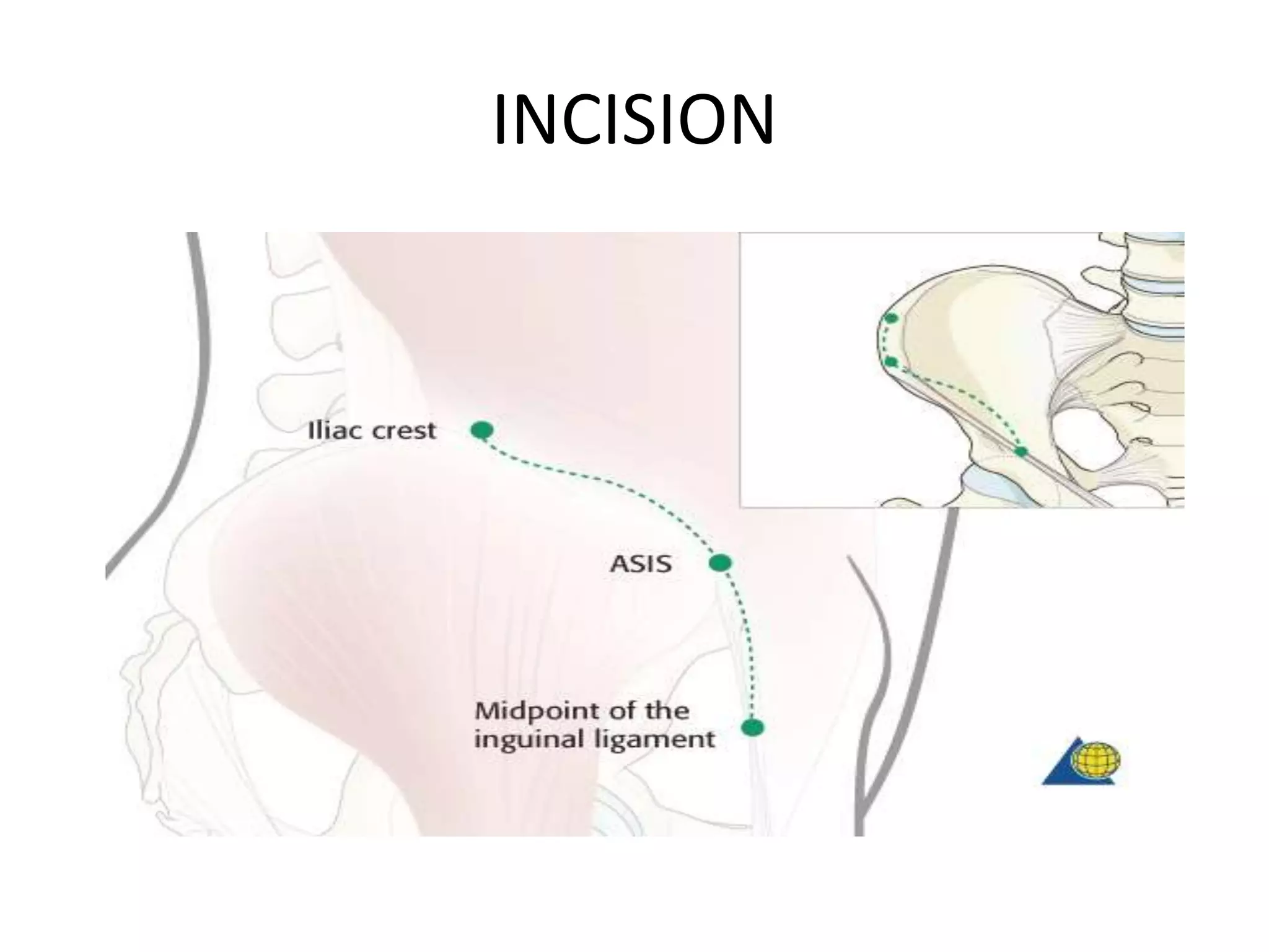
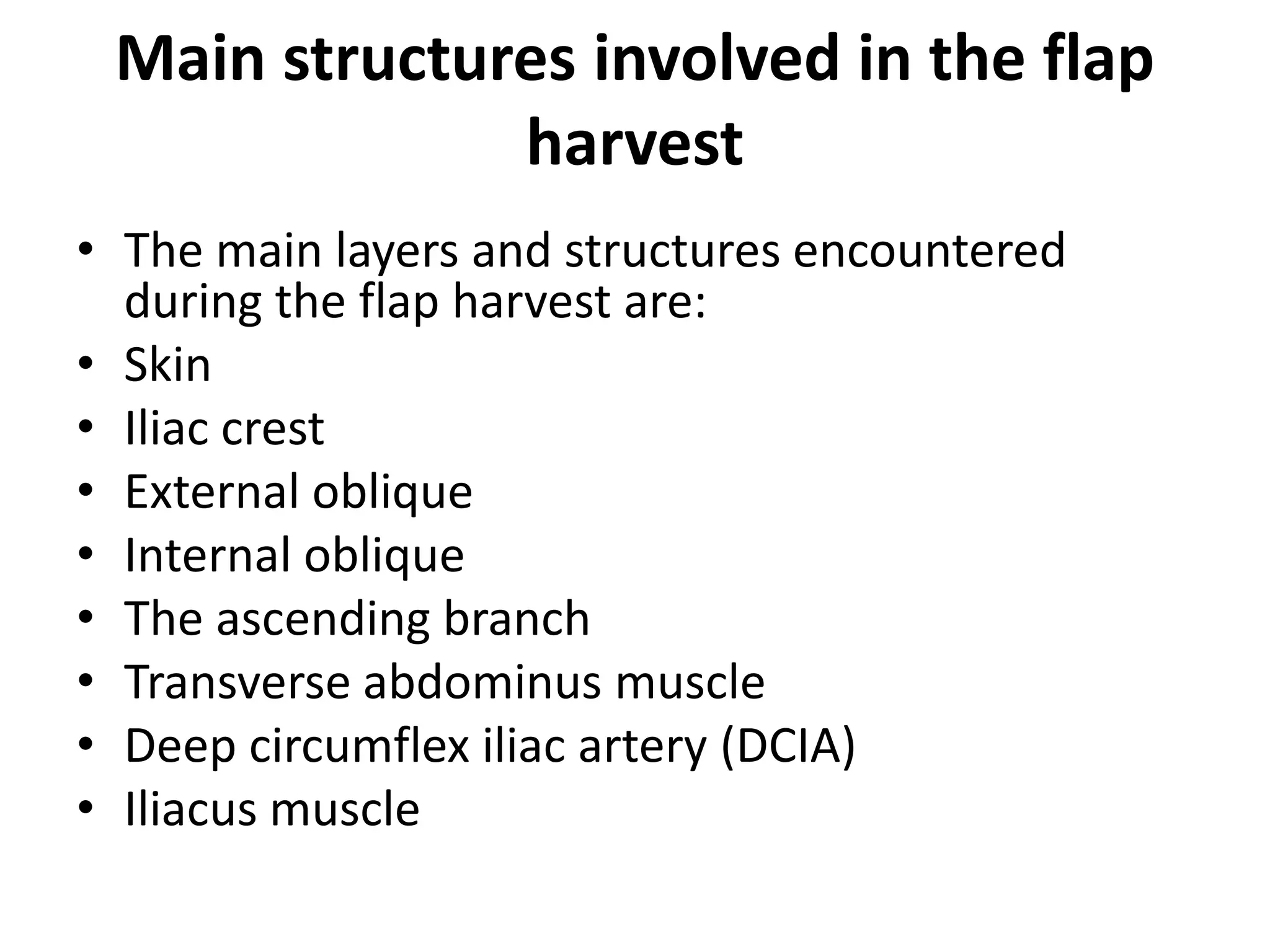
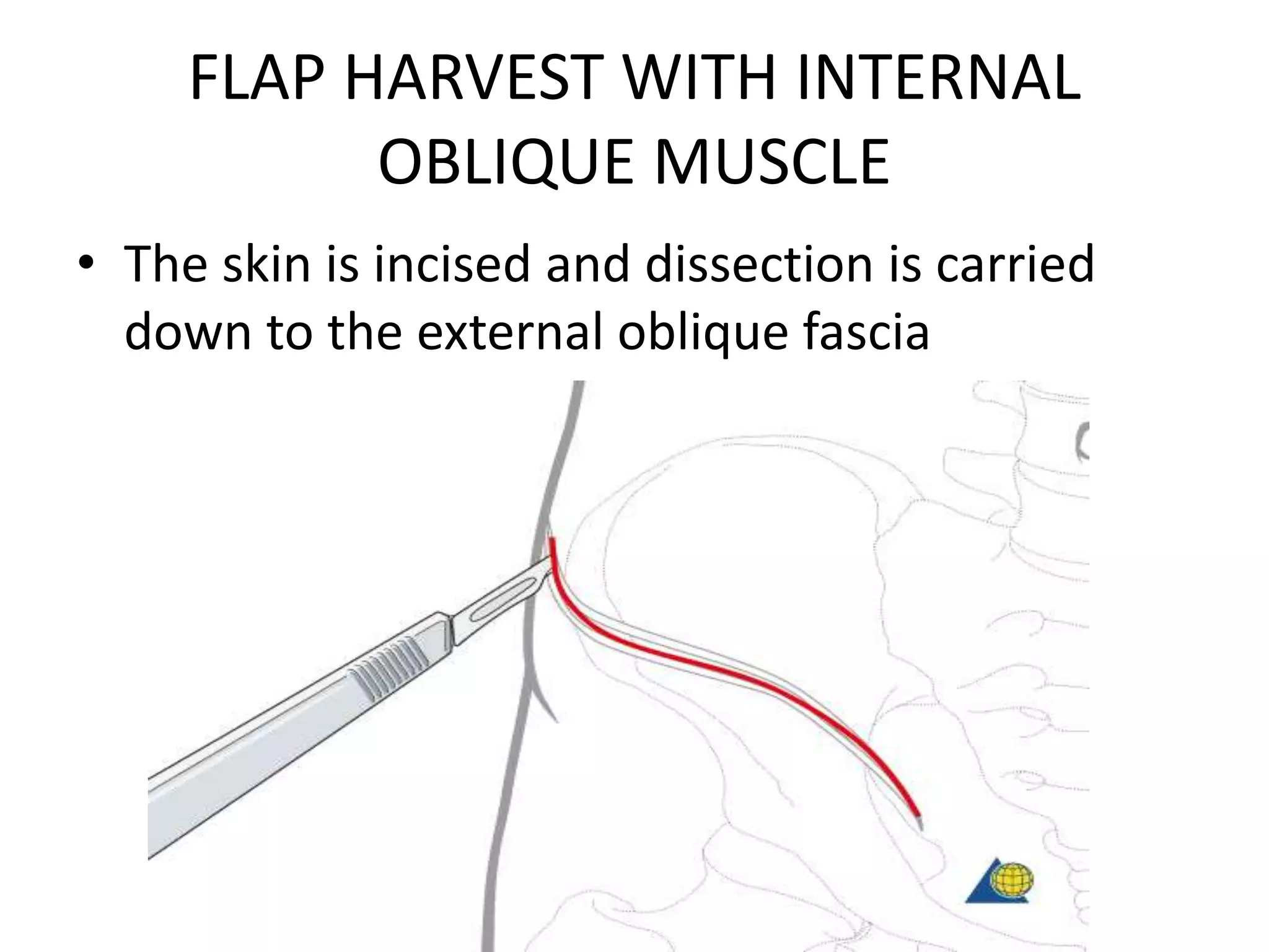
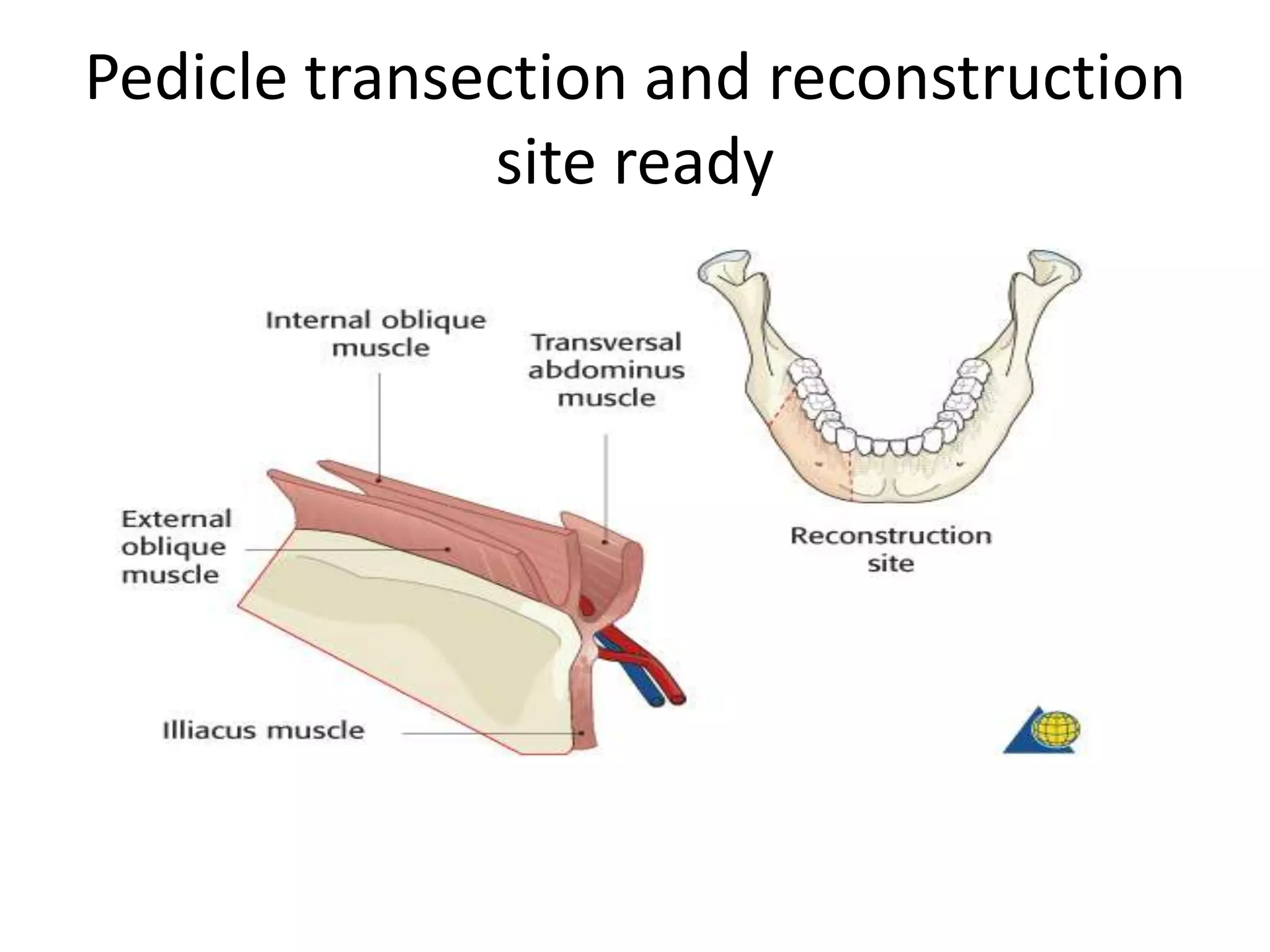
DEEP CIRCUMFLEX ILIAC ARTERY
FLAP
DR JAMEEL KIFAYATULLAH
Oral maxillofacial surgeon Khyber
College of dentistry,Peshawar
DCIA FLAP
Vascular anatomy
• The vascular system will depend on the deep circumflex
iliac artery (DCIA) which has a diameter of 1.5 -2.0
mm.
• The concomitant vein has a diameter of 2.5 – 3.0 mm.
• The length of the pedicle =5 cm.
• The ascending branch is located beneath internal
oblique muscle running superiorly.
• The DCIA is located beneath the transverse abdominis
in the groove between the transverse abdominis and
the iliacus fossa.
DCIA FLAP ANATOMY
Anatomical variations
• There are three main anatomical variations of the
ascending branch, which will have an influence on the
flap harvest.
• In 65 % of the cases, the ascending branch originates
from the DCIA wihin one cm medial to the ASIS.
• In 20 % of the cacses there is no single dominant
ascending branch. The internal oblique is supplied by a
series of small branches of the DCIA.
• In 15 % of the cases ascending branch originates in a
more medial location.
Anatomical variations of ascending
branch
Muscular anatomy
• When dissecting through the external oblique, the
internal oblique and the transverse abdominis muscle
one can readily recognize the muscle layers by the
direction of the muscle fibers.
• The muscle fibers of the external oblique muscle run
inferiomedially.
• The muscle fibers of the internal oblique muscle run
superiomedially perpendicular to the external oblique
muscle fibers.
• The muscle fibers of the transverse abdominus muscle
run medially.
Surgical Landmarks
Design of flap
• The following landmarks are identified and
marked:
• The anterior superior iliac spine (ASIS)
• The midpoint of the inguinal ligament (Femoral
artery is palpable)
• Superior border of the iliac crest
Surgical Landmarks
DCIA FLAP
• The harvested myo osseous flap should
include:
• DCIA
• Iliac crest
• Internal oblique muscle
• Ascending Branch
• Although a relatively large bone flap (up to 18
cm) can be harvested, large bone flaps may
lead to higher risk of donor site morbidity (e.g:
hernia, disfigurement, gait disturbance).
DCIA FLAP
INCISION
• The standard size is around 10 cm in length and up to 5 cm in height.
• A S-shaped incision is used starting at the superior border of the iliac
crest (with the assistant pulling the overlying skin medially, thereby,
end result of surgical scar will be resting laterally on bone edge in
avoiding long term abrasion irritation of clothing), curving medially
through the anterior superior iliac spine and curving inferiorly through
the midpoint of the inguinal ligament.
The exact osteotomy line will depend on the planned reconstruction.
We will here show the harvest of a straight segment(s) used for
mandibular body reconstruction.
INCISION
Main structures involved in the flap
harvest
• The main layers and structures encountered
during the flap harvest are:
• Skin
• Iliac crest
• External oblique
• Internal oblique
• The ascending branch
• Transverse abdominus muscle
• Deep circumflex iliac artery (DCIA)
• Iliacus muscle
Main structures involved in flap
harvest
Patient positioning
• For the harvest the patient is placed supine with the
buttocks elevated on the donor site by using eg. a
bean bag.
FLAP HARVEST WITH INTERNAL
OBLIQUE MUSCLE
• The skin is incised and dissection is carried
down to the external oblique fascia
EXTERNAL OBLIQUE FASCIA
DISSECTION
• The external oblique fascia (as external
oblique running laterally, it is very thin and
almost only fascia can be seen) is incised and
retracted to expose the internal oblique
muscle.
FLAP HARVEST
INTERNAL OBLIQUE MUSCLE
DISSECTION
• If the internal oblique muscle is harvested
together with the bone flap, the maximum internal
oblique muscle that can be harvested, starting 2
cm laterally from the midline and end at the iliac
crest.
• The internal oblique muscle is incised as needed
and raised laterally. The ascending branch of the
DCIA running on the undersurface of the muscle
is identified.
FLAP HARVEST
Flap harvest
• Retrograde dissection is performed along the
ascending branch to the main trunk (External Illiac
Artery). The dissection is then continued until the
bifurcation of the external iliac artery is reached.
• Now, dissect antegradly to identify the DCIA.There is
a variation where the Ascending branch can arise
from the DCIA or directly from External Illiac Artery.
Bifurcation of external iliac artery
Transverse Abdominus dissection
• Dissection is now carried superiolaterally along
the DCIA through and beneath the transverse
abdominal muscle.
• An incision of the transverse abdominal muscle is
along the dissection of DCIA or 2 cm medially to
the iliac crest.
Transverse abdominus dissection
Flap harvesting
• The distal end of the DCIA is ligated.
Iliacus incision
• The iliacus is incised to expose the osteotomy
sites. In order to protect the DCIA, the incision
is made 1-2 cm inferior to the groove where
the DCIA is located.
Iliacus muscle incision
Osteotomy
• The osteotomy is performed according to the
planned outline.
Pedicle transection
• When the reconstruction site is ready, the
pedicle can be transected.
Pedicle transection
Harvest without internal oblique muscle
Skin incision
• The skin is incised and dissection is carried
down to the external oblique fascia.
External oblique fascia incision
• The external oblique fascia is incised and retracted
to expose the internal oblique muscle.
Blunt dissection along external iliac
artery
• The transverse abdominal muscle and the inguinal
ligaments are identified.
• The external iliac artery can be found by palpation
at midpoint of the inguinal ligaments.
• Meticulous blunt dissections are performed along
the external iliac artery. The DCIA is identified by its
course emerging laterally from the external iliac
artery and entering the groove between transverse
abdominous muscle and the iliac fossa
Blunt dissection along external iliac
artery
Transverse abdominus muscle
dissection
• Dissection is now carried superiolaterally along the
DCIA through and beneath the transverse
abdominal muscle.
• An incision of the transverse abdominal muscle is
made from the inferior border to the planned
resection site. The incision is made 2 cm medially to
the iliac crest.
Transverse abdominus muscle
dissection
Ligation of DCIA
• The distal end of the DCIA is ligated.
LIGATION OF DCIA
ILIACUS MUSCLE DISSECTION
The iliacus is incised to expose the the inner
table of Illiac bone prepared for osteotomy. In
order to protect the DCIA, the incision is made
1-2 cm inferior to the illiacus groove where
the DCIA is located.
ILIACUS MUSCLE DISSECTION
OSTEOTOMY
osteotomy
• The osteotomy is performed according to the
planned outline
Pedicle transection and reconstruction
site ready
Pedicle transection
• When the reconstruction site is ready, the
pedicle can be transected.
Closure
closure
• The transverse abdominis muscle is sutured to
the iliacus.
closure
closure
• Harvest without internal oblique:
• The cut edges of the external oblique, internal
oblique and transversus abdominus muscles
are then reapproximated using resorbable
horizontal mattress sutures.
Drain placement
Drain insertion
• A drain is inserted between the transverse
abdominous muscle and internal
oblique/external oblique muscles. the skin
closed in primary fashion. Pressure dressing is
then applied.
Deep circumflex iliac artery flap

Deep circumflex iliac artery flap

  • 1.
    DEEP CIRCUMFLEX ILIACARTERY FLAP DR JAMEEL KIFAYATULLAH Oral maxillofacial surgeon Khyber College of dentistry,Peshawar
  • 2.
    DCIA FLAP Vascular anatomy •The vascular system will depend on the deep circumflex iliac artery (DCIA) which has a diameter of 1.5 -2.0 mm. • The concomitant vein has a diameter of 2.5 – 3.0 mm. • The length of the pedicle =5 cm. • The ascending branch is located beneath internal oblique muscle running superiorly. • The DCIA is located beneath the transverse abdominis in the groove between the transverse abdominis and the iliacus fossa.
  • 3.
  • 4.
    Anatomical variations • Thereare three main anatomical variations of the ascending branch, which will have an influence on the flap harvest. • In 65 % of the cases, the ascending branch originates from the DCIA wihin one cm medial to the ASIS. • In 20 % of the cacses there is no single dominant ascending branch. The internal oblique is supplied by a series of small branches of the DCIA. • In 15 % of the cases ascending branch originates in a more medial location.
  • 5.
    Anatomical variations ofascending branch
  • 6.
    Muscular anatomy • Whendissecting through the external oblique, the internal oblique and the transverse abdominis muscle one can readily recognize the muscle layers by the direction of the muscle fibers. • The muscle fibers of the external oblique muscle run inferiomedially. • The muscle fibers of the internal oblique muscle run superiomedially perpendicular to the external oblique muscle fibers. • The muscle fibers of the transverse abdominus muscle run medially.
  • 7.
    Surgical Landmarks Design offlap • The following landmarks are identified and marked: • The anterior superior iliac spine (ASIS) • The midpoint of the inguinal ligament (Femoral artery is palpable) • Superior border of the iliac crest
  • 8.
  • 9.
    DCIA FLAP • Theharvested myo osseous flap should include: • DCIA • Iliac crest • Internal oblique muscle • Ascending Branch • Although a relatively large bone flap (up to 18 cm) can be harvested, large bone flaps may lead to higher risk of donor site morbidity (e.g: hernia, disfigurement, gait disturbance).
  • 10.
  • 11.
    INCISION • The standardsize is around 10 cm in length and up to 5 cm in height. • A S-shaped incision is used starting at the superior border of the iliac crest (with the assistant pulling the overlying skin medially, thereby, end result of surgical scar will be resting laterally on bone edge in avoiding long term abrasion irritation of clothing), curving medially through the anterior superior iliac spine and curving inferiorly through the midpoint of the inguinal ligament. The exact osteotomy line will depend on the planned reconstruction. We will here show the harvest of a straight segment(s) used for mandibular body reconstruction.
  • 12.
  • 13.
    Main structures involvedin the flap harvest • The main layers and structures encountered during the flap harvest are: • Skin • Iliac crest • External oblique • Internal oblique • The ascending branch • Transverse abdominus muscle • Deep circumflex iliac artery (DCIA) • Iliacus muscle
  • 14.
    Main structures involvedin flap harvest
  • 15.
    Patient positioning • Forthe harvest the patient is placed supine with the buttocks elevated on the donor site by using eg. a bean bag.
  • 16.
    FLAP HARVEST WITHINTERNAL OBLIQUE MUSCLE • The skin is incised and dissection is carried down to the external oblique fascia
  • 17.
    EXTERNAL OBLIQUE FASCIA DISSECTION •The external oblique fascia (as external oblique running laterally, it is very thin and almost only fascia can be seen) is incised and retracted to expose the internal oblique muscle.
  • 18.
  • 19.
    INTERNAL OBLIQUE MUSCLE DISSECTION •If the internal oblique muscle is harvested together with the bone flap, the maximum internal oblique muscle that can be harvested, starting 2 cm laterally from the midline and end at the iliac crest. • The internal oblique muscle is incised as needed and raised laterally. The ascending branch of the DCIA running on the undersurface of the muscle is identified.
  • 20.
  • 21.
    Flap harvest • Retrogradedissection is performed along the ascending branch to the main trunk (External Illiac Artery). The dissection is then continued until the bifurcation of the external iliac artery is reached. • Now, dissect antegradly to identify the DCIA.There is a variation where the Ascending branch can arise from the DCIA or directly from External Illiac Artery.
  • 22.
  • 23.
    Transverse Abdominus dissection •Dissection is now carried superiolaterally along the DCIA through and beneath the transverse abdominal muscle. • An incision of the transverse abdominal muscle is along the dissection of DCIA or 2 cm medially to the iliac crest.
  • 24.
  • 25.
    Flap harvesting • Thedistal end of the DCIA is ligated.
  • 26.
    Iliacus incision • Theiliacus is incised to expose the osteotomy sites. In order to protect the DCIA, the incision is made 1-2 cm inferior to the groove where the DCIA is located.
  • 27.
  • 28.
    Osteotomy • The osteotomyis performed according to the planned outline.
  • 29.
    Pedicle transection • Whenthe reconstruction site is ready, the pedicle can be transected.
  • 30.
  • 31.
  • 32.
    Skin incision • Theskin is incised and dissection is carried down to the external oblique fascia.
  • 33.
    External oblique fasciaincision • The external oblique fascia is incised and retracted to expose the internal oblique muscle.
  • 34.
    Blunt dissection alongexternal iliac artery • The transverse abdominal muscle and the inguinal ligaments are identified. • The external iliac artery can be found by palpation at midpoint of the inguinal ligaments. • Meticulous blunt dissections are performed along the external iliac artery. The DCIA is identified by its course emerging laterally from the external iliac artery and entering the groove between transverse abdominous muscle and the iliac fossa
  • 35.
    Blunt dissection alongexternal iliac artery
  • 36.
    Transverse abdominus muscle dissection •Dissection is now carried superiolaterally along the DCIA through and beneath the transverse abdominal muscle. • An incision of the transverse abdominal muscle is made from the inferior border to the planned resection site. The incision is made 2 cm medially to the iliac crest.
  • 37.
  • 38.
    Ligation of DCIA •The distal end of the DCIA is ligated.
  • 39.
  • 40.
    ILIACUS MUSCLE DISSECTION Theiliacus is incised to expose the the inner table of Illiac bone prepared for osteotomy. In order to protect the DCIA, the incision is made 1-2 cm inferior to the illiacus groove where the DCIA is located.
  • 41.
  • 42.
  • 43.
    osteotomy • The osteotomyis performed according to the planned outline
  • 44.
    Pedicle transection andreconstruction site ready
  • 45.
    Pedicle transection • Whenthe reconstruction site is ready, the pedicle can be transected.
  • 46.
  • 47.
    closure • The transverseabdominis muscle is sutured to the iliacus.
  • 48.
  • 49.
    closure • Harvest withoutinternal oblique: • The cut edges of the external oblique, internal oblique and transversus abdominus muscles are then reapproximated using resorbable horizontal mattress sutures.
  • 50.
  • 51.
    Drain insertion • Adrain is inserted between the transverse abdominous muscle and internal oblique/external oblique muscles. the skin closed in primary fashion. Pressure dressing is then applied.