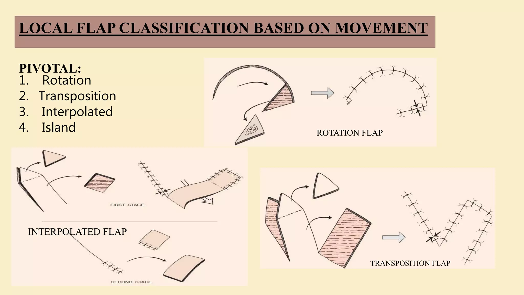
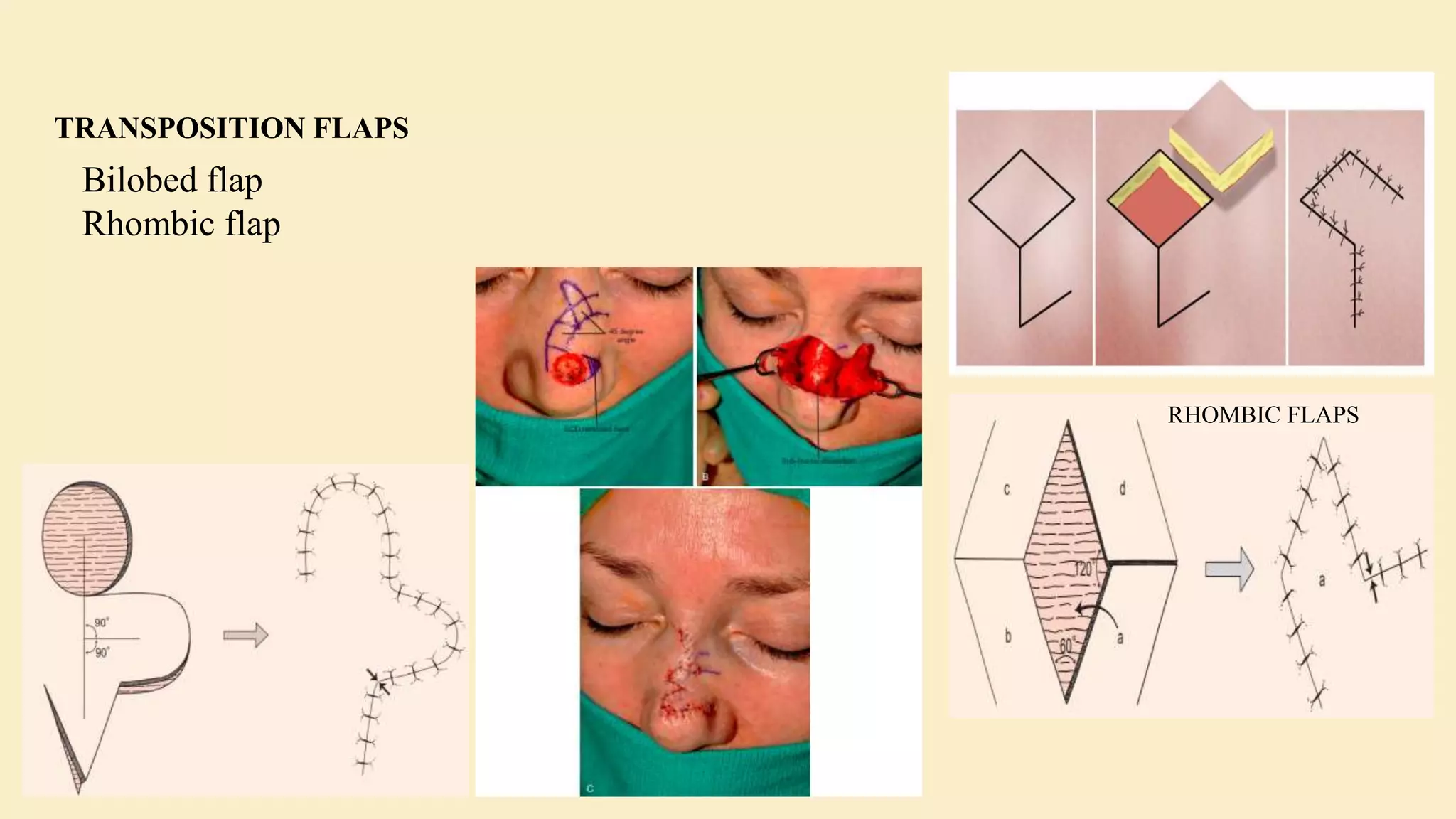
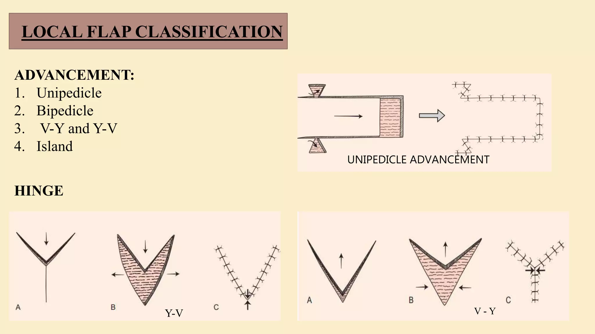
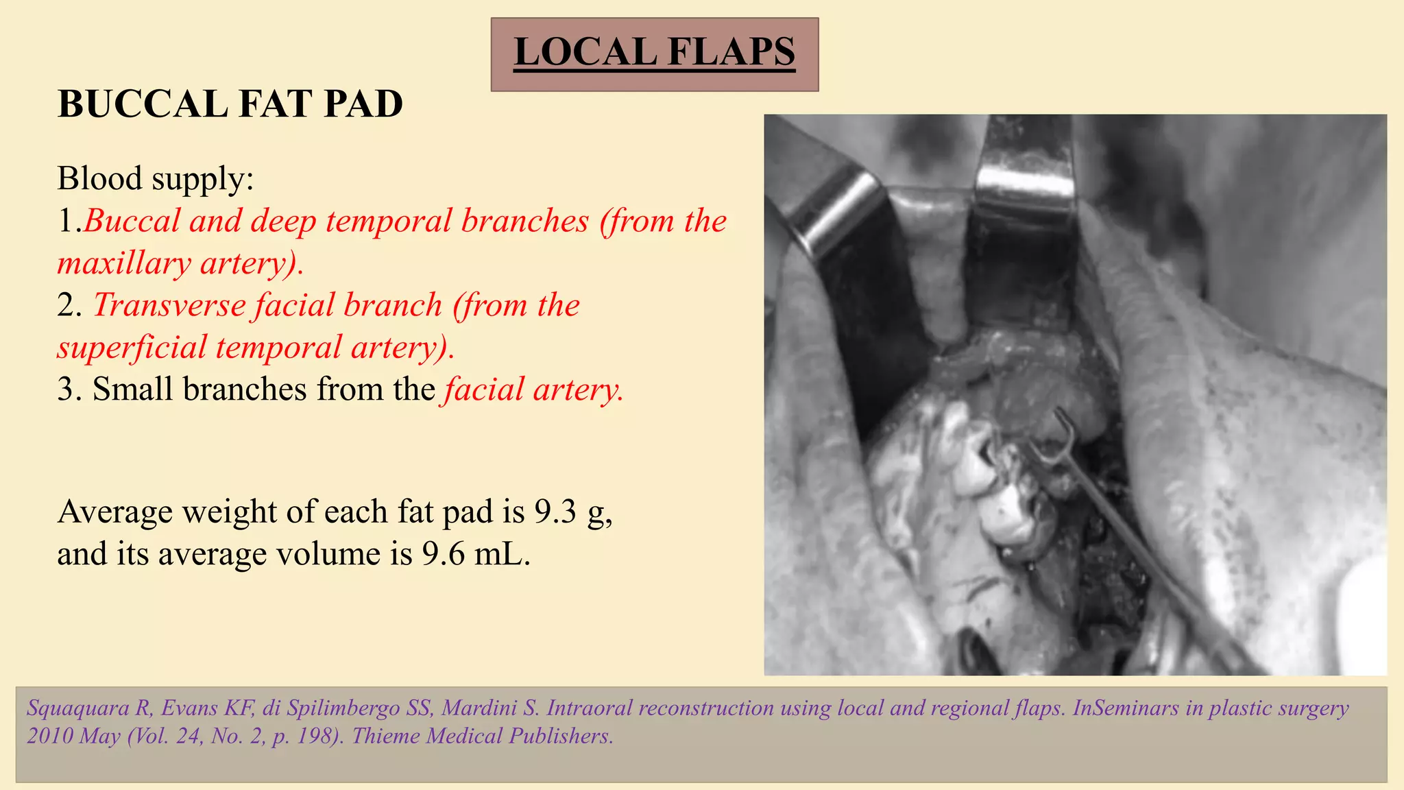
Method and type of flaps used for head and neck reconstruction. Comprising of local regional and free flaps. Indications and pitfalls in each type of flap .
Call Girl Number in Vashi Mumbai📲 9833363713 💞 Full Night Enjoy
FLAPS IN ORAL AND MAXILLOFACIAL SURGERY (monday ppt).pptx
1. FLAPS IN ORAL AND
MAXILLOFACIAL SURGERY
Aastha
Moza
1st Yr. PG
2. OVERVIEW
• BASIC PRINCIPLES
• PHYSIOLOGY OF FLAP
• BIOMECHANICS OF FLAP
• FLAP NOMENCLATURE AND DESIGN
• CLASSIFICATION
• TYPES OF LOCAL FLAPS
• TYPES OF REGIONAL FLAPS
• TYPES OF FREE FLAPS
• MONITORING OF FLAPS
• NOTE ON SKIN GRAFTS
3. BASIC TERMS
• FLAP
• PERFORATORS
• PRIMARY DEFORMITY
• SECONDARY
CORRECTIVE
DEFORMITY
PRINCIPLES OF FLAPS
1. The three-dimensional defect should be carefully assessed.
2. The contralateral side, if normal, should be used as a
model.
3. Donor scars should be hidden or placed in relaxed skin
tension lines – ( local flaps)
4. Donor defects should be minimized and adjacent tissue
should not be distorted.
5. Tissue defects should be replaced with donor material that
is similar in kind.
6. Entire facial aesthetic units should be replaced when
possible
Sykes JM, Murakami CS. Principles of local flaps in head and neck reconstruction. Operative Techniques in Otolaryngology-Head and Neck
Surgery. 1993 Mar 1;4(1):2-10.
6. RECONSTRUCTIVE LADDER / ELEVATOR
Reconstructive surgery is the process of restoring the human body to “whole” following
tumour extirpation, infection, trauma, or congenital or acquired deformity – restoring both
form and function.
Janis JE, Kwon RK, Attinger CE. The new reconstructive ladder: modifications to the traditional
model. Plastic and reconstructive surgery. 2011 Jan 1;127:205S-12S.
7. CLASSIFICATION OF SKIN FLAPS
BASED ON VASCULAR SUPPLY
• AXIAL PATTERN FLAP
• RANDOM PATTERN FLAP
CLASSIFICATION OF FLAP
BASED ON LOCATION:
• LOCAL FLAPS
• REGIONAL FLAPS
• DISTANT / FREE FLAPS
8. CORMACK AND LAMBERTY’S CLASSIFICATION OF
FASCIOCUTANEOUS FLAPS BASED ON VASCULAR ANATOMY
Type A is supplied by multiple fasciocutaneous perforators that enter at the base of the flap
and extend throughout its longitudinal length.
Type B has a single fasciocutaneous perforator, which is of moderate size and is fairly
consistent. This flap may be isolated as an island flap or used as a free flap.
Type C is based on multiple small perforators that run along a fascial septum. The supplying
artery is included with the flap.
Type D is an osteomyocutaneous flap, similar to Type C but including a portion of adjacent
muscle and bone. It may be based proximally or distally on a pedicle or used as a free flap.
9. •TypeA: Direct cutaneous
•TypeB: Direct septocutaneous
•TypeC: Direct cutaneous branch of
a muscular vessel
•TypeD: Perforating cutaneous
branch of a muscular vessel
•TypeE: Septocutaneous perforator
•TypeF: Musculocutaneous
perforator
NAKAJIMA CLASSIFICATION
OF PERFORATING VESSELS
TO FASCIOCUTANEOUS
FLAPS
10. MATHES AND NAHAI’S TRIPARTITE SYSTEM OF FASCIOCUTANEOUS
FLAPS BASED ON THE THREE MAJOR TYPES OF DEEP FASCIAL
PERFORATORS.
TYPE A:Vascular pedicle
travels deep to the fascia for
a variable distance then
pierces the fascia to supply
the skin.
TYPE B: Vascular pedicle
that courses within an
intermuscular septum
TYPE C: Vascular pedicle
that is traveling within the
muscle substance
11. MATHES AND NAHAI CLASSIFICATION FOR MUSCLE FLAPS, ACCORDING TO
THE PREDOMINANT VASCULAR PATTERN FOR THAT TYPE MUSCLE
D, dominant pedicle; M, minor; SS, secondary segmental; S, segmental.
13. (A)Type I: single unbranched
nerve entering muscle
(B)Type II: single nerve that
branches just before
entering muscle.
(C) Type III: multiple
branches from same nerve
trunk.
(D) Type IV: multiple
branches from different nerve
TAYLOR CLASSIFICATION BASED ON THEIR
MODE OF MOTOR INNERVATION
17. BUCCAL FAT PAD
LOCAL FLAPS
Blood supply:
1.Buccal and deep temporal branches (from the
maxillary artery).
2. Transverse facial branch (from the
superficial temporal artery).
3. Small branches from the facial artery.
Average weight of each fat pad is 9.3 g,
and its average volume is 9.6 mL.
Squaquara R, Evans KF, di Spilimbergo SS, Mardini S. Intraoral reconstruction using local and regional flaps. InSeminars in plastic surgery
2010 May (Vol. 24, No. 2, p. 198). Thieme Medical Publishers.
18. LOCAL FLAPS
FACIAL ARTERY MUSCULOMUCOSAL FLAP
1992, Pribaz described an axial musculomucosal flap based on the facial artery.
FAMM flap can be based superiorly (retrograde flow) or inferiorly (anterograde flow). Its average size
is 2.5 7 cm.
Squaquara R, Evans KF, di Spilimbergo SS, Mardini S. Intraoral reconstruction using local and regional flaps. InSeminars in plastic surgery
2010 May (Vol. 24, No. 2, p. 198). Thieme Medical Publishers.
20. PARAMEDIAN FOREHEAD FLAP
Blair in 1925 and Kazanjian in 1946 described the median forehead flap for nasal repair. The
main use of the paramedian forehead flap has been in the reconstruction of partial to total nasal
defects.
Vascular supply to the forehead comes from the paired supraorbital arteries, which divide into
a superficial and a deep branch, and the paired supratrochlear arteries
FOREHEAD FLAP
21. THE TEMPOROPARIETAL FASCIA FLAP
The temporoparietal fascia lies
immediately deep to the skin and
subcutaneous tissue of the scalp
overlying the temporal fossa.
It is continuous with the
superficial musculoaponeurotic
system inferior to the zygomatic
arch and with the galea
aponeurotica above the superior
temporal line.
The dominant blood supply is the
superficial temporal artery, a
terminal branch of the external
carotid artery
22. TEMPORALIS FLAP
The advantage of the temporalis muscle flap
is the ease of access to the muscle, the
moderate quantity of muscle that can be
harvested, and the ability to transfer the
muscle to the oral cavity.
It is a fan-shaped muscle that originates along
the temporal lines of the parietal bone of the
skull.
The muscle lies within the temporal fossa, is
covered by a strong fibrous aponuerotic
sheath, the temporalis fascia, and passes
medial to the zygomatic arch.
main vascular supply from the anterior and
deep temporal arteries, branches from the
second portion of the internal maxillary artery.
23. TONGUE FLAP
• Tongue flaps were introduced for intraoral
reconstruction by Lexer in 1909.
• Lingual artery is the main artery supplying the tongue.
24. PALATAL FLAP
• Palatal island flap remains popular
in reconstructing intraoral defects.
• The palatal flap was initially
described in 1922 by Victor Veau.
• “trilaminar macronet” results in a
dense network, the entire hard
palate mucosa can be elevated on a
single neurovascular pedicle.
Modifications:
• Random rotation-advancement flap
• Submucosal modification
• Palatal flap for skull-based defects
25. REGIONAL FLAPS
PECTORALIS MAJOR MYOCUTANEOUS FLAP
Blood supply : Thoracoacromial artery
originating from Subclavian artery at the
mid – clavicular artery.
MERIT: Quality and quantity of tissue that can be
harvested, muscle coverage in the neck provides
additional protection of the great vessels.
DEMERIT: 1.Rotation of the flap
2.Significant amount of hair growth on the skin in
males.
3.Tunneling of the flap in the neck to reach the defect
creates a bulge over the clavicle and in the neck
26. SUBMENTAL ISLAND FLAP
Chow TL, Kwan WW, Fung SC, Ho LI, Au KL. Reconstruction with submental flap for
aggressive orofacial cancer-an updated series. American journal of otolaryngology.
2018 Nov 1;39(6):693-7.
28. CERVICAL FLAP
• Random blood supply
• Width:length ratio is 1:3
• Flap may be superiorly / posteriorly based and
oriented vertically or transversely.
• 2 types : anterior cervical flap and mutter/
posterior cervical flap
• Blood supply: Occipital artery and Posterior
auricular artery
29. DELTOPECTORAL FLAP
TRAPEZIUS AND
STERNOCLEIDOMASTOID FLAP
Given by Bakamjian in 1960s
Blood supply: Internal Mammary artery through
2nd/ 3rd/4th intercostal spaces.
Workhouse for reconstruction of oropharyngeal
and phayryngoesophageal defects.
Proximal part: Axial blood supply
Distal part: Random relying on subdermal plexus
Owens described the sternocleidomastoid (SCM) flap in
1955 utilizing the entire length of skin overlying the
muscle.
The trapezius muscle is a triangular muscle that covers the
back of the neck and shoulder region and extends
inferiorly in the back.
Blood supply from four sources: transverse cervical artery,
the dorsal scapular artery, the intercostal perforators lying
just off the midline, and the branches from the occipital
30. MICROVASCULAR AND PEDICLE FLAP
FIBULA FLAP
• 1st Microvascular transfer of fibula free
flap by Fujikawa in 1973.
• Blood supply is based on Peroneal
artery.
• Acceptable bone length used for flap is
25 cm.
Hidalgo DA. Fibula free flap mandibular reconstruction. Clinics in
plastic surgery. 1994 Jan 1;21(1):25-35.
31. • Scapular and parascapular
fasiocutaneous free flap is based on
circumflex scapular artery.
• Indicated when massive soft tissue
SCAPULAR FLAP
ANTEROLATERAL THIGH FLAP
• Described by chen and song in
in 1984.
• Fasiocutaneous flap based on
septocutaneous perforators of
descending branch of lateral
circumflex femoral artery –
Profunda femoris artery.
Steel BJ, Cope MR. A brief
history of vascularized free flaps
in the oral and maxillofacial
region. Journal of Oral and
Maxillofacial Surgery. 2015 Apr
1;73(4):786-e1.
Kells AF, Broyles JM, Simoa AF, Lewis VO, Sacks JM. Anterolateral thigh flow-through flap in hand
salvage. Eplasty. 2013;13.
32. ILIAC CREST FLAP/ DEEP CIRCUMFLEX ILIAC ARTERY (DCIA)
• First reported as a bony flap in 1979 by taylor.
• Pitfall : Difficulty in harvest , thin and immobile skin paddle.
• Blood supply is by the Deep circumflex iliac artery.
33. RADIAL FOREARM FREE FLAP
• Radial artery forearm
fasciocutaneous flap developed by
Drs Guofan and yuzhi.
• Radial artery and its vena
comitans will be anastomosed to
facial artery.
• Pitfall : Length of bone available is
limited and only ½ of the
circumference of cortex of bone
can be harvested as flap.
34. RECTUS ABDOMINIS FLAP
• Microvascular pedicles flap with superior and deep inferior epigastric arterial
system with perforating branches traversing through rectus abdominis muscle.
• Provides both external skin and mucosal coverage.
• Mainly used for scalp reconstruction .
35. FLAP MONITORING
• CAPILLARY REFILL
• COLOR
• TEMPERATURE
• TURGOR
• ACOUSTIC DOPPLER SONOGRAPHY
• COLOR DUPLEX ULTRASONOGRAPHY
• FLOW COUPLER
• IMPLANTABLE DOPPLER
• LASER DOPPLER FLOWMETRY
Chao AH, Lamp S. Current
approaches to free flap
monitoring. Plastic Surgical
Nursing. 2014 Apr 1;34(2):52-6.
Poder TG, Fortier PH. Implantable
Doppler in monitoring free flaps: a cost-
effectiveness analysis based on a systematic
review of the literature. European annals of
otorhinolaryngology, head and neck
diseases. 2013 Apr 1;130(2):79-85.
36. FLAP MONITORING
Choudhary AK, Singh AI, Das SI, Singh LO, Singh NS.
Role of flap blood glucose measurement in monitoring of
flap incorporating skin and to detect flap congestion and
flap salvage. Journal of Medical Society. 2020 May
1;34(2):106.
Pafitanis G, Chen HC. The Pinprick Test: Key Considerations in Execution
of Skin Flap Perfusion Testing. Plastic and Reconstructive Surgery Global
Open. 2019 Sep;7(9).
Medtronic glucometer
37. A skin graft is skin that has been
completely detached from its original
donor site and transferred to another site,
where it will develop a new blood supply.
The first recorded successful skin graft
was by Sir Astley Cooper in 1817.
According to
origin:
• Autografts
• Isografts
• Homografts
• Xenografts
According to thickness:
A Line of section of the thin split-thickness (Thiersch) graft.
B Line of section of the split-thickness graft.
C Line of section of the thick split-thickness or three-quarter thickness
graft.
D Line of section of the full-thickness graft.
SKIN GRAFTS
38. BIBLIOGRAPHY
Local flaps in facial reconstruction - Shan R. Baker, 3rd edition
Flaps and reconstructive surgery - Samir Mardini, 2nd Edition
Local and Regional Flaps in Head & Neck Reconstruction A Practical Approach - Rui Fernandes
Mittal G, Agarwal A, Kataria G. Flaps for Oral and Maxillofacial Reconstruction: Review of Literature and a
Clinical Guide to the Clinicians. Asian Journal of Oncology. 2018 Jul;4(02):037-42.
Chow TL, Kwan WW, Fung SC, Ho LI, Au KL. Reconstruction with submental flap for aggressive orofacial
cancer-an updated series. American journal of otolaryngology. 2018 Nov 1;39(6):693-7.
Hidalgo DA. Fibula free flap mandibular reconstruction. Clinics in plastic surgery. 1994 Jan 1;21(1):25-35.
Basic Flap Design Todd A. Schultz.
Steel BJ, Cope MR. A brief history of vascularized free flaps in the oral and maxillofacial region. Journal of
Oral and Maxillofacial Surgery. 2015 Apr 1;73(4):786-e1.