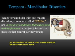
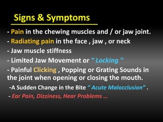
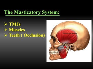
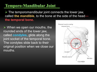
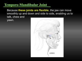
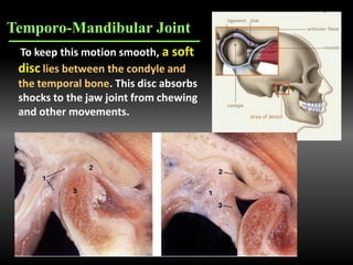
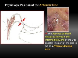
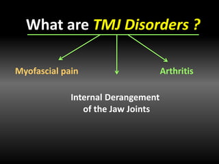
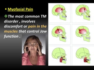
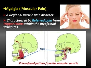
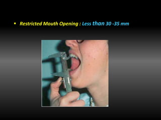
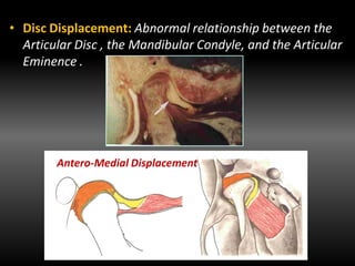
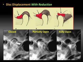
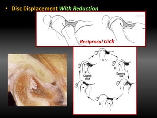
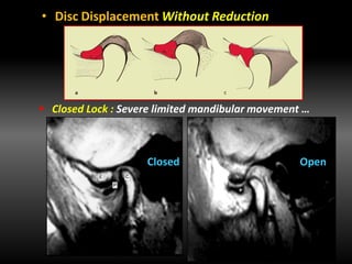
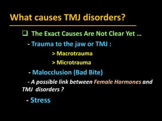
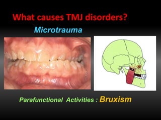
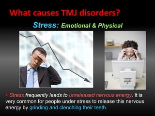
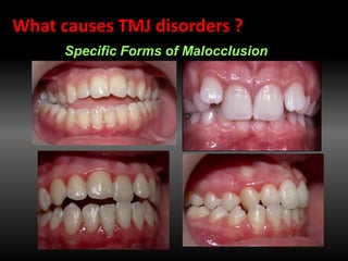
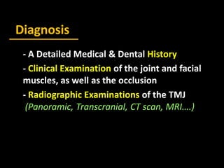
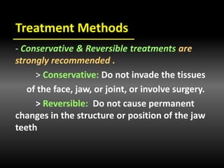
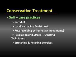
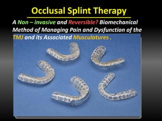
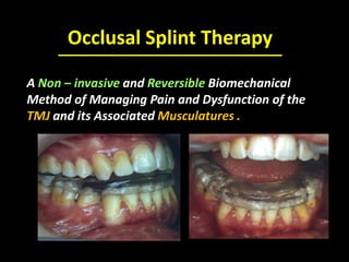
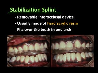
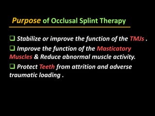
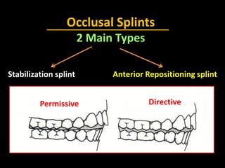
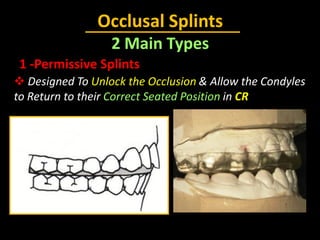
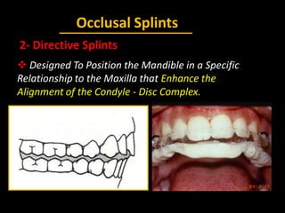
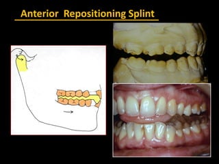
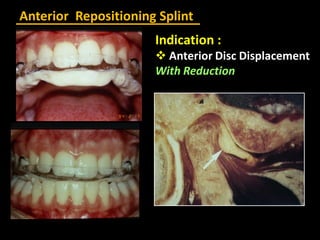
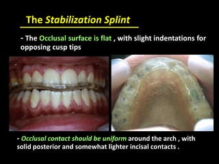
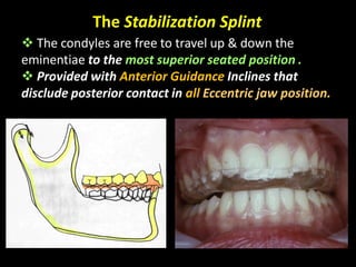
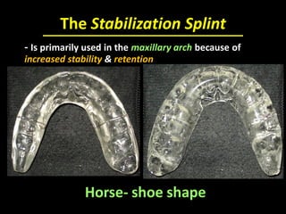
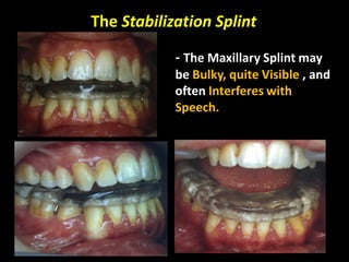
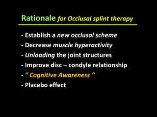
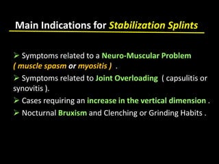
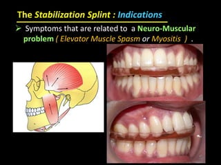
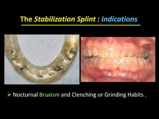
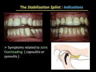
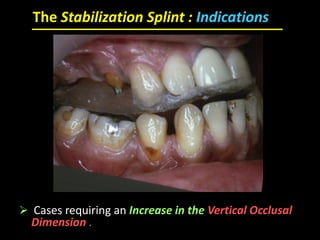
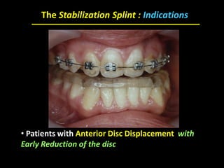
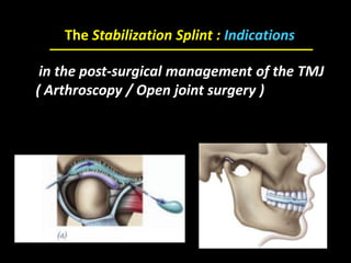
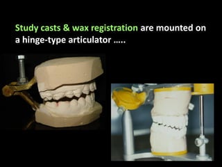
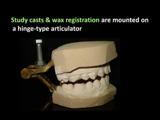
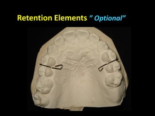
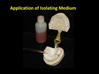
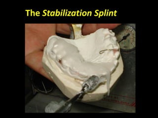
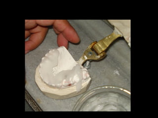
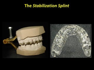
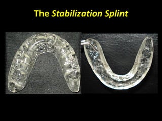
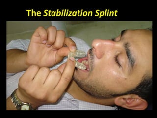
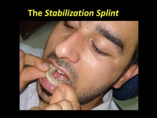
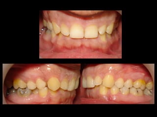
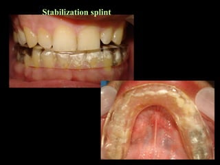
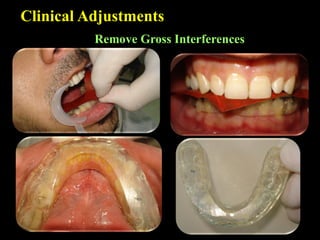
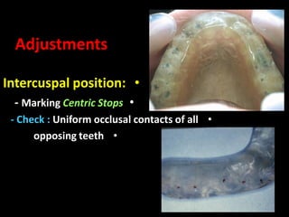
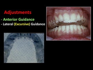
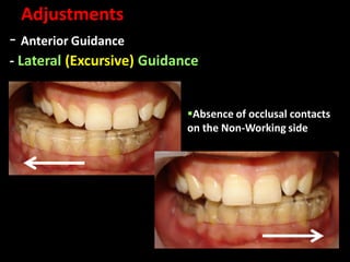
Occlusal Splint Therapy
Dr. Marwan Mouakeh , DDS, Doc.Orhto.Sc
Head of Aleppo Orthodontic Residency Center
Director of the National Committee for
Qualification in Orthodontics
Ministry of Health - Syria
Temporo - Mandibular Disorders
Temporomandibular joint and muscle
disorders, commonly called “TMDs,”
are a group of conditions that cause pain
and dysfunction in the jaw joint and the
muscles that control jaw movement.
U.S. DEPARTMENT OF HEALTH AND HUMAN SERVICES
National Institutes of Health
Temporo - Mandibular Disorders
We don’t know for certain how many people
have TMJ disorders, but some estimates
suggest that over 10 million Americans are
affected. The condition appears to be more
common in women than men .
U.S. DEPARTMENT OF HEALTH AND HUMAN SERVICES
National Institutes of Health
Signs & Symptoms
- Pain in the chewing muscles and / or jaw joint.
- Radiating pain in the face , jaw , or neck
- Jaw muscle stiffness
”- Limited Jaw Movement or “ Locking
- Painful Clicking , Popping or Grating Sounds in
the joint when opening or closing the mouth.
-A Sudden Change in the Bite “ Acute Malocclusion” .
- Ear Pain, Dizziness, Hear Problems …
The Masticatory System:
 TMJs
 Muscles
 Teeth ( Occlusion)
Temporo-Mandibular Joint
 The temporomandibular joint connects the lower jaw,
called the mandible, to the bone at the side of the head—
the temporal bone.
 When we open our mouths, the
rounded ends of the lower jaw,
called condyles, glide along the
joint socket of the temporal bone.
The condyles slide back to their
original position when we close our
mouths.
Temporo-Mandibular Joint
Because these joints are flexible, the jaw can move
smoothly up and down and side to side, enabling us to
talk, chew and
yawn.
To keep this motion smooth, a soft
disc lies between the condyle and
the temporal bone. This disc absorbs
shocks to the jaw joint from chewing
and other movements.
Temporo-Mandibular Joint
Sagittal View
The Articular Disc
A Biconcave Oval Structure Dividing the joint
cavity into 2 distinct compartments.
Parts of the Articular Disc
Anterior Band
Intermediate Zone
Posterior Band
Sup. Attachment
Elastic
Inf. Attachment
Non-Elastic
Physiologic Position of the Articular Disc
1
2
3
 The Absence of Blood
Vessels & Nerves in the
Intermediate Zone of the Disc
Enables this part of the disc to
act as a Pressure-Bearing
Area .
Collateral Discal Ligaments
Frontal View
The Articular Disc
Medial
Distal
The temporomandibular joint is different from the
body’s other joints. The combination of hinge and
sliding motions makes this joint among the most
complicated in the body. Also, the tissues that make up
the temporomandibular joint differ from other load-
bearing joints, like the knee or hip. Because of its
complex movement and unique makeup, the jaw joint
and its controlling muscles can pose a tremendous
challenge to both patients and health care providers
when problems arise.
Masticatory Muscles
 The Primary Movers of the Mandible
Lateral Pterygoid Muscle
Inf. Belly : Depressor
Sup. Belly : Elevator
What are TMJ Disorders ?
Myofascial pain Arthritis
Internal Derangement
of the Jaw Joints
 Myofasial Pain
The most common TM
disorder , involves
discomfort or pain in the
muscles that control Jaw
function .
Myalgia ( Muscular Pain)
 A Regional muscle pain disorder
 Characterized by Referred pain from
Trigger Points within the myofascial
structures
Pain referral pattern from the masseter muscle
TrpS
Myalgia ( Muscular Pain)
 Dull , Deep , and Diffuse pain
 Depressing
 Felt in the morning when related
to Nocturnal Bruxism
 Influenced by functional demands
( chewing…)
Masticatory Muscles Palpation
Lateral Pterygoid Palpation
 Restricted Mouth Opening : Less than 30 -35 mm
 TMJ Internal Derangement
 Involves a Displaced Disc ,
Dislocated Jaw , or Injury to the
Condyle.
• Disc Displacement: Abnormal relationship between the
Articular Disc , the Mandibular Condyle, and the Articular
Eminence .
Antero-Medial Displacement
• Disc Displacement With Reduction
Closed Partially Open Fully Open
• Disc Displacement With Reduction
Reciprocal Click
• Disc Displacement Without Reduction
Closed Lock : Severe limited mandibular movement …
Closed Open
• Disc Displacement Without Reduction
 Closed Lock : Severe limited mandibular movement …
Closed Open
Arthralgia (Articular Pain)
 Loclized in the TMJ Region
 Increased with mandibular
movement.
Origin of pain :
posterior attachment - collateral
ligaments -articular capsule.
 Arthritis
Refers to a Group of Degenerative Inflammatory Joint
Disorders that can affect the Temporomandibular Joint.
Crepitation
Mean Value
%
Symptoms
19TMJ Sounds
11Tiredness,Stiffness of jaw
6Pain on Mandibular function
8Limitation of Mandibular
movement
4Locking
17Frequent Headache
Reported TMD Symptoms in 18 Epidemiologic Studies
“ Carlsson 1984”
Age distribution of 5 samples of patients
with TMDS
A common Peak in the
age distribution of the
patients ,specifically
during the period
between 20 and 40
years .
What causes TMJ disorders?
 The Exact Causes Are Not Clear Yet …
- Trauma to the jaw or TMJ :
> Macrotrauma
> Microtrauma
- Malocclusion (Bad Bite)
- A possible link between Female Hormones and
TMJ disorders ?
- Stress
Acute Trauma to the Neck : Whiplash
What causes TMJ disorders ?
Macrotrauma: Blow, Traumatic extraction ,
Intubation…
Parafunctional Activities : Bruxism
What causes TMJ disorders?
Microtrauma
What causes TMJ disorders?
Stress: Emotional & Physical
 Stress frequently leads to unreleased nervous energy. It is
very common for people under stress to release this nervous
energy by grinding and clenching their teeth.
Specific Forms of Malocclusion
What causes TMJ disorders ?
Balance( in the Masticatory System ) between Destructive
Overloading that leads to CMDs & Homeostatic Adaptability
that promotes normal function .
Diagnosis
- A Detailed Medical & Dental History
- Clinical Examination of the joint and facial
muscles, as well as the occlusion
- Radiographic Examinations of the TMJ
(Panoramic, Transcranial, CT scan, MRI….)
Evolution
How joint and muscles disorders
progress is not clear . Symptoms
worsen and ease over time, but what
causes these changes is not known.
Treatment Methods
- Conservative & Reversible treatments are
strongly recommended .
> Conservative: Do not invade the tissues
of the face, jaw, or joint, or involve surgery.
> Reversible: Do not cause permanent
changes in the structure or position of the jaw
teeth
Conservative Treatment
- Self – care practices
> Soft diet
Moist heat/> Local ice packs
> Rest (avoiding extreme jaw movements)
> Relaxation and Stress – Reducing
Techniques.
> Stretching & Relaxing Exercises.
Conservative Treatment
- Pain Medications :
> Nonsteroidal anti- inflammatory
drugs (NSAIDS) : Ibuprofen
> Corticosteroids Injection??
> Muscle Relaxants
> Anti- Depressants
 Anti- Anxiety Drugs
Conservative Treatment
- Pain Medications :
> Nonsteroidal anti- inflammatory
drugs (NSAIDS) : Ibuprofen
> Anti- Inflammatory Drugs
> Muscle Relaxants
> Anti- depressants
Conservative Treatment
Stabilization Joint Splints
or
Interocclusal Appliances
Irreversible Treatments
- Orthodontics
- Occlusal Adjustment
- Crown & Bridge Works
- Repositioning Splints
-Surgery
Occlusal Splint Therapy
A Non – invasive and Reversible? Biomechanical
Method of Managing Pain and Dysfunction of the
TMJ and its Associated Musculatures .
Conservative Treatment
- Stabilization Splints or
interocclusal Appliances
Irreversible Treatments
- Orthodontics
- Occlusal Adjustment
- Crown & Bridge Works
- Repositioning Splints
-Surgery
Occlusal Splint Therapy
A Non – invasive and Reversible Biomechanical
Method of Managing Pain and Dysfunction of the
TMJ and its Associated Musculatures .
Stabilization Splint
- Removable interocclusal device
- Usually made of hard acrylic resin
- Fits over the teeth in one arch
Purpose of Occlusal Splint Therapy
 Stabilize or improve the function of the TMJs .
 Improve the function of the Masticatory
Muscles & Reduce abnormal muscle activity.
 Protect Teeth from attrition and adverse
traumatic loading .
Occlusal Splints
2 Main Types
Stabilization splint Anterior Repositioning splint
Permissive Directive
Occlusal Splints
2 Main Types
1 -Permissive Splints
 Designed To Unlock the Occlusion & Allow the Condyles
to Return to their Correct Seated Position in CR
Posterior Bite Plane
Full-Coverage” Mandibular”Full-Coverage” Maxillary”
Types of Permissive Occlusal Splints
Anterior Bite Plane
Anterior Maxillary Bite Plane Posterior Maxillary Bite Plane
Anterior Mandibular Bite Plane Posterior Mandibular Bite Plane
Anterior Maxillary Bite Plane Posterior Maxillary Bite Plane
Anterior Mandibular Bite Plane
Occlusal Splints
2- Directive Splints
 Designed To Position the Mandible in a Specific
Relationship to the Maxilla that Enhance the
Alignment of the Condyle - Disc Complex.
Anterior Repositioning Splint
Indication :
 Anterior Disc Displacement
With Reduction
The Stabilization Splint
- Muscle Relaxation S.
- Centric Relation S.
-Michigan S.
- Bruxism Appliance
 Synonyms
 The most commonly used appliance , which is a
hard acrylic splint that provides a temporary & ideal
occlusion .
The Stabilization Splint
- Covers the entire dental arch
- Occludes with all opposing teeth
The Stabilization Splint
- The Occlusal surface is flat , with slight indentations for
opposing cusp tips
- Occlusal contact should be uniform around the arch , with
solid posterior and somewhat lighter incisal contacts .
The Stabilization Splint
 The condyles are free to travel up & down the
eminentiae to the most superior seated position .
 Provided with Anterior Guidance Inclines that
disclude posterior contact in all Eccentric jaw position.
The Stabilization Splint
Horse- shoe shape
- Is primarily used in the maxillary arch because of
increased stability & retention
The Stabilization Splint
- Another advantage of the
maxillary splint is that all of
the opposing mandibular
incisors can easily contact the
surface of the appliance, even
when an excessive overjet is
present .
The Stabilization Splint
- The Maxillary Splint may
be Bulky, quite Visible , and
often Interferes with
Speech.
Rationale for Occlusal splint therapy
- Establish a new occlusal scheme
- Decrease muscle hyperactivity
- Unloading the joint structures
- Improve disc – condyle relationship
- “ Cognitive Awareness “
- Placebo effect
Main Indications for Stabilization Splints
 Symptoms related to a Neuro-Muscular Problem
( muscle spasm or myositis ) .
 Symptoms related to Joint Overloading ( capsulitis or
synovitis ).
 Cases requiring an increase in the vertical dimension .
 Nocturnal Bruxism and Clenching or Grinding Habits .
The Stabilization Splint : Indications
 Symptoms that are related to a Neuro-Muscular
problem ( Elevator Muscle Spasm or Myositis ) .
 Nocturnal Bruxism and Clenching or Grinding Habits .
The Stabilization Splint : Indications
 Symptoms related to Joint
Overloading ( capsulitis or
synovitis ).
The Stabilization Splint : Indications
 Cases requiring an Increase in the Vertical Occlusal
Dimension .
• Patients with Anterior Disc Displacement with
Early Reduction of the disc
The Stabilization Splint : Indications
in the post-surgical management of the TMJ
( Arthroscopy / Open joint surgery )
The Stabilization Splint : Indications
The Stabilization Splint
Clinical & Laboratory Procedures
Impressions & Dental Models
Bite Registration
Dawson’s Method
Habitual Arc of Closure
Bite Registration
Wax Bite: 2 mm thickness between posterior
teeth Bite opening = 4 -6 mm
Wax Bite
Bite Registration
Bite Registration
Wax Bite
Casts preparation
Study casts & wax registration are mounted on
a hinge-type articulator …..
Study casts & wax registration are mounted on
a hinge-type articulator
Retention Elements ” Optional”
Application of Isolating Medium
The Stabilization Splint
Stabilization splint
Clinical Adjustments
Remove Gross Interferences
•Intercuspal position:
•- Marking Centric Stops
•- Check : Uniform occlusal contacts of all
•opposing teeth
Adjustments
- Anterior Guidance
- Lateral (Excursive) Guidance
Adjustments
- Anterior Guidance
- Lateral (Excursive) Guidance
Adjustments
Absence of occlusal contacts
on the Non-Working side
- Follow – up Adjustment Appointments
-Full-time Use vs Part-time Use
- Active Treatment Duration: 3 – 6 months
Postinsertion Instructions
 Primarily At Night
 Static Pain ( Muscular involvement) : Nocturnal use
only .
 Dynamic Pain ( Joint involvement) : Full-time use .
 Acute Cases : Full-time use initially ,then decreased
gradually.
 Nocturnal Bruxism : Continued Night-time use
Use of the Stabilization Splint
- When Symptoms Have Been Significantly Reduced
- Patient is Asymptomatic for a Minimum of 3 Months .
- Discontinue the splint use in a GRADUAL Manner :
 Stop Daytime Use , Then
 Stop Nighttime Use .
The Weaning Process
Instructions for wearing TMD appliances
1- Your appliance is designed to promote healing
and help muscles, which are in spasm, to relax. It is
a treatment device not meant for long – term use.
They must only be worn under the supervision and
guidance of a dentist.
2- Wear the appliance according to the following
instructions:
Day:----------
Night:--------------
Eating:----------------
3- You may experience speech difficulties
initially. Practice reading out loud in front
of a mirror with the appliance in your
mouth to improve your speaking.
4- Avoid hard foods and do not chew gum.
5- Keep the appliance clear by soaking it in
a denture cleanser.
Instructions for wearing TMD appliances
6- Avoid carrying, pushing, or lifting heavy
objects which may strain the muscles in your
face, jaws, neck, and back .
7- Never adjust your appliance.
8- Never wrap the appliance in a tissue or
napkin – it may be lost.
Remember- The More You Wear Your
Appliance, The Faster Your Recovery.
Instructions for wearing TMD appliances
Conclusion
 When Treating Patients With TMDS ,The Overriding Objective is
to TREAT THE PATIENT RATHER THAN THE DISORDER.
 So it is Usual to Utilize More Than One Method of Treatment to
Maximize Any Complementary Effects
Hippocrates
Sometimes Cure, Often
Palliate, and Always Comfort
Thanks For Your Attention
Dr. Marwan Mouakeh

TMD's and occlusal splint therapy

  • 1.
    Occlusal Splint Therapy Dr.Marwan Mouakeh , DDS, Doc.Orhto.Sc Head of Aleppo Orthodontic Residency Center Director of the National Committee for Qualification in Orthodontics Ministry of Health - Syria
  • 2.
  • 3.
    Temporo - MandibularDisorders Temporomandibular joint and muscle disorders, commonly called “TMDs,” are a group of conditions that cause pain and dysfunction in the jaw joint and the muscles that control jaw movement. U.S. DEPARTMENT OF HEALTH AND HUMAN SERVICES National Institutes of Health
  • 4.
    Temporo - MandibularDisorders We don’t know for certain how many people have TMJ disorders, but some estimates suggest that over 10 million Americans are affected. The condition appears to be more common in women than men . U.S. DEPARTMENT OF HEALTH AND HUMAN SERVICES National Institutes of Health
  • 5.
    Signs & Symptoms -Pain in the chewing muscles and / or jaw joint. - Radiating pain in the face , jaw , or neck - Jaw muscle stiffness ”- Limited Jaw Movement or “ Locking - Painful Clicking , Popping or Grating Sounds in the joint when opening or closing the mouth. -A Sudden Change in the Bite “ Acute Malocclusion” . - Ear Pain, Dizziness, Hear Problems …
  • 6.
    The Masticatory System: TMJs  Muscles  Teeth ( Occlusion)
  • 7.
    Temporo-Mandibular Joint  Thetemporomandibular joint connects the lower jaw, called the mandible, to the bone at the side of the head— the temporal bone.  When we open our mouths, the rounded ends of the lower jaw, called condyles, glide along the joint socket of the temporal bone. The condyles slide back to their original position when we close our mouths.
  • 8.
    Temporo-Mandibular Joint Because thesejoints are flexible, the jaw can move smoothly up and down and side to side, enabling us to talk, chew and yawn.
  • 9.
    To keep thismotion smooth, a soft disc lies between the condyle and the temporal bone. This disc absorbs shocks to the jaw joint from chewing and other movements. Temporo-Mandibular Joint
  • 10.
    Sagittal View The ArticularDisc A Biconcave Oval Structure Dividing the joint cavity into 2 distinct compartments.
  • 11.
    Parts of theArticular Disc Anterior Band Intermediate Zone Posterior Band Sup. Attachment Elastic Inf. Attachment Non-Elastic
  • 12.
    Physiologic Position ofthe Articular Disc 1 2 3  The Absence of Blood Vessels & Nerves in the Intermediate Zone of the Disc Enables this part of the disc to act as a Pressure-Bearing Area .
  • 13.
    Collateral Discal Ligaments FrontalView The Articular Disc Medial Distal
  • 14.
    The temporomandibular jointis different from the body’s other joints. The combination of hinge and sliding motions makes this joint among the most complicated in the body. Also, the tissues that make up the temporomandibular joint differ from other load- bearing joints, like the knee or hip. Because of its complex movement and unique makeup, the jaw joint and its controlling muscles can pose a tremendous challenge to both patients and health care providers when problems arise.
  • 15.
    Masticatory Muscles  ThePrimary Movers of the Mandible
  • 16.
    Lateral Pterygoid Muscle Inf.Belly : Depressor Sup. Belly : Elevator
  • 17.
    What are TMJDisorders ? Myofascial pain Arthritis Internal Derangement of the Jaw Joints
  • 18.
     Myofasial Pain Themost common TM disorder , involves discomfort or pain in the muscles that control Jaw function .
  • 19.
    Myalgia ( MuscularPain)  A Regional muscle pain disorder  Characterized by Referred pain from Trigger Points within the myofascial structures Pain referral pattern from the masseter muscle TrpS
  • 20.
    Myalgia ( MuscularPain)  Dull , Deep , and Diffuse pain  Depressing  Felt in the morning when related to Nocturnal Bruxism  Influenced by functional demands ( chewing…)
  • 21.
  • 22.
  • 23.
     Restricted MouthOpening : Less than 30 -35 mm
  • 24.
     TMJ InternalDerangement  Involves a Displaced Disc , Dislocated Jaw , or Injury to the Condyle.
  • 25.
    • Disc Displacement:Abnormal relationship between the Articular Disc , the Mandibular Condyle, and the Articular Eminence . Antero-Medial Displacement
  • 26.
    • Disc DisplacementWith Reduction Closed Partially Open Fully Open
  • 27.
    • Disc DisplacementWith Reduction Reciprocal Click
  • 28.
    • Disc DisplacementWithout Reduction Closed Lock : Severe limited mandibular movement … Closed Open
  • 29.
    • Disc DisplacementWithout Reduction  Closed Lock : Severe limited mandibular movement … Closed Open
  • 30.
    Arthralgia (Articular Pain) Loclized in the TMJ Region  Increased with mandibular movement. Origin of pain : posterior attachment - collateral ligaments -articular capsule.
  • 31.
     Arthritis Refers toa Group of Degenerative Inflammatory Joint Disorders that can affect the Temporomandibular Joint. Crepitation
  • 32.
    Mean Value % Symptoms 19TMJ Sounds 11Tiredness,Stiffnessof jaw 6Pain on Mandibular function 8Limitation of Mandibular movement 4Locking 17Frequent Headache Reported TMD Symptoms in 18 Epidemiologic Studies “ Carlsson 1984”
  • 33.
    Age distribution of5 samples of patients with TMDS A common Peak in the age distribution of the patients ,specifically during the period between 20 and 40 years .
  • 34.
    What causes TMJdisorders?  The Exact Causes Are Not Clear Yet … - Trauma to the jaw or TMJ : > Macrotrauma > Microtrauma - Malocclusion (Bad Bite) - A possible link between Female Hormones and TMJ disorders ? - Stress
  • 35.
    Acute Trauma tothe Neck : Whiplash What causes TMJ disorders ? Macrotrauma: Blow, Traumatic extraction , Intubation…
  • 36.
    Parafunctional Activities :Bruxism What causes TMJ disorders? Microtrauma
  • 37.
    What causes TMJdisorders? Stress: Emotional & Physical  Stress frequently leads to unreleased nervous energy. It is very common for people under stress to release this nervous energy by grinding and clenching their teeth.
  • 38.
    Specific Forms ofMalocclusion What causes TMJ disorders ?
  • 39.
    Balance( in theMasticatory System ) between Destructive Overloading that leads to CMDs & Homeostatic Adaptability that promotes normal function .
  • 40.
    Diagnosis - A DetailedMedical & Dental History - Clinical Examination of the joint and facial muscles, as well as the occlusion - Radiographic Examinations of the TMJ (Panoramic, Transcranial, CT scan, MRI….)
  • 41.
    Evolution How joint andmuscles disorders progress is not clear . Symptoms worsen and ease over time, but what causes these changes is not known.
  • 42.
    Treatment Methods - Conservative& Reversible treatments are strongly recommended . > Conservative: Do not invade the tissues of the face, jaw, or joint, or involve surgery. > Reversible: Do not cause permanent changes in the structure or position of the jaw teeth
  • 43.
    Conservative Treatment - Self– care practices > Soft diet Moist heat/> Local ice packs > Rest (avoiding extreme jaw movements) > Relaxation and Stress – Reducing Techniques. > Stretching & Relaxing Exercises.
  • 44.
    Conservative Treatment - PainMedications : > Nonsteroidal anti- inflammatory drugs (NSAIDS) : Ibuprofen > Corticosteroids Injection?? > Muscle Relaxants > Anti- Depressants  Anti- Anxiety Drugs
  • 45.
    Conservative Treatment - PainMedications : > Nonsteroidal anti- inflammatory drugs (NSAIDS) : Ibuprofen > Anti- Inflammatory Drugs > Muscle Relaxants > Anti- depressants
  • 46.
    Conservative Treatment Stabilization JointSplints or Interocclusal Appliances
  • 47.
    Irreversible Treatments - Orthodontics -Occlusal Adjustment - Crown & Bridge Works - Repositioning Splints -Surgery
  • 48.
    Occlusal Splint Therapy ANon – invasive and Reversible? Biomechanical Method of Managing Pain and Dysfunction of the TMJ and its Associated Musculatures .
  • 49.
    Conservative Treatment - StabilizationSplints or interocclusal Appliances
  • 50.
    Irreversible Treatments - Orthodontics -Occlusal Adjustment - Crown & Bridge Works - Repositioning Splints -Surgery
  • 51.
    Occlusal Splint Therapy ANon – invasive and Reversible Biomechanical Method of Managing Pain and Dysfunction of the TMJ and its Associated Musculatures .
  • 52.
    Stabilization Splint - Removableinterocclusal device - Usually made of hard acrylic resin - Fits over the teeth in one arch
  • 53.
    Purpose of OcclusalSplint Therapy  Stabilize or improve the function of the TMJs .  Improve the function of the Masticatory Muscles & Reduce abnormal muscle activity.  Protect Teeth from attrition and adverse traumatic loading .
  • 54.
    Occlusal Splints 2 MainTypes Stabilization splint Anterior Repositioning splint Permissive Directive
  • 55.
    Occlusal Splints 2 MainTypes 1 -Permissive Splints  Designed To Unlock the Occlusion & Allow the Condyles to Return to their Correct Seated Position in CR
  • 56.
    Posterior Bite Plane Full-Coverage”Mandibular”Full-Coverage” Maxillary” Types of Permissive Occlusal Splints Anterior Bite Plane
  • 57.
    Anterior Maxillary BitePlane Posterior Maxillary Bite Plane Anterior Mandibular Bite Plane Posterior Mandibular Bite Plane
  • 58.
    Anterior Maxillary BitePlane Posterior Maxillary Bite Plane Anterior Mandibular Bite Plane
  • 59.
    Occlusal Splints 2- DirectiveSplints  Designed To Position the Mandible in a Specific Relationship to the Maxilla that Enhance the Alignment of the Condyle - Disc Complex.
  • 60.
  • 61.
    Anterior Repositioning Splint Indication:  Anterior Disc Displacement With Reduction
  • 62.
    The Stabilization Splint -Muscle Relaxation S. - Centric Relation S. -Michigan S. - Bruxism Appliance  Synonyms  The most commonly used appliance , which is a hard acrylic splint that provides a temporary & ideal occlusion .
  • 63.
    The Stabilization Splint -Covers the entire dental arch - Occludes with all opposing teeth
  • 64.
    The Stabilization Splint -The Occlusal surface is flat , with slight indentations for opposing cusp tips - Occlusal contact should be uniform around the arch , with solid posterior and somewhat lighter incisal contacts .
  • 65.
    The Stabilization Splint The condyles are free to travel up & down the eminentiae to the most superior seated position .  Provided with Anterior Guidance Inclines that disclude posterior contact in all Eccentric jaw position.
  • 66.
    The Stabilization Splint Horse-shoe shape - Is primarily used in the maxillary arch because of increased stability & retention
  • 67.
    The Stabilization Splint -Another advantage of the maxillary splint is that all of the opposing mandibular incisors can easily contact the surface of the appliance, even when an excessive overjet is present .
  • 68.
    The Stabilization Splint -The Maxillary Splint may be Bulky, quite Visible , and often Interferes with Speech.
  • 69.
    Rationale for Occlusalsplint therapy - Establish a new occlusal scheme - Decrease muscle hyperactivity - Unloading the joint structures - Improve disc – condyle relationship - “ Cognitive Awareness “ - Placebo effect
  • 70.
    Main Indications forStabilization Splints  Symptoms related to a Neuro-Muscular Problem ( muscle spasm or myositis ) .  Symptoms related to Joint Overloading ( capsulitis or synovitis ).  Cases requiring an increase in the vertical dimension .  Nocturnal Bruxism and Clenching or Grinding Habits .
  • 71.
    The Stabilization Splint: Indications  Symptoms that are related to a Neuro-Muscular problem ( Elevator Muscle Spasm or Myositis ) .
  • 72.
     Nocturnal Bruxismand Clenching or Grinding Habits . The Stabilization Splint : Indications
  • 73.
     Symptoms relatedto Joint Overloading ( capsulitis or synovitis ). The Stabilization Splint : Indications
  • 74.
    The Stabilization Splint: Indications  Cases requiring an Increase in the Vertical Occlusal Dimension .
  • 75.
    • Patients withAnterior Disc Displacement with Early Reduction of the disc The Stabilization Splint : Indications
  • 76.
    in the post-surgicalmanagement of the TMJ ( Arthroscopy / Open joint surgery ) The Stabilization Splint : Indications
  • 77.
    The Stabilization Splint Clinical& Laboratory Procedures
  • 78.
  • 79.
  • 80.
    Habitual Arc ofClosure Bite Registration
  • 81.
    Wax Bite: 2mm thickness between posterior teeth Bite opening = 4 -6 mm
  • 82.
  • 83.
  • 84.
  • 85.
    Study casts &wax registration are mounted on a hinge-type articulator …..
  • 86.
    Study casts &wax registration are mounted on a hinge-type articulator
  • 87.
  • 88.
  • 89.
  • 93.
  • 94.
  • 95.
  • 96.
  • 97.
  • 98.
  • 99.
  • 101.
  • 102.
  • 103.
    •Intercuspal position: •- MarkingCentric Stops •- Check : Uniform occlusal contacts of all •opposing teeth Adjustments
  • 104.
    - Anterior Guidance -Lateral (Excursive) Guidance Adjustments
  • 105.
    - Anterior Guidance -Lateral (Excursive) Guidance Adjustments Absence of occlusal contacts on the Non-Working side
  • 106.
    - Follow –up Adjustment Appointments -Full-time Use vs Part-time Use - Active Treatment Duration: 3 – 6 months Postinsertion Instructions
  • 107.
     Primarily AtNight  Static Pain ( Muscular involvement) : Nocturnal use only .  Dynamic Pain ( Joint involvement) : Full-time use .  Acute Cases : Full-time use initially ,then decreased gradually.  Nocturnal Bruxism : Continued Night-time use Use of the Stabilization Splint
  • 108.
    - When SymptomsHave Been Significantly Reduced - Patient is Asymptomatic for a Minimum of 3 Months . - Discontinue the splint use in a GRADUAL Manner :  Stop Daytime Use , Then  Stop Nighttime Use . The Weaning Process
  • 109.
    Instructions for wearingTMD appliances 1- Your appliance is designed to promote healing and help muscles, which are in spasm, to relax. It is a treatment device not meant for long – term use. They must only be worn under the supervision and guidance of a dentist. 2- Wear the appliance according to the following instructions: Day:---------- Night:-------------- Eating:----------------
  • 110.
    3- You mayexperience speech difficulties initially. Practice reading out loud in front of a mirror with the appliance in your mouth to improve your speaking. 4- Avoid hard foods and do not chew gum. 5- Keep the appliance clear by soaking it in a denture cleanser. Instructions for wearing TMD appliances
  • 111.
    6- Avoid carrying,pushing, or lifting heavy objects which may strain the muscles in your face, jaws, neck, and back . 7- Never adjust your appliance. 8- Never wrap the appliance in a tissue or napkin – it may be lost. Remember- The More You Wear Your Appliance, The Faster Your Recovery. Instructions for wearing TMD appliances
  • 112.
    Conclusion  When TreatingPatients With TMDS ,The Overriding Objective is to TREAT THE PATIENT RATHER THAN THE DISORDER.  So it is Usual to Utilize More Than One Method of Treatment to Maximize Any Complementary Effects
  • 113.
  • 114.
    Thanks For YourAttention Dr. Marwan Mouakeh