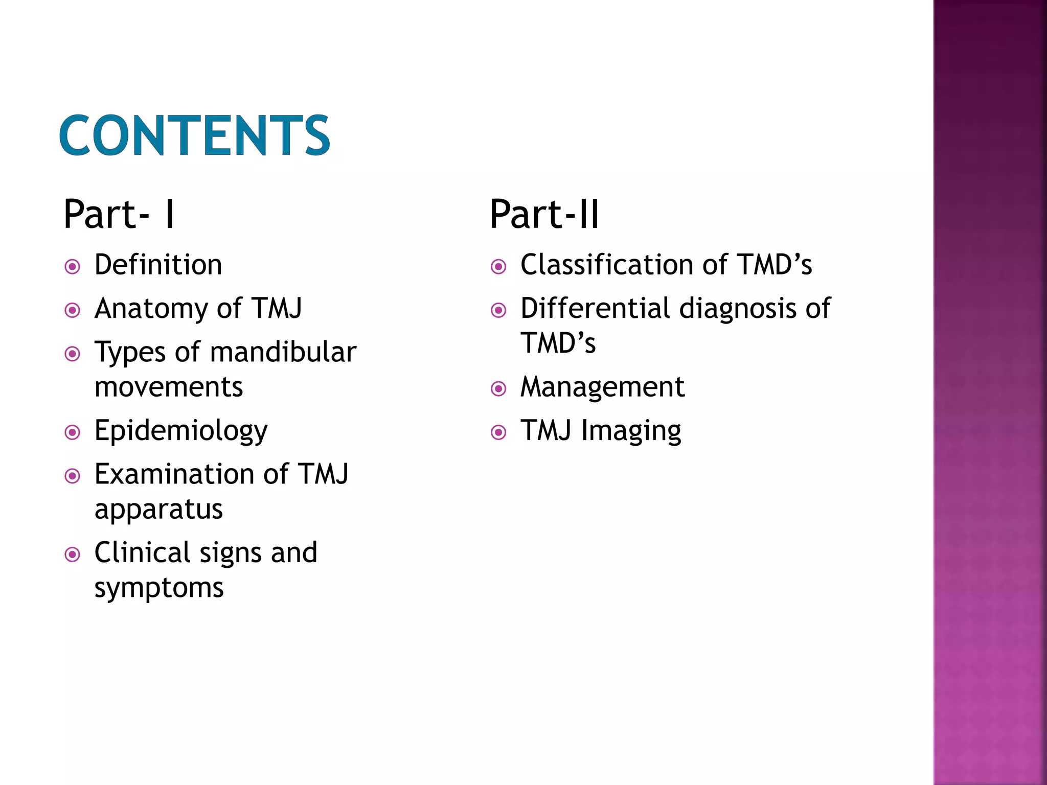
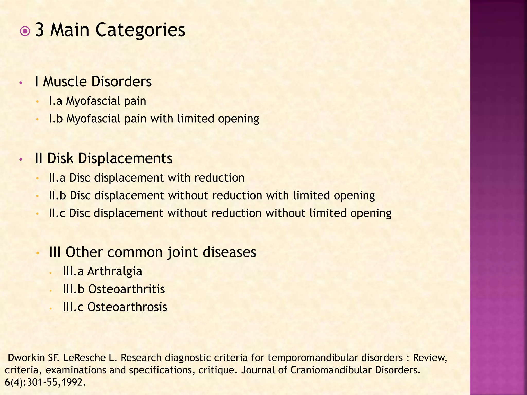
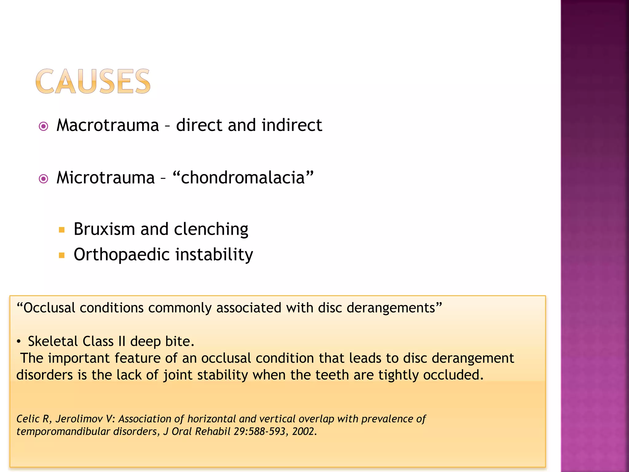
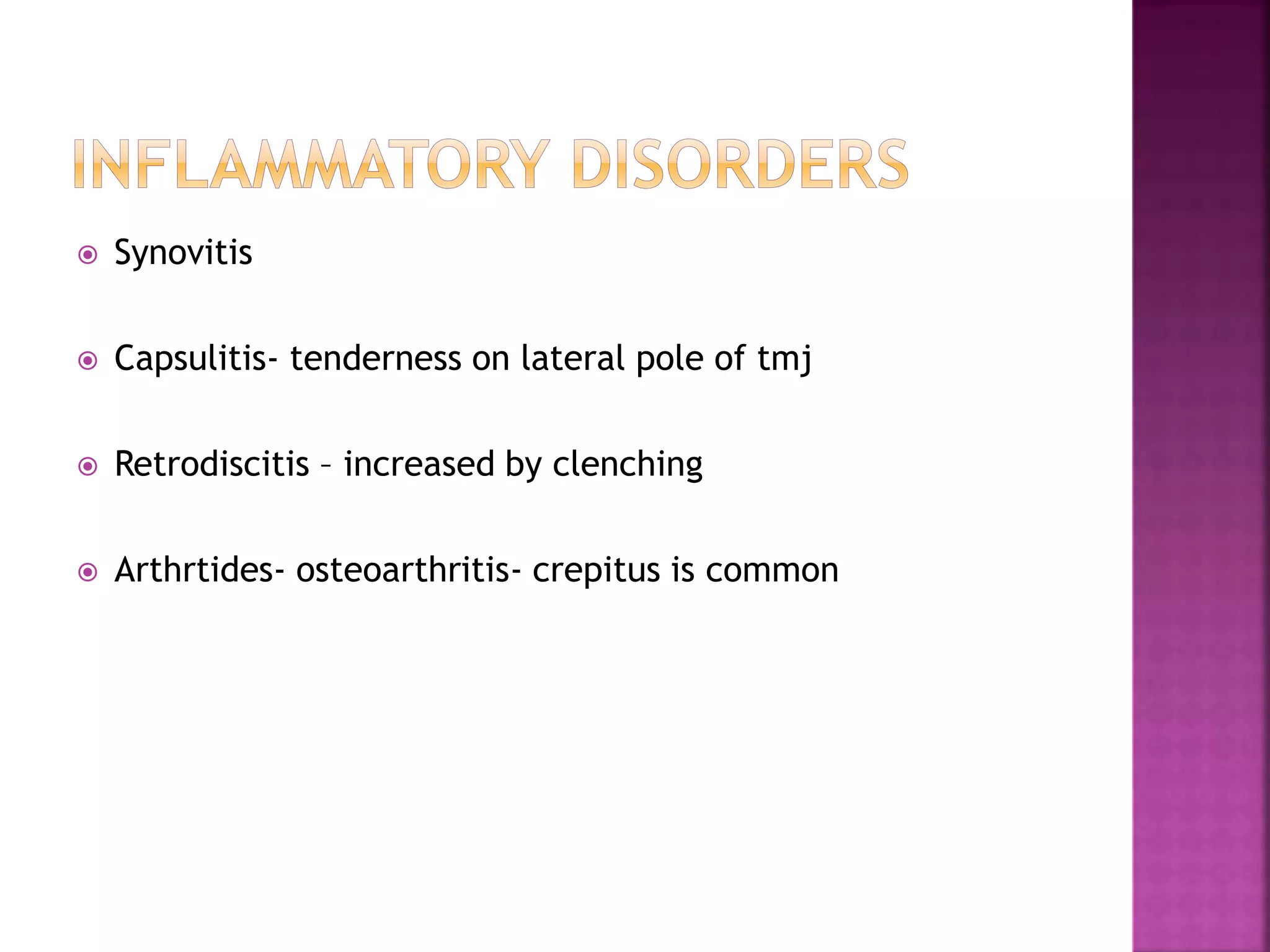
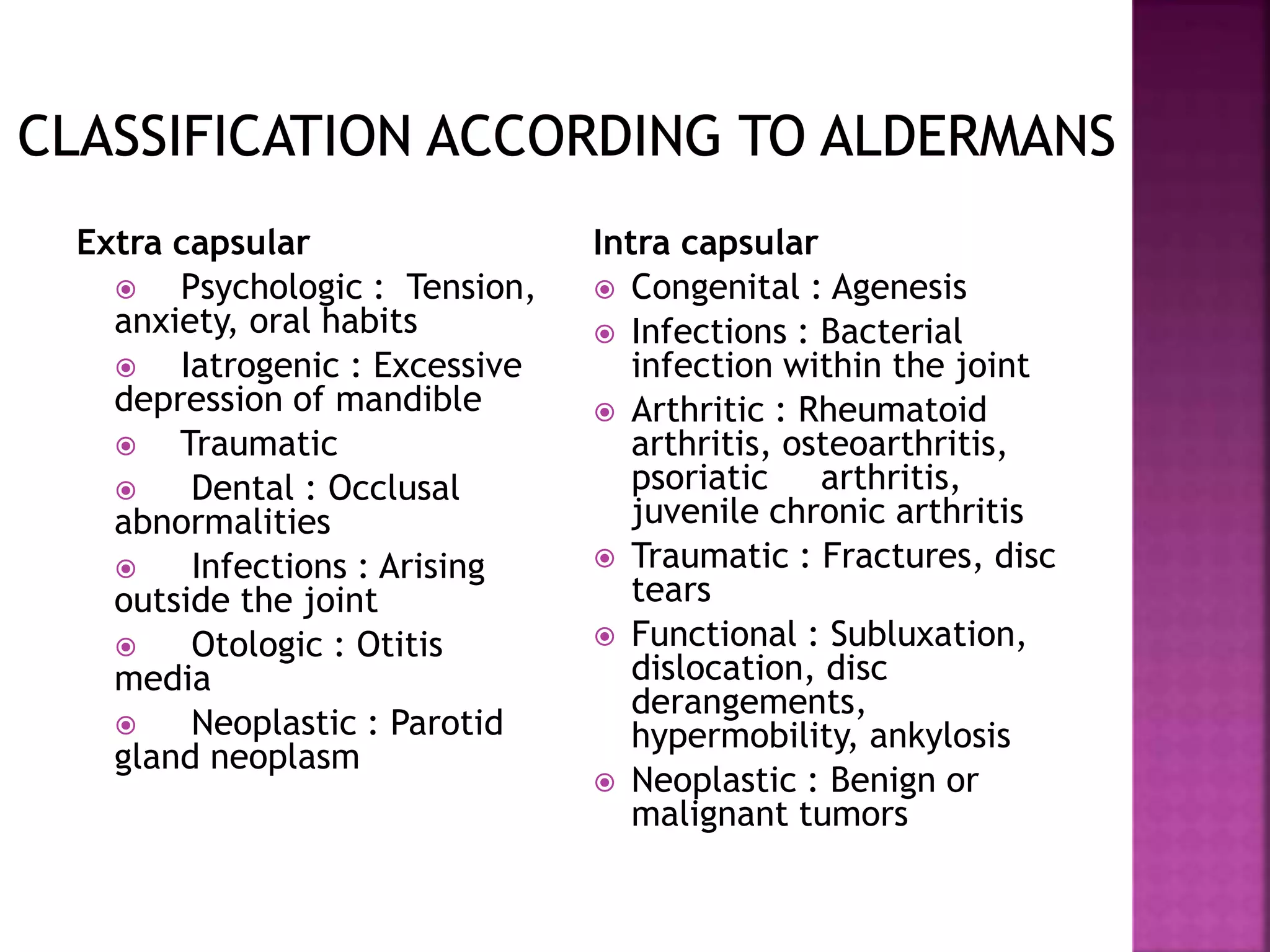
Part- I
 Definition
 Anatomy of TMJ
 Types of mandibular
movements
 Epidemiology
 Examination of TMJ
apparatus
 Clinical signs and
symptoms
Part-II
 Classification of TMD’s
 Differential diagnosis of
TMD’s
 Management
 TMJ Imaging
 1996 NIH Consensus Conference:
 A collection of medical and dental conditions affecting the
TMJ and/or the muscles of mastication as well as contiguous
tissue components.
 3 Main Categories
• I Muscle Disorders
• I.a Myofascial pain
• I.b Myofascial pain with limited opening
• II Disk Displacements
• II.a Disc displacement with reduction
• II.b Disc displacement without reduction with limited opening
• II.c Disc displacement without reduction without limited opening
• III Other common joint diseases
• III.a Arthralgia
• III.b Osteoarthritis
• III.c Osteoarthrosis
Dworkin SF. LeResche L. Research diagnostic criteria for temporomandibular disorders : Review,
criteria, examinations and specifications, critique. Journal of Craniomandibular Disorders.
6(4):301-55,1992.
• Compound joint
• “Ginglymodiarthrodial” joint
 Rotational movement
 Horizontal axis of rotation
 Frontal (vertical) axis of rotation
 Sagittal axis of rotation
 Translational movement
 Around the horizontal axis (hinge axis)
 Around the frontal (vertical) axis
 Around the sagittal axis
 1% - 75% of general population showed at least 1 objective
TMD sign
 5%-33% reported subjective symptoms.
 Age range - 20-40 yrs
 30yrs - Disc Displacement
 50yrs - Inflammatory degenerative disorders
 Screening history and examination
 History taking
 Pain
 Cranial nerve examination
 Eye and ear examination
 Cervical spine examination
 Inspection- assymetry
 Palpation- Soft but firm pressure
 A single firm thrust of 1 or 2 seconds' duration
 Temporalis
 Massetter
 Sternocleidomastoid
 Functional manipulation
 Lateral pterygoid- sup. & inf.
 Medial pterygoid
 Maximum interincisal
distance- 53- 58 mm.
 Lateral movement- <8mm
 Alterations in the opening
pathway
 Extracapsular restrictions
 Intracapsular restrictions
 Stationary and dynamic
movements
 Joint sounds- crepitus and
clicking
 Mobility
 Pdl widening
 Osteosclerosis
 Hypercementosis
 Tooth wear
 Abfractions
 Guiding to CR
 Intercuspal position
- acute malocclusion
- arch intergrity
- VDO
Pain
Dysfunction
Pain
Myalgia
DentitionArthralgia
 Events –
 Local
 Trauma
 Source of constant deep pain
 Systemic
 Stress
 Immunological resistance
 Age
 Diet
 Viral infections
Protective
co-
contraction
Local
muscle
soreness
Centrally
mediated
myalgia
Myofascial
pain
Travell JG, Rinzler SH: The myofascial genesis of pain, Postgrad Med 11:425-434, 1952.
 It occurs to protect the muscles from injury
 Etiology –
 Any change in the sensory input from associated structures
 Feeling of muscle tiredness
 Key to identify – immediately follows an event
 1st response to prolonged co-contraction
 Since it is a source of deep pain – can cause Protective Co-
contraction
 Clinically,
 Tender on palpation
 Increased pain with function
 CNS responds secondary to
 Presence of deep pain input
 Increased emotional stress
 Changes in descending inhibitory system
 Regional myogenous pain condition characterized by
 Local areas of firm , hypersensitive bands of muscle tissue known as
TRIGGER POINTS
 Felt as taut bands when palpated and elicits pain
 Local rise in temperature, increased metabolic demand, reduction of
blood flow
 Unique character of trigger points - source of constant deep pain
Local
 Protracted cause
 Recurrent cause (e.g., bruxism, repeated trauma).
 Therapeutic mismanagement
Systemic
 Continue emotional stress
 Sleep disturbances
 Depression
Clicking Crepitus Dysfunction
 Derangements of the condyle-disc complex
 Structural incompatibility of the articular surfaces
 Inflammatory joint disorders
• Functional disc displacement
• Functional dislocation
- with reduction
- without reduction( closed
lock)
 Macrotrauma – direct and indirect
 Microtrauma – “chondromalacia”
 Bruxism and clenching
 Orthopaedic instability
“Occlusal conditions commonly associated with disc derangements”
• Skeletal Class II deep bite.
The important feature of an occlusal condition that leads to disc derangement
disorders is the lack of joint stability when the teeth are tightly occluded.
Celic R, Jerolimov V: Association of horizontal and vertical overlap with prevalence of
temporomandibular disorders, J Oral Rehabil 29:588-593, 2002.
 Breakdown or adherence of
articular surfaces
 Static loading of joint as in
“clenching”.
 Subluxation (hypermobility)
 Spontaneous dislocation
(open lock)
 Steepness of articular eminence
 Morphology of condyle and fossa
 Joint laxity
 Attachment of sup. lateral pterygoid muscle
 Synovitis
 Capsulitis- tenderness on lateral pole of tmj
 Retrodiscitis – increased by clenching
 Arthrtides- osteoarthritis- crepitus is common
 Headaches
- Common symptom associated with TMD’s
 Otological symptoms
 Ear fullness
 Tinnitus
 Vertigo
 Imaging
 Mounted casts
 EMG
 Mandibular tracking
devices
 Sonography
 Vibration analysis
 Thermography
Part- I
 Definition
 Anatomy of TMJ
 Types of mandibular
movements
 Epidemiology
 Clinical signs and
symptoms and applied
aspects
 Examination of TMJ
apparatus
 Diagnostic aids
 Classification of TMD’s
Part-II
 Differential diagnosis of
TMD’s
 Management
Adapted from McNeill C
Cranial bones
 Congenital and developmental
disorders: Aplasia, hypoplasia,
hyperplasia
 Acquired disorders: Neoplasia,
fracture
Temporomandibular joint
disorders
 Deviation in form, Disk
displacement
 Dislocation
 Inflammatory conditions (synovitis,
capsulitis)
 Arthritides (osteoarthritis,
osteoarthrosis, polyarthritides)
 Ankylosis (fibrous, bony)
 Neoplasia
Masticatory-Muscle Myofacial
pain
 Myositis
 Spasm
 Contracture
Extra capsular
 Psychologic : Tension,
anxiety, oral habits
 Iatrogenic : Excessive
depression of mandible
 Traumatic
 Dental : Occlusal
abnormalities
 Infections : Arising
outside the joint
 Otologic : Otitis
media
 Neoplastic : Parotid
gland neoplasm
Intra capsular
 Congenital : Agenesis
 Infections : Bacterial
infection within the joint
 Arthritic : Rheumatoid
arthritis, osteoarthritis,
psoriatic arthritis,
juvenile chronic arthritis
 Traumatic : Fractures, disc
tears
 Functional : Subluxation,
dislocation, disc
derangements,
hypermobility, ankylosis
 Neoplastic : Benign or
malignant tumors
C/f
 Unilateral or bilateral
 Deviation of the jaw
towards affected side
Primary condylar aplasia and
hypoplasia
 Mandibulofacial dysostosis
(Treacher Collins syndrome)
 Hemifacial microsomia (first
and second branchial arch
syndrome)
 Oculoauriculovertebral
syndrome (Goldenhar
syndrome)
 Oculomandibulodyscephaly
(Hallermann-Streiff syndrome)
 Hurler’s syndrome
Secondary condylar
hypoplasia
 Osteoplasty
 Orthognathic surgery with orthodontics
 Cosmetic surgery
Etiology
 Hormones
 Trauma
 Infection
 Heredity
 Hyper vascularity
Clinical features
 Males, < 20 years of age
 Chin deviated towards unaffected side
 Posterior open bite on the affected side
 Restricted mobility of the enlarged condyle
Differential diagnosis
 Osteochondroma
 Condylar osteoma
Treatment
 Orthodontics + orthognathic surgery before growth completion
 Vertical depression the
center of the condylar head
 Result from an obstructed
blood supply, embryopathy,
trauma.
Radiographic features
 Heart shape anteroposterior
silhouette
 Duplicate condylar head
 Mandibular fossa may
remodels
Clinical features
 Females, children
 A form of condylar hypoplasia
Radiographic features
 Characteristic “toadstool”
appearance
 Shortened condylar neck
 Deep antegonial notch
Differential diagnosis
 Destruction of the anterior
aspect of the condylar head
from rheumatoid arthritis
Treatment
 Intra-articular cortico steroid
injection
 Orthognathic surgery
 Orthodontic therapy
 Abnormality in position & morphology of articular disk
 Lateral and posterior displacements are rare
Etiology – multifactorial
 Disk returns to normal position - with reduction
 Disk remains displaced – non reduction
Clinical features
 Unilateral / bilateral
 Mandibular deviation to the affected side
 Click
 Noises may be absent in long standing cases
 Pain in the preauricular region / headaches
• Closed lock
• 10-15mm opening
• Pain on forced opening
• Deviation – affected
side
• Chronic- no pain and
limited movements
• MRI to confirm
 Occurs at retrodiskal tissue
 Detected by arthrographic
technique
 Dr Fleming Norgaard- 1947
 Single contrast arthrography
 Double contrast radiography
Ravikiran Ongole, Navjyot Panjrath et al Temporomandibular joint Arthrography an
overview Pakistan oral & dent. jr. 22 ( 1) june 2002
 Scar tissue
 Detected by
arthrographic
technique
 MRI with low signal
intensity
Remodelling
• Flattening of condyle
 Degeneration + Proliferation
 Occurs when adaptive capacity is exceeded
 Etiology – unknown, trauma , hypermobility, parafunction
Clinical features
 Any age, > females
 70% old age
 Asymptomatic
 Pain
 Crepitus
 Limited movement
Johansson A, et al: Gender difference in symptoms related to TMJ disorders in a population of
50yr old subjects. J OROFAC PAIN 2008; 17:29-35
Radiographic features
 CT or T1 weighted MRI
 Loss of cortex or erosion
 Ely’s cyst / subchondral bone
cyst
 Bony proliferation at
periphery – osteophyte
 Joint mice
 Glenoid fossa enlarges
K.L. Ferrazzo, L.B. Osorio et al CT Images of a severe
TMJ Osteoarthritis and Differential diagnosis with
other joint disorders. Case Rep Dent. 2013;
2013:242685
Differential diagnosis
 Rheumatoid arthritis
 Osteoma or osteochondroma
Treatment
 Relieve joint stress
 Relieve secondary inflammation
 Increase joint mobility
 Synovial membrane inflammation
Clinical features
 Females
 50% with TMJ involvement
 Any age
 Bilateral, symmetrical
 Swelling, pain, tenderness, stiffness on opening, crepitus
 Open bite
Radiographic features
 Decreased density of
condyle
 Pannus
 Sharpened pencil
appearance
 Entire condyle and
anterior eminence -
destroyed
Differential diagnosis
 Psoriatic arthritis
 Osteoarthritis
Treatment
 Pain relief
 Suppression of inflammation
 Physiotherapy
 Joint replacement surgery
 Juvenile rheumatoid arthritis, Still’s disease
 Chronic inflammatory disease, < 16 years
 Synovial hypertrophy
Clinical features
 Pain and tenderness
 Starts as unilateral, progress to bilateral
 Bird face
 Anterior open bite
 Micrognathia
 Jaw deviated to affected side
Radiographic features
 Decreased density
 Pencil shaped mandibular
condyle
 Impaired mandibular growth
 Deepening of antegonial
notch
Clinical features
 Any age
 No sex predilection
 Unilateral
 Pain
 Inability occlude
 Tender cervical lymph nodes
 Fever
 Mandible deviated towards unaffected side
Radiographic features
 Space between condyle and
roof of mandibular fossa
widened
 Sequestrum and periosteal
new bone formation
 Osseous ankylosis
Treatment
 Antimicrobial therapy
 Drainage of effusion
 Joint rest
 Synovial chondrometaplasia and osteochondromatosis
 Metaplastic formation of multiple cartilaginous nodules
 Nodules may detach
Clinical features
 4th or5th decades
 M:F- 2:1
 Asymptomatic or may complain of preauricular swelling,
pain
 Decreased range of motion
 Crepitus
 Unilateral
Radiographic features
 Joint space widened
 Radiopaque loose bodies
 Erosion through glenoid
fossa
Differential diagnosis
 Chondrocalcinosis
 Osteochondromatosis
 Chondrosarcoma
 Osteosarcoma
 Sarcoma
Treatment
 Removal of the loose bodies
 Resection of abnormal synovial tissue by
arthroscopic surgery
 Open joint surgery
 Pseudogout and calcium pyrophosphate dihydrate
deposition disease
Clinical features
 Unilateral
 Males
 Pain and joint swelling
Radiographic features
 Radiopacities are finer
 Increase in condylar bone
density
 Erosions of the glenoid
fossa –CT
 Soft tissue inflammation
and edema -MRI
Differential diagnosis
 Chondrocalcinosis
 Osteochondromatosis
 Chondrosarcoma
 Osteosarcoma
 Sarcoma
Treatment
 Surgical removal of crystalline deposits
 Steroids, aspirin and NSAIDS
 Colchicine alleviate acute symptoms and for
prophylaxis.
 1934 – Costen’s Syndrome
 1940 – Travell- Muscle spasm could be a source of pain
 1959 – Shwartz-Postulated TMJ dysfunction syndrome
- Psychological make up as a
predisposing factor
- Stress causes clenching and
grinding resulting in spasm
 1969 –Laskin
 Unilateral pain in preauricular region, worse on awakening
 Tenderness on one or more muscles of mastication on palpation
 Limitation or deviation of mandible on opening
 Occlusal disturbances
 Intracapsular disorders
 Emotional turmoil.
 Major and minor trauma
 Poor nutritional status
 Genetic predisposition
 Chronic, focal regional
muscle pain
 Unilateral, may occur in
one or more muscles
 Trigger points are present
 Continuous, dull to sharp
pain in TMJ region
 Present at rest and with
movements
 Pain upon awakening
 History taking
 Examination of face
 Examination of TMJ
 Examination of muscles of mastication & neck muscles
 Intraoral examination
 Examination of other structures
 Voluntary opening tests
 Passive stretch test
 Soft end feel and Hard end feel
 Anesthetic nerve and muscle blocking
 Magnetic resonance spectroscopy
 Thermography and Biopsy
 Electromyography and Jaw tracking
Non-
surgical Surgical
Initial therapy
 Patient re-assurance
 Diet
 Rest
 Thermotherapy
Pharmacological therapy
 Analgesics- NSAIDS and Opioids
 Anxiolytics – Benzodiazepines
 Muscle Relaxants- chloroxazone, cyclobenzaprine,
thiocolchicoside
 Hydrocollator
• Short wave diathermy
• Ultrasound
Laser therapy
0 Increases capillary permeability
Low power Laser therapy
0 Alter cellular function
0 reduces short term pain
Cryotherapy
Acupuncture
Physiotherapy
 Spray and stretch
 Soft pressure and continuous massaging
 Continuous exercises
 Electric galvanic stimulation
 TENS
 Splint therapy
 Directive and permissive splints
 Condylotomy
 High condylectomy
 Menisectomy
 Myotomy
 Arthroscopy
 Botulinim Toxin A injections
• Types
• Clinical features
• Radiographic features
• Treatment
Types
 Osteomas, osteochondromas,
osteoblastomas,
Chondroblastomas,
fibromyxomas
Clinical features
 Grow slowly
 TMJ swelling, pain and
decreased range of motion
 Facial asymmetry, malocclusion
 Deviation of the mandible to the
unaffected side
 Chondrosarcoma, osteogenic
sarcoma, synovial sarcoma,
and fibrosarcoma of the
joint capsule
 Breast, kidney, lung, colon,
prostate and thyroid gland
tumors metastasize to TMJ
Clinical features
 Swelling over TMJ
 Pain
 Limited mandibular opening
 Mandibular deviation
Radiographic features
 Variable degree of bone destruction
with ill defined, irregular margins
 Lack tumor bone formation except
osteogenic sarcoma
 Chondrosarcoma may appear as an
indistinct – resemble pseudo gout
 Nonspecific condylar destruction
 Management of Temporomandibular Disorders and Occlusion by Jeffrey P.
Okeson 6th edition
 Manual of Temporomandibular Disorders – Edward F. Wright
 Daniele Manfredini, DDS,Fabio Piccotti, DDS,Luca Guarda-Nardini, et al
Research diagnostic criteria for temporomandibular disorders: a
systematic review of axis I epidemiologic findings Oral Surg Oral Med Oral
Pathol Oral Radiol Endod 2011;112: 453-462)
 Shirish Ingawale, Tarun GoswamI et al. Temporomandibular Joint:
Disorders, Treatments, and Biomechanics. Annals of Biomedical
Engineering, Vol. 37, No. 5, May 2009 ( 2009) pp. 976–996
 Thomas CA, Okeson JP: Evaluation of lateral pterygoid muscle symptoms
using a common palpation technique and a method of functional
manipulation, Cranio 5:125-129,1987.
 Celic R, Jerolimov V: Association of horizontal and vertical overlap with
prevalence of temporomandibular disorders, J Oral Rehabil 29:588-593,
2002.
 Oral and Maxillofacial Pathology by Neville, Damn, Allen – 3rd edition
THANK YOU

Temporomandibular joint disorders

  • 2.
    Part- I  Definition Anatomy of TMJ  Types of mandibular movements  Epidemiology  Examination of TMJ apparatus  Clinical signs and symptoms Part-II  Classification of TMD’s  Differential diagnosis of TMD’s  Management  TMJ Imaging
  • 3.
     1996 NIHConsensus Conference:  A collection of medical and dental conditions affecting the TMJ and/or the muscles of mastication as well as contiguous tissue components.
  • 4.
     3 MainCategories • I Muscle Disorders • I.a Myofascial pain • I.b Myofascial pain with limited opening • II Disk Displacements • II.a Disc displacement with reduction • II.b Disc displacement without reduction with limited opening • II.c Disc displacement without reduction without limited opening • III Other common joint diseases • III.a Arthralgia • III.b Osteoarthritis • III.c Osteoarthrosis Dworkin SF. LeResche L. Research diagnostic criteria for temporomandibular disorders : Review, criteria, examinations and specifications, critique. Journal of Craniomandibular Disorders. 6(4):301-55,1992.
  • 5.
    • Compound joint •“Ginglymodiarthrodial” joint
  • 7.
     Rotational movement Horizontal axis of rotation  Frontal (vertical) axis of rotation  Sagittal axis of rotation  Translational movement
  • 8.
     Around thehorizontal axis (hinge axis)
  • 9.
     Around thefrontal (vertical) axis
  • 10.
     Around thesagittal axis
  • 12.
     1% -75% of general population showed at least 1 objective TMD sign  5%-33% reported subjective symptoms.  Age range - 20-40 yrs  30yrs - Disc Displacement  50yrs - Inflammatory degenerative disorders
  • 13.
     Screening historyand examination  History taking  Pain
  • 14.
     Cranial nerveexamination  Eye and ear examination  Cervical spine examination
  • 15.
     Inspection- assymetry Palpation- Soft but firm pressure  A single firm thrust of 1 or 2 seconds' duration  Temporalis  Massetter  Sternocleidomastoid
  • 16.
     Functional manipulation Lateral pterygoid- sup. & inf.  Medial pterygoid
  • 17.
     Maximum interincisal distance-53- 58 mm.  Lateral movement- <8mm  Alterations in the opening pathway  Extracapsular restrictions  Intracapsular restrictions
  • 18.
     Stationary anddynamic movements  Joint sounds- crepitus and clicking
  • 19.
     Mobility  Pdlwidening  Osteosclerosis  Hypercementosis  Tooth wear  Abfractions
  • 20.
     Guiding toCR  Intercuspal position - acute malocclusion - arch intergrity - VDO
  • 21.
  • 22.
  • 23.
     Events – Local  Trauma  Source of constant deep pain  Systemic  Stress  Immunological resistance  Age  Diet  Viral infections
  • 24.
  • 25.
     It occursto protect the muscles from injury  Etiology –  Any change in the sensory input from associated structures  Feeling of muscle tiredness  Key to identify – immediately follows an event
  • 26.
     1st responseto prolonged co-contraction  Since it is a source of deep pain – can cause Protective Co- contraction  Clinically,  Tender on palpation  Increased pain with function
  • 27.
     CNS respondssecondary to  Presence of deep pain input  Increased emotional stress  Changes in descending inhibitory system
  • 28.
     Regional myogenouspain condition characterized by  Local areas of firm , hypersensitive bands of muscle tissue known as TRIGGER POINTS  Felt as taut bands when palpated and elicits pain  Local rise in temperature, increased metabolic demand, reduction of blood flow  Unique character of trigger points - source of constant deep pain
  • 29.
    Local  Protracted cause Recurrent cause (e.g., bruxism, repeated trauma).  Therapeutic mismanagement Systemic  Continue emotional stress  Sleep disturbances  Depression
  • 30.
  • 31.
     Derangements ofthe condyle-disc complex  Structural incompatibility of the articular surfaces  Inflammatory joint disorders
  • 32.
    • Functional discdisplacement • Functional dislocation - with reduction - without reduction( closed lock)
  • 33.
     Macrotrauma –direct and indirect  Microtrauma – “chondromalacia”  Bruxism and clenching  Orthopaedic instability “Occlusal conditions commonly associated with disc derangements” • Skeletal Class II deep bite. The important feature of an occlusal condition that leads to disc derangement disorders is the lack of joint stability when the teeth are tightly occluded. Celic R, Jerolimov V: Association of horizontal and vertical overlap with prevalence of temporomandibular disorders, J Oral Rehabil 29:588-593, 2002.
  • 34.
     Breakdown oradherence of articular surfaces  Static loading of joint as in “clenching”.  Subluxation (hypermobility)  Spontaneous dislocation (open lock)
  • 35.
     Steepness ofarticular eminence  Morphology of condyle and fossa  Joint laxity  Attachment of sup. lateral pterygoid muscle
  • 36.
     Synovitis  Capsulitis-tenderness on lateral pole of tmj  Retrodiscitis – increased by clenching  Arthrtides- osteoarthritis- crepitus is common
  • 38.
     Headaches - Commonsymptom associated with TMD’s  Otological symptoms  Ear fullness  Tinnitus  Vertigo
  • 40.
     Imaging  Mountedcasts  EMG  Mandibular tracking devices  Sonography  Vibration analysis  Thermography
  • 41.
    Part- I  Definition Anatomy of TMJ  Types of mandibular movements  Epidemiology  Clinical signs and symptoms and applied aspects  Examination of TMJ apparatus  Diagnostic aids  Classification of TMD’s Part-II  Differential diagnosis of TMD’s  Management
  • 42.
    Adapted from McNeillC Cranial bones  Congenital and developmental disorders: Aplasia, hypoplasia, hyperplasia  Acquired disorders: Neoplasia, fracture Temporomandibular joint disorders  Deviation in form, Disk displacement  Dislocation  Inflammatory conditions (synovitis, capsulitis)  Arthritides (osteoarthritis, osteoarthrosis, polyarthritides)  Ankylosis (fibrous, bony)  Neoplasia Masticatory-Muscle Myofacial pain  Myositis  Spasm  Contracture
  • 43.
    Extra capsular  Psychologic: Tension, anxiety, oral habits  Iatrogenic : Excessive depression of mandible  Traumatic  Dental : Occlusal abnormalities  Infections : Arising outside the joint  Otologic : Otitis media  Neoplastic : Parotid gland neoplasm Intra capsular  Congenital : Agenesis  Infections : Bacterial infection within the joint  Arthritic : Rheumatoid arthritis, osteoarthritis, psoriatic arthritis, juvenile chronic arthritis  Traumatic : Fractures, disc tears  Functional : Subluxation, dislocation, disc derangements, hypermobility, ankylosis  Neoplastic : Benign or malignant tumors
  • 45.
    C/f  Unilateral orbilateral  Deviation of the jaw towards affected side
  • 46.
    Primary condylar aplasiaand hypoplasia  Mandibulofacial dysostosis (Treacher Collins syndrome)  Hemifacial microsomia (first and second branchial arch syndrome)  Oculoauriculovertebral syndrome (Goldenhar syndrome)  Oculomandibulodyscephaly (Hallermann-Streiff syndrome)  Hurler’s syndrome Secondary condylar hypoplasia
  • 47.
     Osteoplasty  Orthognathicsurgery with orthodontics  Cosmetic surgery
  • 48.
    Etiology  Hormones  Trauma Infection  Heredity  Hyper vascularity Clinical features  Males, < 20 years of age  Chin deviated towards unaffected side  Posterior open bite on the affected side  Restricted mobility of the enlarged condyle
  • 49.
    Differential diagnosis  Osteochondroma Condylar osteoma Treatment  Orthodontics + orthognathic surgery before growth completion
  • 50.
     Vertical depressionthe center of the condylar head  Result from an obstructed blood supply, embryopathy, trauma. Radiographic features  Heart shape anteroposterior silhouette  Duplicate condylar head  Mandibular fossa may remodels
  • 51.
    Clinical features  Females,children  A form of condylar hypoplasia Radiographic features  Characteristic “toadstool” appearance  Shortened condylar neck  Deep antegonial notch
  • 52.
    Differential diagnosis  Destructionof the anterior aspect of the condylar head from rheumatoid arthritis Treatment  Intra-articular cortico steroid injection  Orthognathic surgery  Orthodontic therapy
  • 53.
     Abnormality inposition & morphology of articular disk  Lateral and posterior displacements are rare Etiology – multifactorial  Disk returns to normal position - with reduction  Disk remains displaced – non reduction
  • 54.
    Clinical features  Unilateral/ bilateral  Mandibular deviation to the affected side  Click  Noises may be absent in long standing cases  Pain in the preauricular region / headaches
  • 56.
    • Closed lock •10-15mm opening • Pain on forced opening • Deviation – affected side • Chronic- no pain and limited movements • MRI to confirm
  • 57.
     Occurs atretrodiskal tissue  Detected by arthrographic technique  Dr Fleming Norgaard- 1947  Single contrast arthrography  Double contrast radiography Ravikiran Ongole, Navjyot Panjrath et al Temporomandibular joint Arthrography an overview Pakistan oral & dent. jr. 22 ( 1) june 2002
  • 58.
     Scar tissue Detected by arthrographic technique  MRI with low signal intensity Remodelling • Flattening of condyle
  • 59.
     Degeneration +Proliferation  Occurs when adaptive capacity is exceeded  Etiology – unknown, trauma , hypermobility, parafunction Clinical features  Any age, > females  70% old age  Asymptomatic  Pain  Crepitus  Limited movement Johansson A, et al: Gender difference in symptoms related to TMJ disorders in a population of 50yr old subjects. J OROFAC PAIN 2008; 17:29-35
  • 60.
    Radiographic features  CTor T1 weighted MRI  Loss of cortex or erosion  Ely’s cyst / subchondral bone cyst  Bony proliferation at periphery – osteophyte  Joint mice  Glenoid fossa enlarges K.L. Ferrazzo, L.B. Osorio et al CT Images of a severe TMJ Osteoarthritis and Differential diagnosis with other joint disorders. Case Rep Dent. 2013; 2013:242685
  • 61.
    Differential diagnosis  Rheumatoidarthritis  Osteoma or osteochondroma Treatment  Relieve joint stress  Relieve secondary inflammation  Increase joint mobility
  • 62.
     Synovial membraneinflammation Clinical features  Females  50% with TMJ involvement  Any age  Bilateral, symmetrical  Swelling, pain, tenderness, stiffness on opening, crepitus  Open bite
  • 63.
    Radiographic features  Decreaseddensity of condyle  Pannus  Sharpened pencil appearance  Entire condyle and anterior eminence - destroyed
  • 64.
    Differential diagnosis  Psoriaticarthritis  Osteoarthritis Treatment  Pain relief  Suppression of inflammation  Physiotherapy  Joint replacement surgery
  • 65.
     Juvenile rheumatoidarthritis, Still’s disease  Chronic inflammatory disease, < 16 years  Synovial hypertrophy Clinical features  Pain and tenderness  Starts as unilateral, progress to bilateral  Bird face  Anterior open bite  Micrognathia  Jaw deviated to affected side
  • 66.
    Radiographic features  Decreaseddensity  Pencil shaped mandibular condyle  Impaired mandibular growth  Deepening of antegonial notch
  • 67.
    Clinical features  Anyage  No sex predilection  Unilateral  Pain  Inability occlude  Tender cervical lymph nodes  Fever  Mandible deviated towards unaffected side
  • 68.
    Radiographic features  Spacebetween condyle and roof of mandibular fossa widened  Sequestrum and periosteal new bone formation  Osseous ankylosis
  • 69.
    Treatment  Antimicrobial therapy Drainage of effusion  Joint rest
  • 70.
     Synovial chondrometaplasiaand osteochondromatosis  Metaplastic formation of multiple cartilaginous nodules  Nodules may detach Clinical features  4th or5th decades  M:F- 2:1  Asymptomatic or may complain of preauricular swelling, pain  Decreased range of motion  Crepitus  Unilateral
  • 71.
    Radiographic features  Jointspace widened  Radiopaque loose bodies  Erosion through glenoid fossa
  • 72.
    Differential diagnosis  Chondrocalcinosis Osteochondromatosis  Chondrosarcoma  Osteosarcoma  Sarcoma Treatment  Removal of the loose bodies  Resection of abnormal synovial tissue by arthroscopic surgery  Open joint surgery
  • 73.
     Pseudogout andcalcium pyrophosphate dihydrate deposition disease Clinical features  Unilateral  Males  Pain and joint swelling
  • 74.
    Radiographic features  Radiopacitiesare finer  Increase in condylar bone density  Erosions of the glenoid fossa –CT  Soft tissue inflammation and edema -MRI
  • 75.
    Differential diagnosis  Chondrocalcinosis Osteochondromatosis  Chondrosarcoma  Osteosarcoma  Sarcoma Treatment  Surgical removal of crystalline deposits  Steroids, aspirin and NSAIDS  Colchicine alleviate acute symptoms and for prophylaxis.
  • 76.
     1934 –Costen’s Syndrome  1940 – Travell- Muscle spasm could be a source of pain  1959 – Shwartz-Postulated TMJ dysfunction syndrome - Psychological make up as a predisposing factor - Stress causes clenching and grinding resulting in spasm
  • 77.
     1969 –Laskin Unilateral pain in preauricular region, worse on awakening  Tenderness on one or more muscles of mastication on palpation  Limitation or deviation of mandible on opening
  • 78.
     Occlusal disturbances Intracapsular disorders  Emotional turmoil.  Major and minor trauma  Poor nutritional status  Genetic predisposition
  • 79.
     Chronic, focalregional muscle pain  Unilateral, may occur in one or more muscles  Trigger points are present  Continuous, dull to sharp pain in TMJ region  Present at rest and with movements  Pain upon awakening
  • 81.
     History taking Examination of face  Examination of TMJ  Examination of muscles of mastication & neck muscles  Intraoral examination  Examination of other structures
  • 82.
     Voluntary openingtests  Passive stretch test  Soft end feel and Hard end feel  Anesthetic nerve and muscle blocking  Magnetic resonance spectroscopy  Thermography and Biopsy
  • 83.
  • 84.
  • 85.
    Initial therapy  Patientre-assurance  Diet  Rest  Thermotherapy Pharmacological therapy  Analgesics- NSAIDS and Opioids  Anxiolytics – Benzodiazepines  Muscle Relaxants- chloroxazone, cyclobenzaprine, thiocolchicoside
  • 87.
     Hydrocollator • Shortwave diathermy • Ultrasound
  • 88.
    Laser therapy 0 Increasescapillary permeability Low power Laser therapy 0 Alter cellular function 0 reduces short term pain Cryotherapy Acupuncture
  • 89.
    Physiotherapy  Spray andstretch  Soft pressure and continuous massaging  Continuous exercises  Electric galvanic stimulation  TENS
  • 90.
     Splint therapy Directive and permissive splints
  • 91.
     Condylotomy  Highcondylectomy  Menisectomy  Myotomy  Arthroscopy  Botulinim Toxin A injections
  • 92.
    • Types • Clinicalfeatures • Radiographic features • Treatment
  • 93.
    Types  Osteomas, osteochondromas, osteoblastomas, Chondroblastomas, fibromyxomas Clinicalfeatures  Grow slowly  TMJ swelling, pain and decreased range of motion  Facial asymmetry, malocclusion  Deviation of the mandible to the unaffected side
  • 94.
     Chondrosarcoma, osteogenic sarcoma,synovial sarcoma, and fibrosarcoma of the joint capsule  Breast, kidney, lung, colon, prostate and thyroid gland tumors metastasize to TMJ Clinical features  Swelling over TMJ  Pain  Limited mandibular opening  Mandibular deviation Radiographic features  Variable degree of bone destruction with ill defined, irregular margins  Lack tumor bone formation except osteogenic sarcoma  Chondrosarcoma may appear as an indistinct – resemble pseudo gout  Nonspecific condylar destruction
  • 95.
     Management ofTemporomandibular Disorders and Occlusion by Jeffrey P. Okeson 6th edition  Manual of Temporomandibular Disorders – Edward F. Wright  Daniele Manfredini, DDS,Fabio Piccotti, DDS,Luca Guarda-Nardini, et al Research diagnostic criteria for temporomandibular disorders: a systematic review of axis I epidemiologic findings Oral Surg Oral Med Oral Pathol Oral Radiol Endod 2011;112: 453-462)  Shirish Ingawale, Tarun GoswamI et al. Temporomandibular Joint: Disorders, Treatments, and Biomechanics. Annals of Biomedical Engineering, Vol. 37, No. 5, May 2009 ( 2009) pp. 976–996  Thomas CA, Okeson JP: Evaluation of lateral pterygoid muscle symptoms using a common palpation technique and a method of functional manipulation, Cranio 5:125-129,1987.  Celic R, Jerolimov V: Association of horizontal and vertical overlap with prevalence of temporomandibular disorders, J Oral Rehabil 29:588-593, 2002.  Oral and Maxillofacial Pathology by Neville, Damn, Allen – 3rd edition
  • 96.

Editor's Notes

  • #14 Concomitant symptoms- parasthesia, anaesthesia or hyperesthesia
  • #17 Inf LP- protrude the jaw against resistance, releived by keeping tongue blade between teeth and asked to clench Sup LP- clenching increases the pain Medial pterygoid- pain on wide opening
  • #18 Extracapsular restrictions often create a deflection of the incisal path during opening. The direction of the deflection depends on the location of the muscle that causes the restriction. If the restricting muscle is lateral to the joint (as with the masseter), the deflection during opening will be to the ipsilateral side. If the muscle is medial (as with the medial pterygoid), the deflection will be to the contralateral side.
  • #21 Arch integrity- loss of teeth
  • #25 These positional changes create certain acute malocclusions. Myospasms are also characterized by firm muscles as noted by palpation. Chronic myositis and fibromyalgia, This occurs because 42% of patients with fibromyalgia also report TMD-like symptoms.
  • #30 Protracted cause- If the clinician fails to eliminate the cause of an acute myalgic disorder, a more chronic condition is likely to develop.
  • #33 the opening click can occur at any time during that movement depending on disc-condyle morphology, muscle pull, and the pull of the superior retrodiscal lamina. The closing click almost always occurs near the closed or intercuspal position (ICP). The mouth cannot be opened maximally because the position of the disc does not allow full translation of the condyle . Typically the initial opening will be only 25 to 30 mm interincisally, which represents the maximum rotation of the joint.
  • #34 Direct trauma- accidents and iatrogenic Elongation of discal ligaments Indirect trauma- whiplash(sudden extension of cervical spine) Chondromalacia- excessive loading of tmj beyond functional limit causes collagen fragmentation and degradation in turn causing softening of articular surfaces Hypoxia/perfusion theory
  • #35 Weeping and boundary lubrication by synovial fluid
  • #46 There are many causes of the various unilateral and bilateral growth disturbances of the mandibular condyle and its related structures (Sarnat 1969; Thoma 1969). Any alteration in the condyle’s size or shape affects the TMJ. A more general disturbance, deriving from changes in the mandibular condyle, results in facial abnormalities. Such a disturbance in the development of the mandibular condyle and fossa may occur in utero late in the first trimester. This may result in aplasia or hypoplasia of the mandibular condyle and its associated soft tissues (Thoma 1969; Buch). Meanwhile, a disturbance of the mandibular condyle during the normal growth period may result in condylar hyperplasia, which is caused by abnormal local growth stimulation (Buchbinder & Kaplan 1991; Tank et al. 1998). binder & Kaplan 1991).
  • #79 tiologic factors also include:  Whip lash injury from an auto accident,  Wrestling blow,  Trauma from falling, and unexpectedly biting into a hard object.
  • #88 300mhz- diathermy