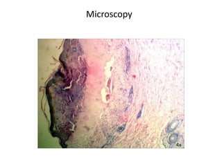
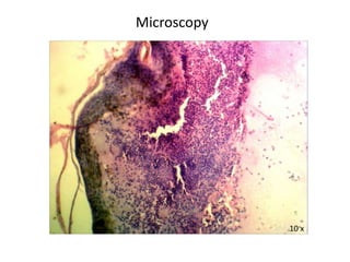
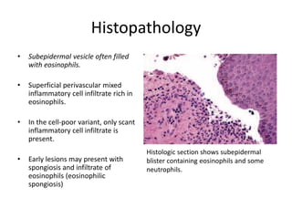
• Age/Sex : 80/Female
• Hospital OP/ IP No: A18009538
• Biopsy No: 349/18
• Date Of Receiving Specimen : 10/02/2018
• Clinical Diagnosis : Bullous pemphigoid.
• Nature of Specimen : A 6mm punch biopsy
including a vessel from abdomen.
.
Gross Examination
Received a single skin covered punch biopsy specimen
measuring 0.5x0.5cm. All embedded in one block.
Microscopy
4x
Microscopy
10 x
Microscopy
40 x
Microscopy
10 x
Microscopy
40 x
Microscopy
Section studied shows epidermis with subepidermal bulla.
Bullous cavity is filled with plasma, moderate infiltration of
neutrophils and eosinophils along with nuclear debris.
Adjacent epidermis shows basal cell vacuolation and mild
spongiosis. Dermis beneath the bulla shows dense
perivascular infiltrate composed of lymphocytes, eosinophils,
few plasma cells and melanophages.
Impression
Features are consistent with Bullous pemphigoid.
DISCUSSION
Bullous pemphigoid
• Common subepidermal, blistering, autoimmune disease of skin due to IgG
antibodies to the hemidesmosomal antigens Bullous pemphigoid antigen
1 and 2.
• Affects elderly patients and presents as large, tense bullae involving trunk,
extremities, and intertriginous areas.
• Nikolsky sign is negative.
• Oral lesions are present in about one third of the patients.
Histopathology
• Subepidermal vesicle often filled
with eosinophils.
• Superficial perivascular mixed
inflammatory cell infiltrate rich in
eosinophils.
• In the cell-poor variant, only scant
inflammatory cell infiltrate is
present.
• Early lesions may present with
spongiosis and infiltrate of
eosinophils (eosinophilic
spongiosis)
Histologic section shows subepidermal
blister containing eosinophils and some
neutrophils.
Techniques for Diagnosis
• Direct immunofluorescence studies
- a linear deposition of C3 and IgG at
the dermoepidermal junction.
• Salt-split skin immunofluorescence
shows that the pemphigoid
antibodies are localized to the roof
of the blister in most cases
Differential Diagnosis
Herpes gestationis
• Presents as intensely pruritic lesions
on the abdomen and extremities of
pregnant women in second and third
trimesters.
• Perivascular infiltrate composed of
lymphocytes and eosinophils in zones
of erythema and edema.
• There is marked papillary dermal
edema and eosinophilic spongiosis
• Focal necrosis of the basal
keratinocytes leading to the
subpeidermal blister.
Epidermolysis bullosa acquisita
• Presents as blisters developing on acral
areas that heal with scarring.
• Histologic and immunofluorescence
changes may be identical to that of
bullous pemphigoid.
• Eosinophils are fewer in number, and
lymphocytes and neutrophils may
predominate.
• Immunofluorescence of salt-split skin
shows the localization of IgG
antibodies to the floor of the blister
Porphyria cutanea tarda
• Subepidermal blister with minimal
inflammatory cell infiltrate.
• The dermal papillae extend into the
blister cavity with a festooning
appearance.
• PAS-positive eosinophilic deposits
around the blood vessels of the
papillary dermis are characteristic.

Bullous pemphigoid

  • 1.
    • Age/Sex :80/Female • Hospital OP/ IP No: A18009538 • Biopsy No: 349/18 • Date Of Receiving Specimen : 10/02/2018 • Clinical Diagnosis : Bullous pemphigoid. • Nature of Specimen : A 6mm punch biopsy including a vessel from abdomen. .
  • 2.
    Gross Examination Received asingle skin covered punch biopsy specimen measuring 0.5x0.5cm. All embedded in one block.
  • 3.
  • 4.
  • 5.
  • 6.
  • 7.
  • 8.
    Microscopy Section studied showsepidermis with subepidermal bulla. Bullous cavity is filled with plasma, moderate infiltration of neutrophils and eosinophils along with nuclear debris. Adjacent epidermis shows basal cell vacuolation and mild spongiosis. Dermis beneath the bulla shows dense perivascular infiltrate composed of lymphocytes, eosinophils, few plasma cells and melanophages.
  • 9.
    Impression Features are consistentwith Bullous pemphigoid.
  • 10.
  • 11.
    Bullous pemphigoid • Commonsubepidermal, blistering, autoimmune disease of skin due to IgG antibodies to the hemidesmosomal antigens Bullous pemphigoid antigen 1 and 2. • Affects elderly patients and presents as large, tense bullae involving trunk, extremities, and intertriginous areas. • Nikolsky sign is negative. • Oral lesions are present in about one third of the patients.
  • 12.
    Histopathology • Subepidermal vesicleoften filled with eosinophils. • Superficial perivascular mixed inflammatory cell infiltrate rich in eosinophils. • In the cell-poor variant, only scant inflammatory cell infiltrate is present. • Early lesions may present with spongiosis and infiltrate of eosinophils (eosinophilic spongiosis) Histologic section shows subepidermal blister containing eosinophils and some neutrophils.
  • 13.
    Techniques for Diagnosis •Direct immunofluorescence studies - a linear deposition of C3 and IgG at the dermoepidermal junction. • Salt-split skin immunofluorescence shows that the pemphigoid antibodies are localized to the roof of the blister in most cases
  • 14.
  • 15.
    Differential Diagnosis Herpes gestationis •Presents as intensely pruritic lesions on the abdomen and extremities of pregnant women in second and third trimesters. • Perivascular infiltrate composed of lymphocytes and eosinophils in zones of erythema and edema. • There is marked papillary dermal edema and eosinophilic spongiosis • Focal necrosis of the basal keratinocytes leading to the subpeidermal blister.
  • 16.
    Epidermolysis bullosa acquisita •Presents as blisters developing on acral areas that heal with scarring. • Histologic and immunofluorescence changes may be identical to that of bullous pemphigoid. • Eosinophils are fewer in number, and lymphocytes and neutrophils may predominate. • Immunofluorescence of salt-split skin shows the localization of IgG antibodies to the floor of the blister
  • 17.
    Porphyria cutanea tarda •Subepidermal blister with minimal inflammatory cell infiltrate. • The dermal papillae extend into the blister cavity with a festooning appearance. • PAS-positive eosinophilic deposits around the blood vessels of the papillary dermis are characteristic.