
Recommended
More Related Content
Similar to approach-to-fractures-managment-in-elderly-1-_1_.ppt
Similar to approach-to-fractures-managment-in-elderly-1-_1_.ppt (20)
Recently uploaded
Recently uploaded (20)
approach-to-fractures-managment-in-elderly-1-_1_.ppt
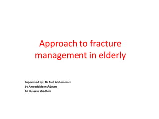
- 1. Approach to fracture management in elderly Supervised by : Dr Zaid Alshemmari By Ameedaldeen Adnan Ali Hussein khadhim
- 2. Objectives 1. Know the importance of fracture in elderly 2. Risk factor that contribute to these fractures 3. Common fracture in elderly 4. How you can manage these fractures 5. Impact of fracture of their life
- 3. introduction Older adults, adults 65 years and older, have both an increased rate of trauma and an increased predisposition to injury from even minimal force. This makes older adults a high-risk population for traumatic fracture from high- or low-impact mechanisms. One of the most common changes is their bones undergo several changes decrease in bone density, also known as osteoporosis. Also due to other physiologic changes of ageing as well as common comorbidities, Difficulties with gait, vision, and proprioception contribute to falls.
- 4. introduction Multiple medical conditions also increase the risk of fracture. Any condition requiring chronic glucocorticoid use, such as inflammatory bowel disease, celiac disease, chronic obstructive pulmonary disease, and rheumatoid arthritis, decreases BMD. treatment of low bone mineral density (BMD) has been shown to significantly reduce fracture rates. The most common fractures in older adults are vertebral fractures from compression or trauma, followed by hip and distal radius fractures.
- 5. 75 years female present with back pain after falling.
- 6. Vertebral fracture Spinal fractures in the elderly are the most common manifestation of osteoporosis and often occur from low energy mechanism or even in the absence of trauma. Cervical spine fractures are quite common, especially at C2. Compression fracture of the vertebral body is common, especially in elderly. A compression fracture : is defined as a collapsing in vertebral bone in the spine and decreased in height at least 15-20 % as a result of pressure or degeneration of the spinal bones. Osteoporosis is the underlying cause of many lumbar fractures, especially in postmenopausal women. Osteoporotic spinal fractures are unique in that they may occur without apparent trauma. However, a thorough diagnostic workup is always required to rule out spinal malignancy.
- 7. Vertebral fracture Etiology 1.Trauma 2.Osteoporosis/Osteopenia (bone density loss) 3.Osteomyelitis (Bone infection) 4.Pathologic Fractures (Primary or Metastatic Tumors) 5.Prolonged use of corticosteroids Clinical features compression fracture should be suspected in any patient older than 50 years with acute onset of sudden low back pain. Midline back pain is the hallmark symptom of lumbar compression fractures. The pain is axial, no radiating, aching, or stabbing in quality and may be severe and disabling. The location of the pain corresponds to the fracture site. In elderly patients with severe osteoporosis, there may be no pain at all as the fracture occurs spontaneously.
- 8. investigation Perform a complete blood cell count with differential, prostate-specific antigen testing and ESR. Radiography is the standard imaging study for spine fractures. Anteroposterior and lateral views Computed tomography (CT) scanning is an invaluable tool to evaluate the complexity of fractures seen on radiographs Magnetic resonance imaging (MRI) is required when the patient describes lower extremity motor or sensory loss. Radicular pain is another indication for MRI. Also, when canal compromise is suspected. When malignancy is strongly suspected, a vertebral biopsy is indicated.
- 9. treatment In the past, treatment options for lumbar fractures were quite limited, with bracing and rest prescribed most often. While many patients improved with this regimen. some did not and were left with chronic, disabling pain so they may need surgery vertebroplasty and kyphoplasty. Vertebroplasty involves injecting a form of cement polymer into the fractured vertebral body. Kyphoplasty is similar to vertebroplasty, except a balloon is used to expand the volume of the fractured segment prior to introducing the cement polymer. Elderly patients with osteoporotic compression fractures are often treated with TLSO bracing and rehabilitation. To facilitate progress in the rehabilitation program
- 10. complications Early mobilization is extremely important to decrease the frequency of secondary medical complications. Complications can occur in young adults and in elderly patients. Osteoporotic lumbar fractures Many of these patients have comorbid medical illnesses such as heart disease, lung disease, or diabetes. Often, a period of bed rest can worsen these and other conditions. Other common complications that can occur during bed rest include pneumonia, deep vein thrombosis, pulmonary embolism, skin breakdown, and gastric ulceration. Prolonged bed rest in an elderly individual can worsen underlying osteoporosis and increase the risk of additional fractures.
- 11. complications Pathologic fractures All patients with compression fractures require a thorough examination to make certain the fracture is not a secondary manifestation of a systemic illness. Traumatic injuries As in nontraumatic injuries, early mobilization is important in patients with traumatic injuries, to prevent secondary complications These patients can also have neurologic injury affecting the bowel and bladder, in addition to the complications listed above. Therefore, programs for catheterization and bowel evacuation are required.
- 12. 60 years female Presents to the emergency department after a fall.
- 14. LOW-ENERGY DORSALLY DISPLACED FRACTURES (‘COLLES’ FRACTURE’) Define as a transverse fracture of the radius just above the wrist, with dorsal displacement of the distal fragment Clinical features We can recognize the most common fracture pattern by the ‘dinner-fork’ deformity. In patients with less deformity may have pain on movement, swelling, ecchymoses, tenderness.
- 15. Treatment UNDISPLACED FRACTURES (minimally displaced) dorsal splint for a day or two until the swelling resolved, then the cast is completed for 5 weeks. An X-ray is taken at 10–14 day to exclude slippingn DISPLACED FRACTURES Displaced fractures must be reduced under anaesthesia If it is satisfactory, a dorsal plaster slab is applied, extending from just below the elbow to the metacarpal necks. The position is then checked by X-ray. It is held in position by a crepe bandage.
- 16. Treatment IMPACTED OR FRAGMENTED LOW-ENERGY DISTAL RADIUS FRACTURES With substantial impaction or fragmentation in osteoporotic bone, manipulation and plaster immobilization alone may be insufficient. The fracture can sometimes be reduced and held with percutaneous wires or a volar locking plate
- 17. Complications of distal radius fractures: EARLY 1. Circulatory problems 2. Nerve injury 3. Complex regional pain syndrome (CRPS) 4. Associated injuries of the carpus 5. Redisplacement LATE 1. Malunion 2. Delayed union and non-union 3. Tendon rupture 4. Carpal instability 5. Secondary osteoarthritis
- 18. Fracture neck of femur The fracture usually results from a fall directly onto the greater trochanter. In younger individuals,the usual cause is a fall from a height or a blow sustained in a road accident; these patients often have multiple injuries and in 20% there is an associated fracture of the femoral shaft. However, this injury is most commonly seen in elderly osteoporotic people; here less force is required – perhaps no more than catching a toe in the carpet and twisting the hip into external rotation.
- 19. Garden’s classification Stage I is an incomplete impacted fracture. Stage II is a complete but undisplaced fracture . Stage III is a complete fracture with moderate displacement Stage IV is a severely displaced fracture. Left untreated, a benign-looking Stage I fracture may rapidly disintegrate to Stage IV. With displaced fractures there is an increased risk of damage to the femoral head blood supply and thus a significant incidence of avascular necrosis.
- 20. Special features There is usually a history of a fall, followed by pain in the hip. If the fracture is displaced, the patient lies with the limb in lateral rotation and the leg looks short. X-rays Two questions must be answered: is there a fracture ? is it displaced? Usually the break is obvious, but an impacted fracture can be missed by the unwary. Displacement is judged by the abnormal shape of the bone images and the degree of mismatch of the trabecular lines in the femoral head and neck and the innominate (supraacetabular) bone. This assessment is important because impacted or undisplaced fractures do well after internal fixation, whereas displaced fractures have a high rate of nonunion and avascular necrosis.
- 21. Treatment Operative treatment is almost mandatory. Why ? Displaced fractures will not unite without internal fixation, and in any case elderly people should be got up and kept active without delay if pulmonary complications and bed sores are to be prevented. Impacted fractures can be left to unite, but there is always a risk that they may become displaced, even while lying in bed, so fixation is safer. When should the operation be performed? young patients operation is urgent: interruption of the blood supply will produce irreversible cellular changes after 12 hours and the way to prevent this is to obtain accurate reduction and internal fixation as soon as possible. In older patients also the delay of treatment carries a high risk of complications.especially in the very elderly, who are often ill .
- 22. What if operation is considered too dangerous? Lying in bed on traction may be even more dangerous! And leaving the fracture untreated too painful. The patient least fit for operation may need it most. Prophylaxis against thromboembolism is very important . The principles are accurate reduction, secure fixation and early activity. Under anaesthesia the fracture is manipulated and reduction is checked by x-ray. If it is satisfactory, the fracture is securely fixed with cannulated screws, or with a sliding (‘dynamic’) compression screw which attaches to the femoral shaft. Impacted fractures can be fixed as they lie. What if the fracture cannot be accurately reduced? In patients over 60 years old partial or total hip replacement should be seriously considered. In patients under 60 years it is worth trying open reduction rather than replace the joint. From the first day the patient should sit up in bed or in a chair. Walking with crutches is encouraged as soon as possible.
- 23. Dynamic hip screw Total hip replacement
- 24. Complications 1/ General complications There is a high incidence of general complications in these elderly patients. Thromboembolism, pneumonia and bed sores are constant dangers. 2/ Avascular necrosis. 3/ Non-union 4/ Osteoarthritis Subarticular bone necrosis or femoral head collapse may lead, after several years, to secondary osteoarthritis. If the symptoms warrant it, the joint should be replaced.
- 25. Intertrochanteric Fractures As with femoral neck fractures, these injuries are common in elderly, osteoporotic women. However, in sharp contrast to the intracapsular neck fractures, the extracapsular intertrochanteric fractures usually unite quite easily and seldom cause avascular necrosis. Intertrochanteric fractures can be classified according to the degree of comminution, and thus the degree of instability . ( Kyle classification )
- 26. Clinical features Following a fall the patient is in pain and unable to stand. The limb is shortened and lies in external rotation more than with a neck femur . Swelling and ecchymosis over the greater trochanter may be seen later . X-rays The fracture usually runs diagonally from the greater to the lesser trochanter; it may be comminuted and severely displaced, but in some cases the crack can hardly be seen
- 27. Treatment These fractures are almost always treated by early internal fixation – not because they fail to unite with conservative treatment (they unite quite readily). but (1) to obtain the best possible position . (2) to get the patient up and walking as soon as possible. The common type of intertrochanteric fracture can be reduced under x-ray control and then fixed with a compression screw and plate. The patient is allowed to weightbear early, using crutches until the fracture has united (8–12 weeks). Severely comminuted and ‘reverse’ fractures are unstable and require more complex fixation, similar to the devices used for subtrochanteric fractures .
- 28. Complications 1/ General complications Early complications are the same as with femoral neck fractures, reflecting the fact that most of these patients are in poor health. 2/ Failure of fixation Screws may cut out of the osteoporotic bone. Reduction is poor. Fixation device is incorrectly positioned. reduction and fixation may have to be re-done. 3/ Malunion Varus and external rotation deformities are common. Fortunately they are seldom severe and rarely interfere with function.
- 29. Subtrochanteric hip fractures These fractures occur between the inferior margin of the lesser trochanter and 5 cm below this point, 10-30 ٪of hip fractures . Subtrochanteric fractures have several features which make them interesting (and challenging to treat) 1/ Blood loss is greater than with femoral neck or trochanteric fractures – the region is covered with anastomosing branches of the medial and lateral circumflex femoral arteries which come off the profunda femoris trunk. 2/ There may be subtle extensions of the fracture into the intertrochanteric region, which may affect choice of treatment. 3/ The proximal part is abducted and externally rotated by the gluteal muscles, and flexed by the psoas. The shaft of the femur has to be brought into a position to match the proximal part or else a malunion is created by internal fixation .
- 30. Classification Russel-Taylor classification used to differentiate between fractures that would amenable to an intramedullary nail (type I) and those that required some form of a lateral fixed angle device (type II) Type 1: Fractures not involving the piriformis fossa of the femur. Subdivided into: Type 1-A: Fractures that extend below the lesser trochanter of the femur; and Type 1-B: Fractures that involve the lesser trochanter of the femur. Type 2: Fractures that do involve the piriformis fossa of the femur. Subdivided into: Type 2-A: Fracture patterns with a stable medial buttress; and Type 2-B: Fracture patterns with no medial femoral cortex stability
- 31. Diagnosis Simple X-rays (anteroposterior and lateral) of the hip diagnose fracture in the majority of cases.In subtrochanteric fractures, the fracture is through or below the lesser trochanter. It may be transverse, oblique or spiral, and it is frequently comminuted. upper fragment is flexed and appears short; the shaft is adducted and is displaced proximally.
- 32. Treatment A subtrochanteric fracture can be corrected and aligned with non-operative and operative methods. Skeletal traction may be applied under local anesthesia to : 1/Stabilize and prepare elderly patient with multiple medical problems for surgery. 2/decrease blood loss. Surgery is usually the main treatment for subtrochanteric fractures.(open or close reduction with internal fixation). Surgical options include external fixation, intramedullary fixation or by using 95° plates and DHS.
- 33. Complications Early These patients, most of whom are elderly, are prone to general complications such as deep vein thrombosis, pulmonary embolism, pneumonia ,bed sores . Late 1) varus and malunion or nonUnion . 2) most common complications Limp or limited hip rotation due to malunion . 3) Nail or screw fixation failure 4) Wound infection