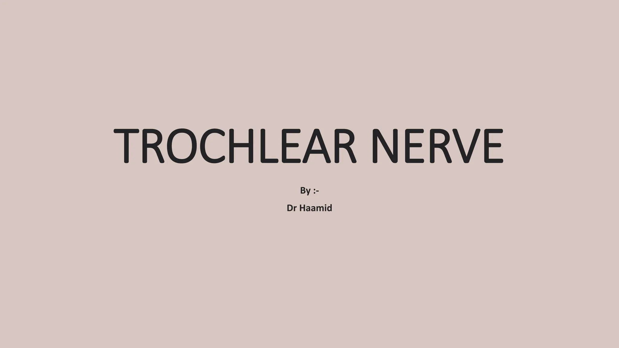
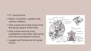
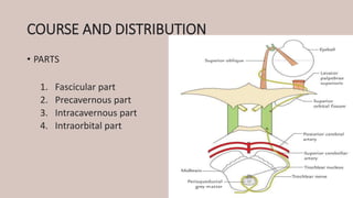
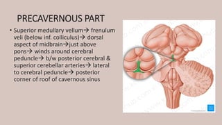
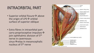
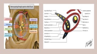
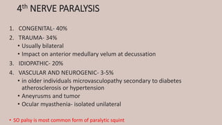
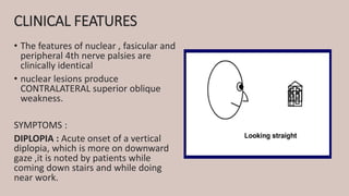
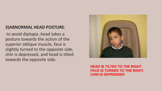
TROCHLEAR NERVE
By :-
Dr Haamid
• 4TH cranial nerve
• Motor in function, supplies only
superior oblique
• Only cranial nerve that arises from
the dorsal aspect of the brain
• Only cranial nerve to cross
completely to the other side (arises
from the contralateral nucleus)
• Longest and thinnest of all cranial
nerves
FUNCTIONAL COMPONENTS
• SOMATIC EFFERENT- movement of the eyeball through superior
oblique muscle
• GENERAL SOMATIC AFFERENT- proprioceptive impulses from superior
oblique relayed in mesencephalic nucleus of trigeminal nerve
NUCLEUS
• Midbrain- Ventromedial part of
central gray matter at the level
of inferior colliculus
• Caudal to and continuous with
3rd nerve nucleus complex
• Belongs to somatic efferent
column of nuclei and closely
related to medial longitudinal
bundle
CONNECTIONS OF THE NUCLEUS
• CEREBRAL CORTEX-
• Motor cortex (precentral gyrus) corticonuclear tracts
• Visual cortex superior colliculus & tactobulbar tract
• Frontal eye fields
• Nuclei of 3rd, 6th & 8th cranial nerves  medial longitudinal bundle
• Superior colliculi  descenginf predorsal bundle
• Vertical & torsional gaze centres
• Cerebellum vestibular nuclei
COURSE AND DISTRIBUTION
• PARTS
1. Fascicular part
2. Precavernous part
3. Intracavernous part
4. Intraorbital part
FASCICULAR PART
• Nucles efferent fibres
posteriorly around the
aqueductal gray matter
anterior medullary velum
decussate completely
PRECAVERNOUS PART
• Superior medullary vellum frenulum
veli (below inf. colliculus) dorsal
aspect of midbrainjust above
pons winds around cerebral
peduncle b/w posterior cerebral &
superior cerebellar arteries lateral
to cerebral peduncle posterior
corner of roof of cavernous sinus
INTRACAVERNOUS PART
• Cavernous sinus lateral wall
below oculomotor + above 1st
div of trigeminal ant part
crosses over 3rd nerve lateral
part of superior orbital fissure
INTRAORBITAL PART
• Superior orbital fissure above
the origin of LPS orbital
surface of superior oblique
• Extra fibres in intraorbital part
carry proprioceptive impulses
join ophthalmic division of 5th
nerve in cavernouos
sinusrelay in mesencephalic
nucleus of 5th nerve
SUPERIOR OBLIQUE MUSCLE
• PRIMARY POSITION OF GAZE
• Primary action- INTORSION (along A-P axis)
• Secondary action- DEPRESSION (along horizontal
axis)
• Tertiary action- ABDUCTION ( along vertical axis)
• GLOBE 51° ADDUCTED
• Axis of muscle rotation coincides with optical axis
• DEPRESSION only
• GLOBE 39° ABDUCTED
• Optical axis and line of pull of muscle mae an angle
of 90°
• INTORSION only
4th NERVE PARALYSIS
1. CONGENITAL- 40%
2. TRAUMA- 34%
• Usually bilateral
• Impact on anterior medullary velum at decussation
3. IDIOPATHIC- 20%
4. VASCULAR AND NEUROGENIC- 3-5%
• in older individuals microvasculopathy secondary to diabetes
atherosclerosis or hypertension
• Aneyrusms and tumor
• Ocular myasthenia- isolated unilateral
• SO palsy is most common form of paralytic squint
CLINICAL FEATURES
• The features of nuclear , fasicular and
peripheral 4th nerve palsies are
clinically identical
• nuclear lesions produce
CONTRALATERAL superior oblique
weakness.
SYMPTOMS :
DIPLOPIA : Acute onset of a vertical
diplopia, which is more on downward
gaze ,it is noted by patients while
coming down stairs and while doing
near work.
SIGNS :
1)HYPERTROPIA – the involved eye is
higher as a result of weakness of the
superior oblique muscle, which
becomes more prominent when the
head is tilted towards the ipsilateral
shoulder
2)RESTRICTED OCULAR MOVEMENTS:
there is limitation of depression on
adduction.
3)ABNORMAL HEAD POSTURE:
to avoid diplopia ,head takes a
posture towards the action of the
superior oblique muscle, face is
slightly turned to the opposite side,
chin is depressed, and head is tilted
towards the opposite side.
HEAD IS TILTED TO THE RIGHT.
FACE IS TURNED TO THE RIGHT.
CHIN IS DEPRESSED
THANK YOU

Trochlear nerve by Dr Haamid.pptx. learn about cranial nerved

  • 1.
  • 2.
    • 4TH cranialnerve • Motor in function, supplies only superior oblique • Only cranial nerve that arises from the dorsal aspect of the brain • Only cranial nerve to cross completely to the other side (arises from the contralateral nucleus) • Longest and thinnest of all cranial nerves
  • 3.
    FUNCTIONAL COMPONENTS • SOMATICEFFERENT- movement of the eyeball through superior oblique muscle • GENERAL SOMATIC AFFERENT- proprioceptive impulses from superior oblique relayed in mesencephalic nucleus of trigeminal nerve
  • 4.
    NUCLEUS • Midbrain- Ventromedialpart of central gray matter at the level of inferior colliculus • Caudal to and continuous with 3rd nerve nucleus complex • Belongs to somatic efferent column of nuclei and closely related to medial longitudinal bundle
  • 6.
    CONNECTIONS OF THENUCLEUS • CEREBRAL CORTEX- • Motor cortex (precentral gyrus) corticonuclear tracts • Visual cortex superior colliculus & tactobulbar tract • Frontal eye fields • Nuclei of 3rd, 6th & 8th cranial nerves  medial longitudinal bundle • Superior colliculi  descenginf predorsal bundle • Vertical & torsional gaze centres • Cerebellum vestibular nuclei
  • 7.
    COURSE AND DISTRIBUTION •PARTS 1. Fascicular part 2. Precavernous part 3. Intracavernous part 4. Intraorbital part
  • 8.
    FASCICULAR PART • Nuclesefferent fibres posteriorly around the aqueductal gray matter anterior medullary velum decussate completely
  • 9.
    PRECAVERNOUS PART • Superiormedullary vellum frenulum veli (below inf. colliculus) dorsal aspect of midbrainjust above pons winds around cerebral peduncle b/w posterior cerebral & superior cerebellar arteries lateral to cerebral peduncle posterior corner of roof of cavernous sinus
  • 11.
    INTRACAVERNOUS PART • Cavernoussinus lateral wall below oculomotor + above 1st div of trigeminal ant part crosses over 3rd nerve lateral part of superior orbital fissure
  • 12.
    INTRAORBITAL PART • Superiororbital fissure above the origin of LPS orbital surface of superior oblique • Extra fibres in intraorbital part carry proprioceptive impulses join ophthalmic division of 5th nerve in cavernouos sinusrelay in mesencephalic nucleus of 5th nerve
  • 14.
    SUPERIOR OBLIQUE MUSCLE •PRIMARY POSITION OF GAZE • Primary action- INTORSION (along A-P axis) • Secondary action- DEPRESSION (along horizontal axis) • Tertiary action- ABDUCTION ( along vertical axis) • GLOBE 51° ADDUCTED • Axis of muscle rotation coincides with optical axis • DEPRESSION only • GLOBE 39° ABDUCTED • Optical axis and line of pull of muscle mae an angle of 90° • INTORSION only
  • 15.
    4th NERVE PARALYSIS 1.CONGENITAL- 40% 2. TRAUMA- 34% • Usually bilateral • Impact on anterior medullary velum at decussation 3. IDIOPATHIC- 20% 4. VASCULAR AND NEUROGENIC- 3-5% • in older individuals microvasculopathy secondary to diabetes atherosclerosis or hypertension • Aneyrusms and tumor • Ocular myasthenia- isolated unilateral • SO palsy is most common form of paralytic squint
  • 16.
    CLINICAL FEATURES • Thefeatures of nuclear , fasicular and peripheral 4th nerve palsies are clinically identical • nuclear lesions produce CONTRALATERAL superior oblique weakness. SYMPTOMS : DIPLOPIA : Acute onset of a vertical diplopia, which is more on downward gaze ,it is noted by patients while coming down stairs and while doing near work.
  • 17.
    SIGNS : 1)HYPERTROPIA –the involved eye is higher as a result of weakness of the superior oblique muscle, which becomes more prominent when the head is tilted towards the ipsilateral shoulder 2)RESTRICTED OCULAR MOVEMENTS: there is limitation of depression on adduction.
  • 18.
    3)ABNORMAL HEAD POSTURE: toavoid diplopia ,head takes a posture towards the action of the superior oblique muscle, face is slightly turned to the opposite side, chin is depressed, and head is tilted towards the opposite side. HEAD IS TILTED TO THE RIGHT. FACE IS TURNED TO THE RIGHT. CHIN IS DEPRESSED
  • 19.