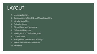
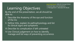
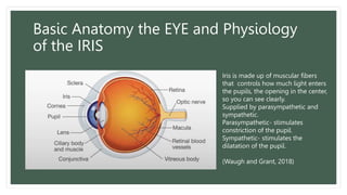
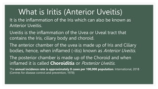
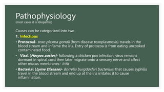
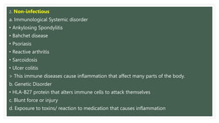
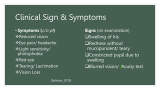
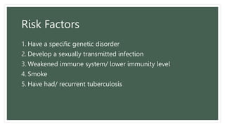
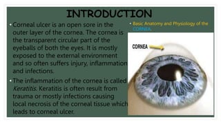
LUTHERAN SCHOOL
OF NURSING
Medical and Surgical Nursing 1
LAYOUT
1. Learning objectives
2. Basic Anatomy of the EYE and Physiology of Iris
3. Introduction of Irits
4. Pathophysiology
5. Clinical Signs and Symptoms
6. Differential Diagnosis
7. Investigation to confirm Diagnosis
8. Complications
9. Management (Medical and Nursing)
10. Health Education and Promotion
11. Reference
Learning Objectives
By the end of the presentation, we all should be
able to;
I. Describe the Anatomy of the eye and function
of the Iris
II. Define Iritis, explain its pathophysiology and list
its clinical signs and symptoms
III.Describe its complication if left untreated
IV.Use Clinical judgement on how to identify,
manage and list ways of preventing occurrence.
Learning Issues: (Iritis & Corneal Ulcer)
1.Clinical Signs and Symptoms
2. Management- (Standard Rx Book)
Basic Anatomy the EYE and Physiology
of the IRIS
Iris is made up of muscular fibers
that controls how much light enters
the pupils, the opening in the center,
so you can see clearly.
Supplied by parasympathetic and
sympathetic.
Parasympathetic- stimulates
constriction of the pupil.
Sympathetic- stimulates the
dilatation of the pupil.
(Waugh and Grant, 2018)
What is Iritis (Anterior Uveitis)
It is the inflammation of the Iris which can also be known as
Anterior Uveitis.
Uveitis is the inflammation of the Uvea or Uveal tract that
contains the Iris, ciliary body and choroid.
The anterior chamber of the uvea is made up of Iris and Ciliary
bodies, hence, when inflamed (-itis) known as Anterior Uveitis.
The posterior chamber is made up of the Choroid and when
inflamed it is called Choroiditis or Posterior Uveitis.
The annual incidence rate is approximately 8 cases per 100,000 population. International, 2018
(Centres for disease control and prevention, 1978)
Pathophysiology
(most cases it is Idiopathic)
Causes can be categorized into two
1. Infectious
• Protozoal- toxo plasma gondii (from disease toxoplasmosis) travels in the
blood stream and inflame the iris. Entry of protozoa is from eating uncooked
contaminated food.
• Viral (Herpes zoster)- following a chicken pox infection, virus remains
dormant in spinal cord then later migrate onto a sensory nerve and affect
other mucus membranes- Iritis
• Bacterial (Lyme Disease)- Borrelia burgdorferi bacterium that causes syphilis
travel in the blood stream and end up at the iris irritates it to cause
inflammation.
2. Non-infectious
a. Immunological Systemic disorder
• Ankylosing Spondylitis
• Bahchet disease
• Psoriasis
• Reactive arthritis
• Sarcoidosis
• Ulcer colitis
> This immune diseases cause inflammation that affect many parts of the body.
b. Genetic Disorder
• HLA-B27 protein that alters immune cells to attack themselves
c. Blunt force or injury
d. Exposure to toxins/ reaction to medication that causes inflammation
Clinical Sign & Symptoms
• Symptoms (c/o pt)
Reduced vision
Eye pain/ headache
Light sensitivity/
photophobia
Red eye
Tearing/ Lacrimation
Vision Loss
Signs (on examination)
Swelling of Iris
Redness without
mucopurulent/ teary
Constricted pupil due to
swelling
Blurred vision/ Acuity test
(Seltman, 2019)
Risk Factors
1. Have a specific genetic disorder
2. Develop a sexually transmitted infection
3. Weakened immune system/ lower immunity level
4. Smoke
5. Have had/ recurrent tuberculosis
Differential
Diagnosis
1.Conjunctivitis
2.Acute closure
Glaucoma
3. Keratitis
Investigation
• Interrogation- Ask if required, of any known
diseases or condition if not stated during
initial assessment
• Examination may include:
oA vision acuity test
oPressure reading
oPenlight and Slit lamp
*if Doctor suspects to be secondary, meaning another health
problem, may order
Blood tests,
Imaging tests (Chest x-ray)
Test fluid inside the eye
Management
Medical Treatment
Optimal Rx is dependent on the determination of cause. Iritis or uveitis in general is a
potentially blinding condition which requires prompt referral(administer loading dose
then refer to Hospital).
I. Admit and treat as inpatient
II. Look for signs for other disease and treat accordingly
III. Dilate the pupil using Atropine 1% eye ointment b.d until improve then use daily
optically then pad the eye (if nil discharge)
IV. Analgesics / Antihistamine- Aspirin 600mg (2 tabs) q.i.d
V. Antibiotic compound- Chloramphenicol eye ointment q.i.d 5/7
VI. Doctors might order Anti-inflammatory eye ointment- Hydrocortisone q.i.d 10/7 or
until improved
VII.Should inflammation persist, doctor may run blood works to confirm causative agents.
Nursing Care Process
Nursing Dx Goal/ Plan Intervention
Fear and Anxiety
related to
reduced vision
and red eye.
Awareness and
acceptance of
Condition.
1. Explain to patient the condition and treatment and possible
course of disease.
2. Advise on coping strategies- treatment
3. Advise on safety aspects related to reduced vision
Pain and
photophobia
Alleviation of Pain 1. Explain the cause of the pain.
2. Encourage compliance with topical treatment
3. Advise on the use of analgesics
4. Advise on wearing sun glasses
Potential
complications of
acute secondary
glaucoma
The early recognition
and treatment of
complication
1. Ensure patient is fully aware pain, further reduction in vision, or
in conjunctival injection requires further medical consultation
2. Advise patient to seek urgent medical help if signs of complication
appears.
Possibility of
reoccurrence
The recognition and
early treatment of
reoccurrence
1. Ensure the patient is aware of the signs of reoccurrence and the
need for early treatment.
2. Advise on the means of seeking urgent medical consultation should
reoccurrence be suspected.
3. Advise not to use old medication prior to seeking medical help.
Complication
• Cataracts. Development of a clouding of the lens of your eye (cataract) is a
possible complication, especially if you've had a long period of inflammation.
• An irregular pupil. Scar tissue can cause the iris to stick to the underlying lens
or the cornea, making the pupil irregular in shape and the iris sluggish in its
reaction to light.
• Glaucoma. Recurrent iritis can result in glaucoma, a serious eye condition
characterized by increased pressure inside the eye and possible vision loss.
• Calcium deposits on the cornea. This causes degeneration of your cornea
and could decrease your vision.
• Swelling within the retina. Swelling and fluid-filled cysts that develop in the
retina at the back of the eye can blur or decrease your central vision.
Health Education and Promotion
• Apply a warm compress to your eye.
• Use eye drops as prescribed. With the appropriate treatment strategy, supervised by an
ophthalmologist.
• Wear sunglasses to decrease light sensitivity and discomfort.
• If your eye is dilated or if you have been given an eye patch, avoid driving and any hazardous
activities.
• know when to go see a doctor
• Foods observed to reduce or 'turn down' the inflammatory response are: olive oil, leafy green
vegetables, fatty fish, and some fruits, nuts, herbs and spices. (Natural Eye Care, 1999)
Summary
• How does the Iris Function?
• What is Iritis?
• Who are responsible for the inflammation?
• List the type of investigation done to confirm medical diagnosis?
• What are the drugs given to reduce swelling and pain.
• When should we administer antibiotics
• And what can be assumed for further testing should inflammation persist?
• What is the main complication of Iritis
• What are some ways we can avoid this inflammation?
Kagutoki👌🤞
Any questions👀🤞
TOPIC:
Corneal Ulcer
OUTLINE
1. Learning outcome 12. Therapeutic Management
2. Introduction 13. Nursing Management
3. Epidemiology 14. Summary
4. Etiology(cause)
5. Pathophysiology
6. Risk/ Pre-disposing factors
7. Signs and Symptoms
8. Differential Diagnosis
9. Investigations
10.Provisional Diagnosis
11.Diagram of Corneal
12.Complications
LEARNING OUTCOME
By the end of this presentation we will be able to:
1. Understand more about Corneal Ulcer, its Cause and its Pathophysiology.
2. Know the contributing factors or risk factors that leads to corneal ulcer.
3. Identify the signs and symptoms (clinical manifestation) of corneal ulcer.
4. Make differential diagnosis based on some possible clinical manifestations of
corneal Ulcer.
5. Know and understand the proper different investigations done for accurate
diagnosis of corneal ulcer.
6. Know the complications of Corneal Ulcer.
7. Identify the Therapeutic Management and Nursing Management of Corneal
Ulcer.
INTRODUCTION
• Corneal ulcer is an open sore in the
outer layer of the cornea. The cornea is
the transparent circular part of the
eyeballs of both the eyes. It is mostly
exposed to the external environment
and so often suffers injury, inflammation
and infections.
• The inflammation of the cornea is called
Keratitis. Keratitis is often result from
trauma or mostly infections causing
local necrosis of the corneal tissue which
leads to corneal ulcer.
• Basic Anatomy and Physiology of the
CORNEA.
EPIDEMIOLOGY
• The magnitude of blindness (all causes) in the countries of the South-East Asia
Region varies from 3 000 people per million population in communities with
good economy and health care to over 10 000 per million in low-income
settings.
• Corneal ulcer is a common cause of blindness in low-income settings being
responsible for 5-20% of all blindness.
• In warm, humid areas the relative proportion of fungal to bacterial ulcers
approaches 50:50, while in cool dry climates most ulcers are due to bacteria.
• The major bacterial causes are streptococcus; pseudomonas and staphylococcus.
• The major fungal isolates are fusarium and aspergillus species. Candida is
relatively uncommon.
AETIOLOGY(CAUSE)
Some causative agents which leads of Corneal Ulcer includes;
• Viruses (herpes simplex, chlamydia)
• Bacteria (streptococcus; pseudomonas and staphylococcus.)
• Fungi (aspergillus, fusarium)
• Protozoa (acanthamoeba)
• Vitamin A deficiency
PATHOPHYSIOLOGY
• Corneal ulcer is the corneal epithelial defect with underlying
inflammation usually due to the invasions by Bacteria, Fungi, Viruses
or Acanthamoeba. It can be initiated by mechanical trauma,
nutritional deficiency and uncontrolled inflammation which can lead
to cause corneal necrosis. During the inflammation of the cornea, the
inflammatory cells infiltrate the different corneal layers in response to
noxious stimuli, either infectious exogenous agents or self antigens.
The inflammatory reaction may result in the supperative melting of
the corneal epithelium and stroma resulting in the formation of the
ulcer.
Diagram Of Corneal
Ulcer.
RISK FACTORS
• Contact Lenses- wearing contact lenses increases the risk of infectious and non-infectious keratitis.
The risk stems from not disinfecting lenses properly, wearing contact lenses while swimming,
wearing them longer than recommended & etc.
• Reduce immunity- when the immune system is compromised due to disease or medications.
• Warm climate- living in a warm climate and when a plant material gets into the eye. Plant material
can scratch the corneal epithelium and the chemical from the plant can cause inflammation which
may lead to an infection.
• Corticosteroids- the use of corticosteroids eye drops to treat eye disorders can increase the risk
developing keratitis or worsen the existing keratitis.
• Eye injury- if one of the cornea has been damaged from an injury in the past, the vulnerability of
developing keratitis is high.
• Vitamin A deficiency- if the body lacks vitamin A, the eyes will be at high risk of developing eye
problem and other eye complications.
SIGNS AND SYMPTOMS
• Eye redness
• Eye pain
• Excess tears or other discharge from the
eyes
• Difficulty opening the eyelid because of
the pain or irritation
• Blurred vision
• Decreased or diminished vision
• Irritation and feeling of discomfort in the
eyes
• Sensitivity to light (Photophobia)
• A feeling that something is in the eye
DIFFERENTIAL DIAGNOSIS
The differential diagnosis of includes ;
• Conjunctivitis
• Eye trauma/ Eye injury
• Foreign body in the eye
• Trachoma
DIAGNOSTIC EVALUATION
Accurate Diagnosis of Corneal Ulcer are made possible through
the following evaluations;
• Eye exam- The exam will include an effort to determine how well one
can see(visual acuity)
• Penlight exam- This is to examine the eyes using a penlight to check
the pupils reaction, size and other factors by applying a stain to help
identify the extent and character of surface irregularities and ulcer of
the cornea.
• Fluorescein Dye- To see the extent of the damage.
Cont.…
• Slit-lamp exam- This instrument uses an intense of light, a slit-like
beam to illuminate the cornea, iris, lens and space between the iris
and the cornea. The light to view these structure with high
magnification to detect the character and extent of Keratitis as well the
effects it may have on other structures of the eyes.
• Laboratory Analysis- Taking a sample of the tears or some cells from
the cornea for laboratory analysis to determine the cause of Keratitis
to help develop treatment and management of the condition.
PROVISIONAL DIAGNOSIS
• Corneal Ulcer
Therapeutic & Nursing
Management
• Viral infection (Herpes simplex keratitis)
Acyclovir (Zovirax) 3% ointment 5 times a day.
Acyclovir 200-400mg ‘o’ Dly 5-10 days
Eye irrigation of any discharge particularly with sodium chloride 0.9% or cooled
boil water.
Symptomatic relief can be provided by: Cool compress and or simple eye lubricants
or gel.
• Bacterial infection
Chloramphenicol eye drop 0.5% topical 1 drop dly 3/7
Chloramphenicol ointment 1% topical Nocte 3/7
Analgesic- Paracetamol 1g ‘o’ PRN/QID adult, 125-250mg children
• Fungal infection
Topical 0.2% Fluconazole dly until improvement.
Ketoconazole 200mg dly
COMPLICATIONS
Potential complications of keratitis and corneal ulcer includes;
• Chronic corneal inflammation
• Chronic or recurrent viral infection of the cornea
• Open sores on the cornea
• Corneal swelling and scaring
• Temporary or permanent reduction of vision
• Possible complete blindness
Health education and
promotion
• Avoid eye makeup.
• DO NOT wear contact lenses at all, especially while asleep.
• Take pain medicines.
• Wear protective glasses.
• Wear sunglasses when you're outside.
• Wearing goggles or safety glasses when dust or other particles could fly
into your eye.
• Carrots contain vitamin A, which helps with night vision and protects the
eyes by helping to absorb light. Increased levels of vitamin A mean your
eyes can absorb more energy and become more sensitive in dim
light. Eating carrots can also help prevent cataracts, and corneal ulcers.
SUMMARY
• Corneal ulcer is an open sore on the outer layer of the cornea. The underlying
defect of epithelial tissue of the cornea being damaged due to inflammation by
different causative agents.
• Corneal ulcer is a secondary condition results from the inflammation of the
cornea.
• The inflammation of the cornea is known as Keratitis
• Keratitis and corneal ulcer are caused mostly by Bacteria, Virus, Fungi, and at
times Protozoa through trauma, eye injury or any foreign body in the eyes.
• Corneal Ulcers are managed based on its causative agents, and i.e. through
Antimicrobial, Antiviral, Antifungal and Antiprotozoal drugs.
End Of
Presentation
Reference
Centers for Disease Control and Prevention, 1978, Principles of Epidemiology: Lesson 3 - Section
2, United States; https://www.cdc.gov/csels/dsepd/ss1978/lesson3/section2.html
Grossman. M and Edson. M, (2019), Natural Eye Care: Your Guide to Healthy Vision and Healing,
2019 Accessed: https://www.naturaleyecare.com/about.asp
Perry, J.P and Tullo, A. B, (1995), Care of the ophthalmic patient: a guide for nurses and health
professional, (2nd edn.) London: Chapman & Hall.
Seltman. W, (2019), Eye health, WebMD editorial contributors: https://www.webmd.com/eye-
health/iritis
The Mayo Clinic, (2019), Mayo Clinic Family Health Book, (5th edn.)
https://www.mayoclinic.org/diseases-conditions/iritis/symptoms-causes/syc-20354961
Waugh. A and Grant. A, 2018, Ross and Wilson Anatomy and Physiology in health and illness (13th
edn.) Elsevier.

Iritis and Corneal Ulcer Presentation.pptx

  • 1.
    LUTHERAN SCHOOL OF NURSING Medicaland Surgical Nursing 1
  • 2.
    LAYOUT 1. Learning objectives 2.Basic Anatomy of the EYE and Physiology of Iris 3. Introduction of Irits 4. Pathophysiology 5. Clinical Signs and Symptoms 6. Differential Diagnosis 7. Investigation to confirm Diagnosis 8. Complications 9. Management (Medical and Nursing) 10. Health Education and Promotion 11. Reference
  • 3.
    Learning Objectives By theend of the presentation, we all should be able to; I. Describe the Anatomy of the eye and function of the Iris II. Define Iritis, explain its pathophysiology and list its clinical signs and symptoms III.Describe its complication if left untreated IV.Use Clinical judgement on how to identify, manage and list ways of preventing occurrence. Learning Issues: (Iritis & Corneal Ulcer) 1.Clinical Signs and Symptoms 2. Management- (Standard Rx Book)
  • 4.
    Basic Anatomy theEYE and Physiology of the IRIS Iris is made up of muscular fibers that controls how much light enters the pupils, the opening in the center, so you can see clearly. Supplied by parasympathetic and sympathetic. Parasympathetic- stimulates constriction of the pupil. Sympathetic- stimulates the dilatation of the pupil. (Waugh and Grant, 2018)
  • 5.
    What is Iritis(Anterior Uveitis) It is the inflammation of the Iris which can also be known as Anterior Uveitis. Uveitis is the inflammation of the Uvea or Uveal tract that contains the Iris, ciliary body and choroid. The anterior chamber of the uvea is made up of Iris and Ciliary bodies, hence, when inflamed (-itis) known as Anterior Uveitis. The posterior chamber is made up of the Choroid and when inflamed it is called Choroiditis or Posterior Uveitis. The annual incidence rate is approximately 8 cases per 100,000 population. International, 2018 (Centres for disease control and prevention, 1978)
  • 6.
    Pathophysiology (most cases itis Idiopathic) Causes can be categorized into two 1. Infectious • Protozoal- toxo plasma gondii (from disease toxoplasmosis) travels in the blood stream and inflame the iris. Entry of protozoa is from eating uncooked contaminated food. • Viral (Herpes zoster)- following a chicken pox infection, virus remains dormant in spinal cord then later migrate onto a sensory nerve and affect other mucus membranes- Iritis • Bacterial (Lyme Disease)- Borrelia burgdorferi bacterium that causes syphilis travel in the blood stream and end up at the iris irritates it to cause inflammation.
  • 7.
    2. Non-infectious a. ImmunologicalSystemic disorder • Ankylosing Spondylitis • Bahchet disease • Psoriasis • Reactive arthritis • Sarcoidosis • Ulcer colitis > This immune diseases cause inflammation that affect many parts of the body. b. Genetic Disorder • HLA-B27 protein that alters immune cells to attack themselves c. Blunt force or injury d. Exposure to toxins/ reaction to medication that causes inflammation
  • 8.
    Clinical Sign &Symptoms • Symptoms (c/o pt) Reduced vision Eye pain/ headache Light sensitivity/ photophobia Red eye Tearing/ Lacrimation Vision Loss Signs (on examination) Swelling of Iris Redness without mucopurulent/ teary Constricted pupil due to swelling Blurred vision/ Acuity test (Seltman, 2019)
  • 9.
    Risk Factors 1. Havea specific genetic disorder 2. Develop a sexually transmitted infection 3. Weakened immune system/ lower immunity level 4. Smoke 5. Have had/ recurrent tuberculosis
  • 10.
  • 11.
    Investigation • Interrogation- Askif required, of any known diseases or condition if not stated during initial assessment • Examination may include: oA vision acuity test oPressure reading oPenlight and Slit lamp
  • 12.
    *if Doctor suspectsto be secondary, meaning another health problem, may order Blood tests, Imaging tests (Chest x-ray) Test fluid inside the eye
  • 13.
    Management Medical Treatment Optimal Rxis dependent on the determination of cause. Iritis or uveitis in general is a potentially blinding condition which requires prompt referral(administer loading dose then refer to Hospital). I. Admit and treat as inpatient II. Look for signs for other disease and treat accordingly III. Dilate the pupil using Atropine 1% eye ointment b.d until improve then use daily optically then pad the eye (if nil discharge) IV. Analgesics / Antihistamine- Aspirin 600mg (2 tabs) q.i.d V. Antibiotic compound- Chloramphenicol eye ointment q.i.d 5/7 VI. Doctors might order Anti-inflammatory eye ointment- Hydrocortisone q.i.d 10/7 or until improved VII.Should inflammation persist, doctor may run blood works to confirm causative agents. Nursing Care Process
  • 14.
    Nursing Dx Goal/Plan Intervention Fear and Anxiety related to reduced vision and red eye. Awareness and acceptance of Condition. 1. Explain to patient the condition and treatment and possible course of disease. 2. Advise on coping strategies- treatment 3. Advise on safety aspects related to reduced vision Pain and photophobia Alleviation of Pain 1. Explain the cause of the pain. 2. Encourage compliance with topical treatment 3. Advise on the use of analgesics 4. Advise on wearing sun glasses Potential complications of acute secondary glaucoma The early recognition and treatment of complication 1. Ensure patient is fully aware pain, further reduction in vision, or in conjunctival injection requires further medical consultation 2. Advise patient to seek urgent medical help if signs of complication appears. Possibility of reoccurrence The recognition and early treatment of reoccurrence 1. Ensure the patient is aware of the signs of reoccurrence and the need for early treatment. 2. Advise on the means of seeking urgent medical consultation should reoccurrence be suspected. 3. Advise not to use old medication prior to seeking medical help.
  • 15.
    Complication • Cataracts. Developmentof a clouding of the lens of your eye (cataract) is a possible complication, especially if you've had a long period of inflammation. • An irregular pupil. Scar tissue can cause the iris to stick to the underlying lens or the cornea, making the pupil irregular in shape and the iris sluggish in its reaction to light. • Glaucoma. Recurrent iritis can result in glaucoma, a serious eye condition characterized by increased pressure inside the eye and possible vision loss. • Calcium deposits on the cornea. This causes degeneration of your cornea and could decrease your vision. • Swelling within the retina. Swelling and fluid-filled cysts that develop in the retina at the back of the eye can blur or decrease your central vision.
  • 16.
    Health Education andPromotion • Apply a warm compress to your eye. • Use eye drops as prescribed. With the appropriate treatment strategy, supervised by an ophthalmologist. • Wear sunglasses to decrease light sensitivity and discomfort. • If your eye is dilated or if you have been given an eye patch, avoid driving and any hazardous activities. • know when to go see a doctor • Foods observed to reduce or 'turn down' the inflammatory response are: olive oil, leafy green vegetables, fatty fish, and some fruits, nuts, herbs and spices. (Natural Eye Care, 1999)
  • 17.
    Summary • How doesthe Iris Function? • What is Iritis? • Who are responsible for the inflammation? • List the type of investigation done to confirm medical diagnosis? • What are the drugs given to reduce swelling and pain. • When should we administer antibiotics • And what can be assumed for further testing should inflammation persist? • What is the main complication of Iritis • What are some ways we can avoid this inflammation?
  • 18.
  • 19.
  • 20.
    OUTLINE 1. Learning outcome12. Therapeutic Management 2. Introduction 13. Nursing Management 3. Epidemiology 14. Summary 4. Etiology(cause) 5. Pathophysiology 6. Risk/ Pre-disposing factors 7. Signs and Symptoms 8. Differential Diagnosis 9. Investigations 10.Provisional Diagnosis 11.Diagram of Corneal 12.Complications
  • 21.
    LEARNING OUTCOME By theend of this presentation we will be able to: 1. Understand more about Corneal Ulcer, its Cause and its Pathophysiology. 2. Know the contributing factors or risk factors that leads to corneal ulcer. 3. Identify the signs and symptoms (clinical manifestation) of corneal ulcer. 4. Make differential diagnosis based on some possible clinical manifestations of corneal Ulcer. 5. Know and understand the proper different investigations done for accurate diagnosis of corneal ulcer. 6. Know the complications of Corneal Ulcer. 7. Identify the Therapeutic Management and Nursing Management of Corneal Ulcer.
  • 22.
    INTRODUCTION • Corneal ulceris an open sore in the outer layer of the cornea. The cornea is the transparent circular part of the eyeballs of both the eyes. It is mostly exposed to the external environment and so often suffers injury, inflammation and infections. • The inflammation of the cornea is called Keratitis. Keratitis is often result from trauma or mostly infections causing local necrosis of the corneal tissue which leads to corneal ulcer. • Basic Anatomy and Physiology of the CORNEA.
  • 23.
    EPIDEMIOLOGY • The magnitudeof blindness (all causes) in the countries of the South-East Asia Region varies from 3 000 people per million population in communities with good economy and health care to over 10 000 per million in low-income settings. • Corneal ulcer is a common cause of blindness in low-income settings being responsible for 5-20% of all blindness. • In warm, humid areas the relative proportion of fungal to bacterial ulcers approaches 50:50, while in cool dry climates most ulcers are due to bacteria. • The major bacterial causes are streptococcus; pseudomonas and staphylococcus. • The major fungal isolates are fusarium and aspergillus species. Candida is relatively uncommon.
  • 24.
    AETIOLOGY(CAUSE) Some causative agentswhich leads of Corneal Ulcer includes; • Viruses (herpes simplex, chlamydia) • Bacteria (streptococcus; pseudomonas and staphylococcus.) • Fungi (aspergillus, fusarium) • Protozoa (acanthamoeba) • Vitamin A deficiency
  • 25.
    PATHOPHYSIOLOGY • Corneal ulceris the corneal epithelial defect with underlying inflammation usually due to the invasions by Bacteria, Fungi, Viruses or Acanthamoeba. It can be initiated by mechanical trauma, nutritional deficiency and uncontrolled inflammation which can lead to cause corneal necrosis. During the inflammation of the cornea, the inflammatory cells infiltrate the different corneal layers in response to noxious stimuli, either infectious exogenous agents or self antigens. The inflammatory reaction may result in the supperative melting of the corneal epithelium and stroma resulting in the formation of the ulcer.
  • 26.
  • 27.
    RISK FACTORS • ContactLenses- wearing contact lenses increases the risk of infectious and non-infectious keratitis. The risk stems from not disinfecting lenses properly, wearing contact lenses while swimming, wearing them longer than recommended & etc. • Reduce immunity- when the immune system is compromised due to disease or medications. • Warm climate- living in a warm climate and when a plant material gets into the eye. Plant material can scratch the corneal epithelium and the chemical from the plant can cause inflammation which may lead to an infection. • Corticosteroids- the use of corticosteroids eye drops to treat eye disorders can increase the risk developing keratitis or worsen the existing keratitis. • Eye injury- if one of the cornea has been damaged from an injury in the past, the vulnerability of developing keratitis is high. • Vitamin A deficiency- if the body lacks vitamin A, the eyes will be at high risk of developing eye problem and other eye complications.
  • 28.
    SIGNS AND SYMPTOMS •Eye redness • Eye pain • Excess tears or other discharge from the eyes • Difficulty opening the eyelid because of the pain or irritation • Blurred vision • Decreased or diminished vision • Irritation and feeling of discomfort in the eyes • Sensitivity to light (Photophobia) • A feeling that something is in the eye
  • 29.
    DIFFERENTIAL DIAGNOSIS The differentialdiagnosis of includes ; • Conjunctivitis • Eye trauma/ Eye injury • Foreign body in the eye • Trachoma
  • 30.
    DIAGNOSTIC EVALUATION Accurate Diagnosisof Corneal Ulcer are made possible through the following evaluations; • Eye exam- The exam will include an effort to determine how well one can see(visual acuity) • Penlight exam- This is to examine the eyes using a penlight to check the pupils reaction, size and other factors by applying a stain to help identify the extent and character of surface irregularities and ulcer of the cornea. • Fluorescein Dye- To see the extent of the damage.
  • 31.
    Cont.… • Slit-lamp exam-This instrument uses an intense of light, a slit-like beam to illuminate the cornea, iris, lens and space between the iris and the cornea. The light to view these structure with high magnification to detect the character and extent of Keratitis as well the effects it may have on other structures of the eyes. • Laboratory Analysis- Taking a sample of the tears or some cells from the cornea for laboratory analysis to determine the cause of Keratitis to help develop treatment and management of the condition.
  • 32.
  • 33.
    Therapeutic & Nursing Management •Viral infection (Herpes simplex keratitis) Acyclovir (Zovirax) 3% ointment 5 times a day. Acyclovir 200-400mg ‘o’ Dly 5-10 days Eye irrigation of any discharge particularly with sodium chloride 0.9% or cooled boil water. Symptomatic relief can be provided by: Cool compress and or simple eye lubricants or gel. • Bacterial infection Chloramphenicol eye drop 0.5% topical 1 drop dly 3/7 Chloramphenicol ointment 1% topical Nocte 3/7 Analgesic- Paracetamol 1g ‘o’ PRN/QID adult, 125-250mg children • Fungal infection Topical 0.2% Fluconazole dly until improvement. Ketoconazole 200mg dly
  • 34.
    COMPLICATIONS Potential complications ofkeratitis and corneal ulcer includes; • Chronic corneal inflammation • Chronic or recurrent viral infection of the cornea • Open sores on the cornea • Corneal swelling and scaring • Temporary or permanent reduction of vision • Possible complete blindness
  • 35.
    Health education and promotion •Avoid eye makeup. • DO NOT wear contact lenses at all, especially while asleep. • Take pain medicines. • Wear protective glasses. • Wear sunglasses when you're outside. • Wearing goggles or safety glasses when dust or other particles could fly into your eye. • Carrots contain vitamin A, which helps with night vision and protects the eyes by helping to absorb light. Increased levels of vitamin A mean your eyes can absorb more energy and become more sensitive in dim light. Eating carrots can also help prevent cataracts, and corneal ulcers.
  • 36.
    SUMMARY • Corneal ulceris an open sore on the outer layer of the cornea. The underlying defect of epithelial tissue of the cornea being damaged due to inflammation by different causative agents. • Corneal ulcer is a secondary condition results from the inflammation of the cornea. • The inflammation of the cornea is known as Keratitis • Keratitis and corneal ulcer are caused mostly by Bacteria, Virus, Fungi, and at times Protozoa through trauma, eye injury or any foreign body in the eyes. • Corneal Ulcers are managed based on its causative agents, and i.e. through Antimicrobial, Antiviral, Antifungal and Antiprotozoal drugs.
  • 37.
  • 38.
    Reference Centers for DiseaseControl and Prevention, 1978, Principles of Epidemiology: Lesson 3 - Section 2, United States; https://www.cdc.gov/csels/dsepd/ss1978/lesson3/section2.html Grossman. M and Edson. M, (2019), Natural Eye Care: Your Guide to Healthy Vision and Healing, 2019 Accessed: https://www.naturaleyecare.com/about.asp Perry, J.P and Tullo, A. B, (1995), Care of the ophthalmic patient: a guide for nurses and health professional, (2nd edn.) London: Chapman & Hall. Seltman. W, (2019), Eye health, WebMD editorial contributors: https://www.webmd.com/eye- health/iritis The Mayo Clinic, (2019), Mayo Clinic Family Health Book, (5th edn.) https://www.mayoclinic.org/diseases-conditions/iritis/symptoms-causes/syc-20354961 Waugh. A and Grant. A, 2018, Ross and Wilson Anatomy and Physiology in health and illness (13th edn.) Elsevier.

Editor's Notes

  • #4 Learning Issues
  • #11 They all have red eye, some sensitivity to light, and some degree of pain
  • #12 A good clinical assessment can already give you a sure clinical diagnosis of Iritis but the trick is the causative agent
  • #14 Since Iritis is often idiopathic, our aim is to reduce inflammation, pain and sensitivity to light and avoid any complication that may lead to Vision loss. Atropine 1% is used for mydriasis (widening of the pupils) and cycloplegic (paralysis of the iris muscle and ciliary muscles) to allow a fixed dilatation of the pupil. Always advise patients to avoid working/ driving- causes s
  • #15 Nursing care process basically involves relieving pain, create room for acceptance, ensure compliance to treatment and prevent reoccurrence.
  • #17 Help soothe the eye and decrease inflammation Use them as directed. Your provider may give you detailed instructions to follow. Demonstrate- Some doctors recommend pressing the finger against the inner corner of the eye to keep the medication from going into the tear duct. Common sense with eye pad A transient burning sensation may occur upon instillation. You have severe eye pain and a headache even after RX, vision suddenly gets worse, nausea or are vomiting, see halos or rainbows around lights, or have questions or concerns about your condition or care.