Downloaded 1,022 times


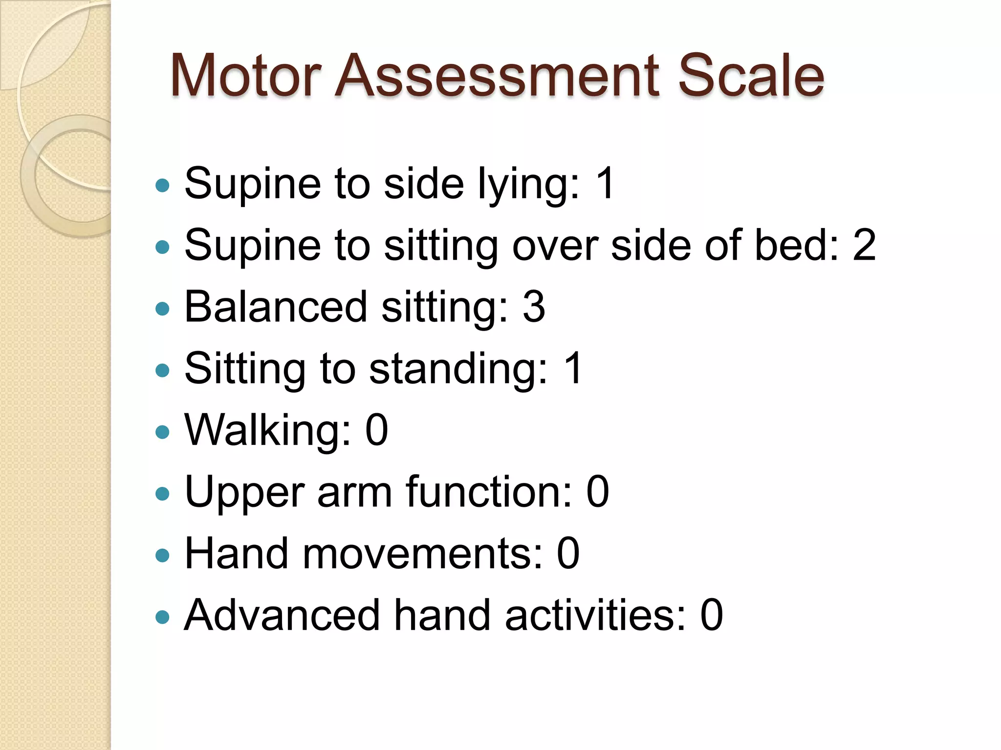


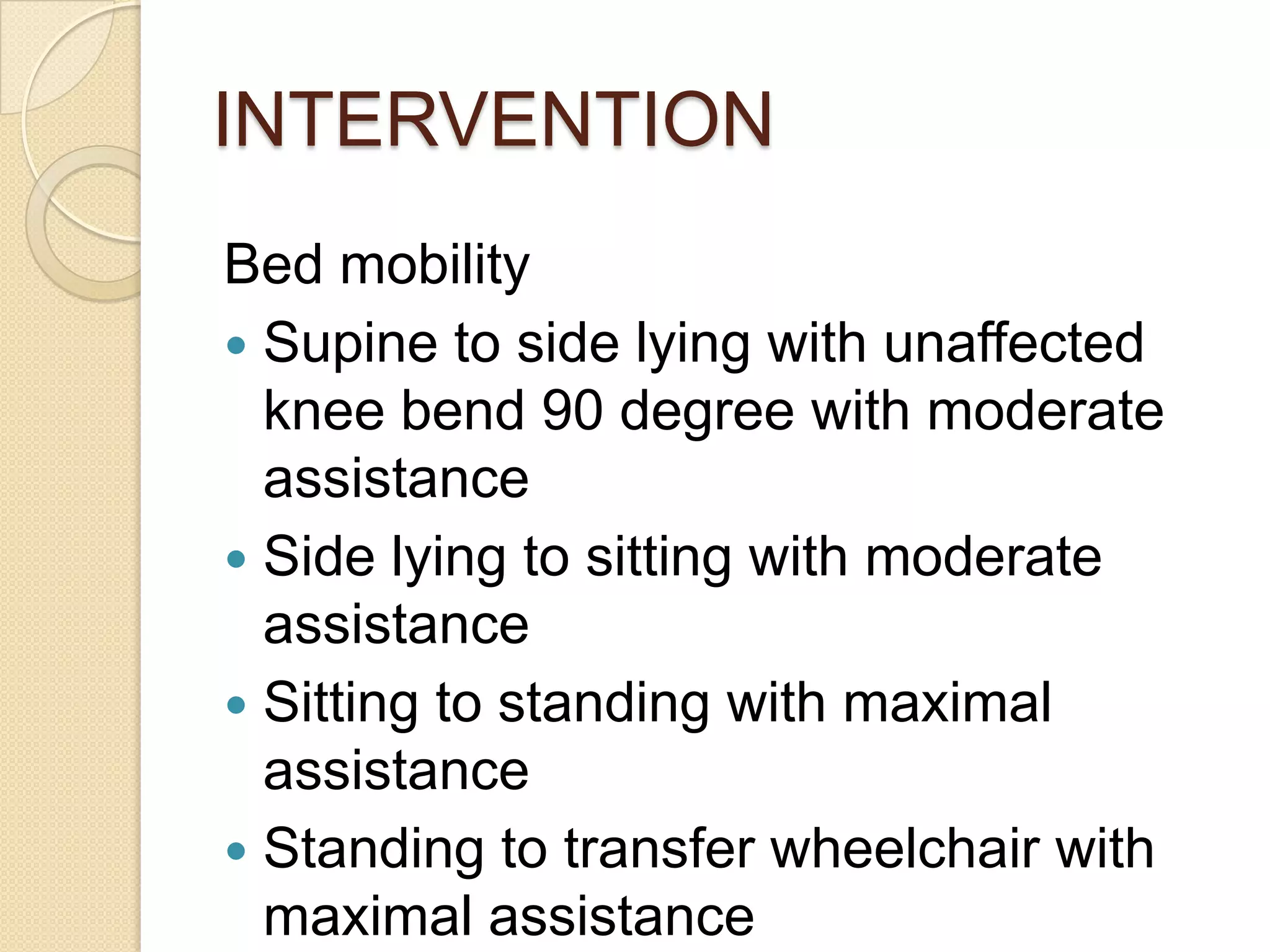
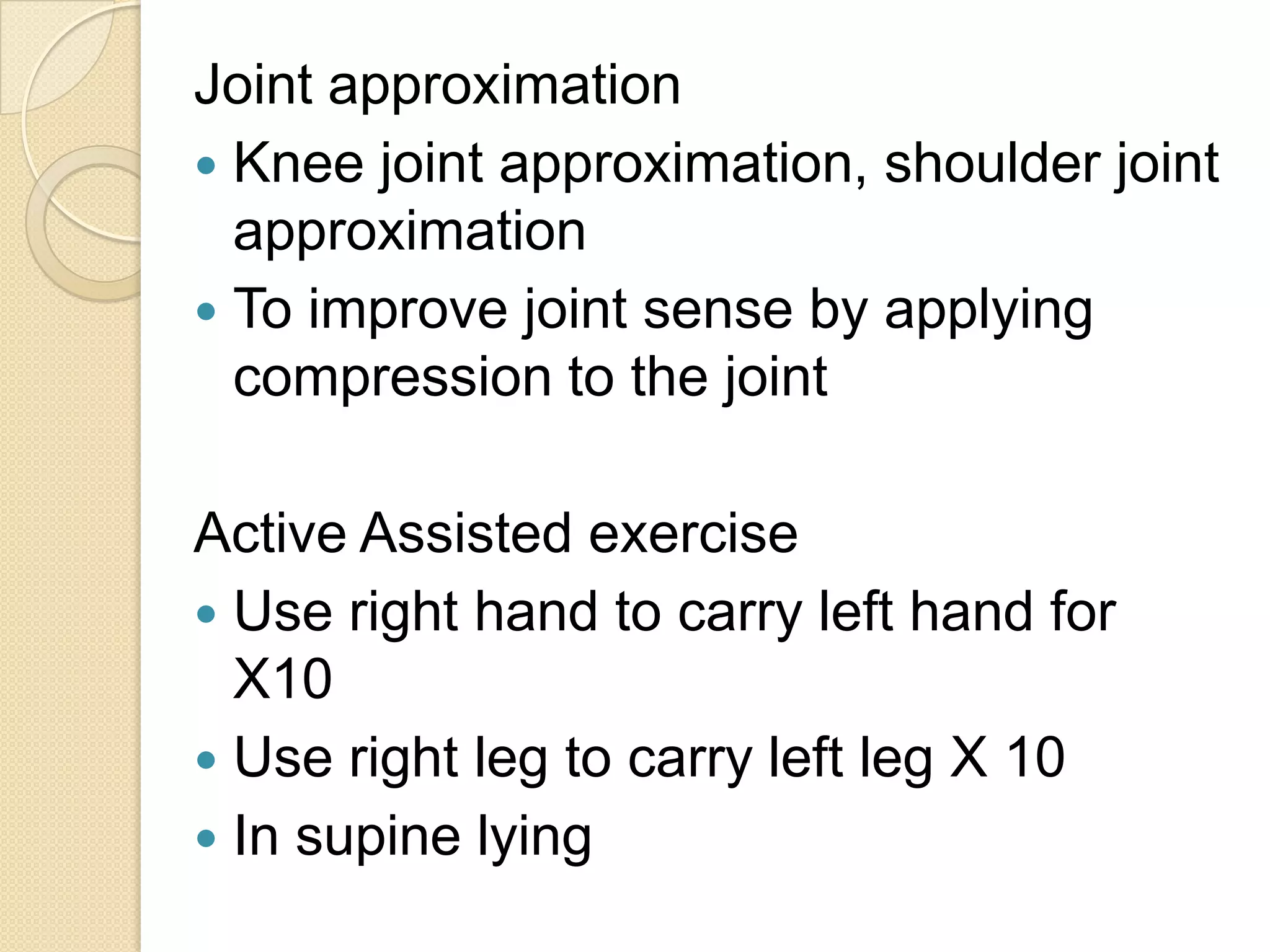


This document provides information about stroke including its causes, symptoms, diagnosis, and treatment. It begins with an introduction defining stroke as the interruption of blood flow to the brain. It then discusses the two main types of stroke: ischemic (caused by blockage) and hemorrhagic (caused by bleeding). Symptoms vary depending on the area of brain affected but can include paralysis, weakness, sensory loss, and speech problems. Stroke is diagnosed using CT scans or MRI. Treatment involves medications to prevent clots like aspirin, and sometimes surgery to repair blood vessels. Physiotherapy focuses on improving mobility, balance, and function.


![Stroke [uncensored] - by MHR Corporation](https://cdn.slidesharecdn.com/ss_thumbnails/mhr4-stroke-101129110104-phpapp01-thumbnail.jpg?width=640&height=640&fit=bounds)


![Stroke_Overview cause study[1] r.pptxyyyyh](https://cdn.slidesharecdn.com/ss_thumbnails/strokeoverviewcausestudy1r-250911133650-1b8cffa7-thumbnail.jpg?width=640&height=640&fit=bounds)


