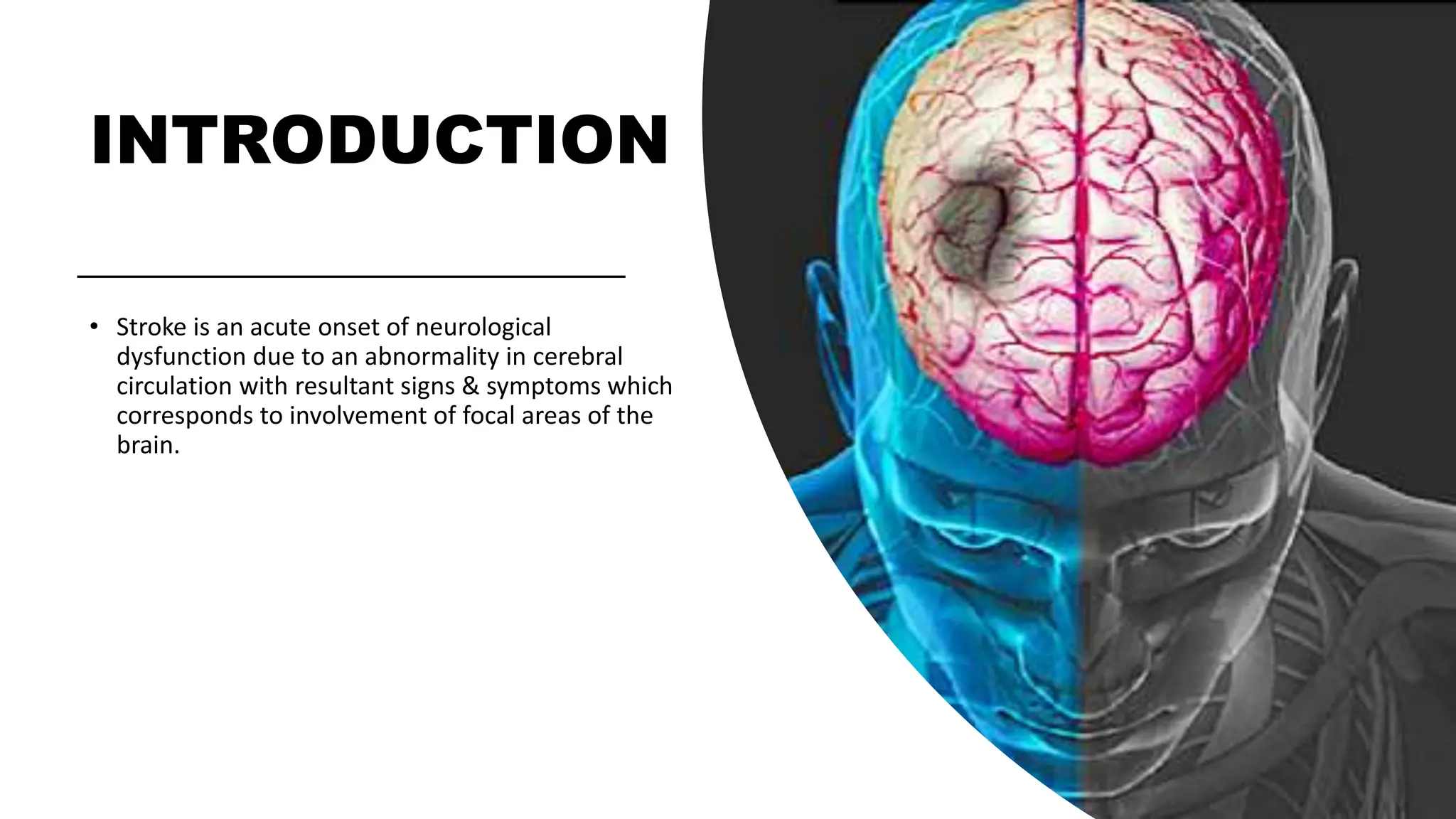
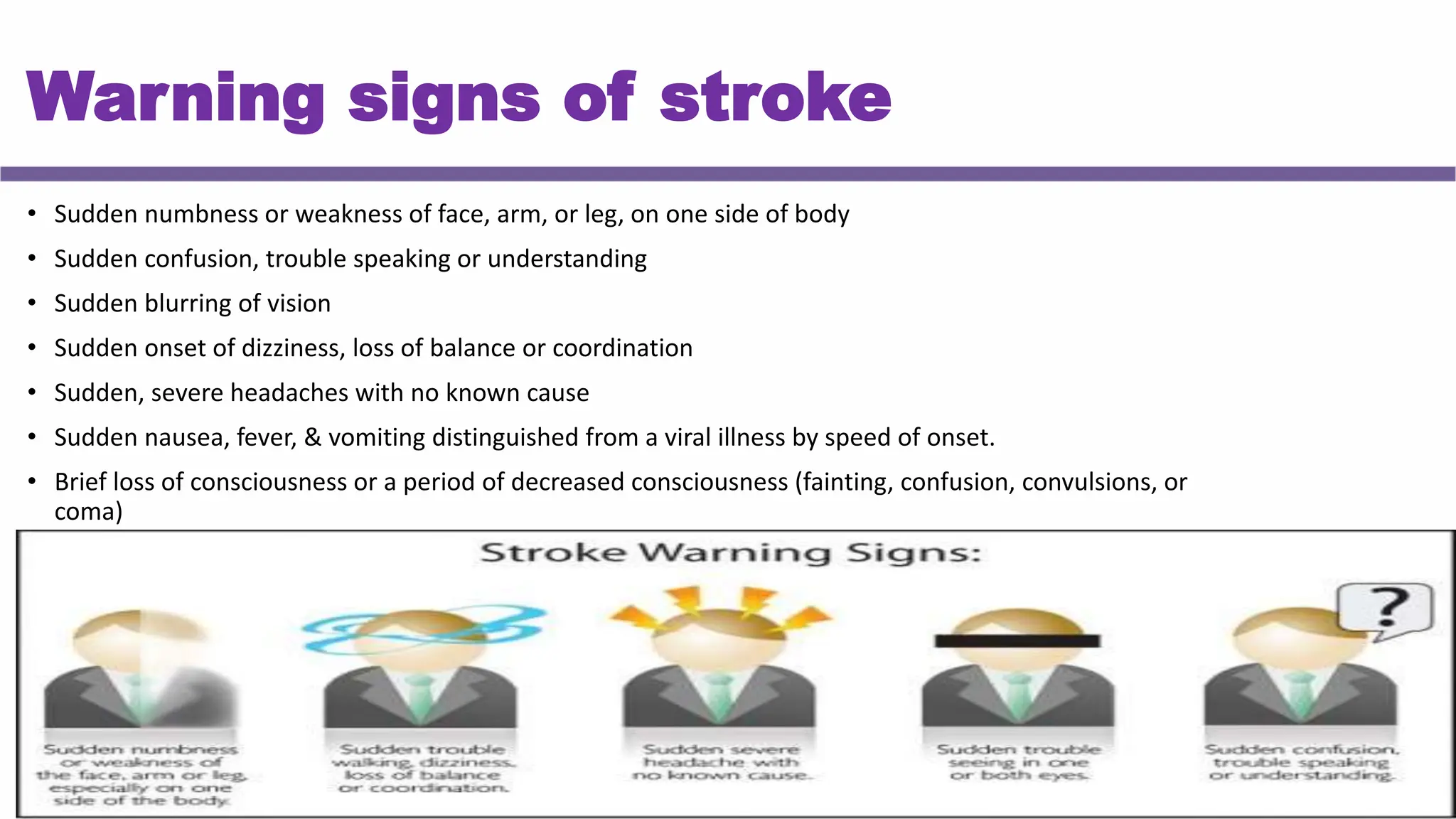
STROKE ASSESSMENT AND MANAGEMENT
Dr. Madiha Anees (PT)
Asst. Prof/VP RCRS
MS-PT , T-DPT, BS-PT
INTRODUCTION
• Stroke is an acute onset of neurological
dysfunction due to an abnormality in cerebral
circulation with resultant signs & symptoms which
corresponds to involvement of focal areas of the
brain.
ACC. TO WHO
• It is defined as the sudden onset of
neurological deficits due to an
abnormality in cerebral circulation with
the signs and symptoms lasting for more
than 24 hours or longer.
Transient ischemic attack
• It is defined as the sudden onset of
neurological deficits due to an
abnormality in cerebral circulation with
the signs and symptoms lasting for less
than 24 hours.
Epidemiology
• Third leading cause of death.
• The incidence of stroke is about 1.25 times greater for males than females.
• Most common cause of disability among adults.
Risk factors
MODIFIABLE
NON MODIFIABLE
• Ageing & gender
• Positive family history
• Heart disease
• Diabetes mellitus
• Hypertension
• Peripheral arterial disease
• Blood pathology
• Hyperlipidemia
• Smoking
• Obesity
• Lack of physical exercise or sedentary life style
• Diet & excess alcohol consumption
• Oral contraceptives
• Infection (meningeal infection)
• Psychological factors
Pathophysiology
Warning signs of stroke
• Sudden numbness or weakness of face, arm, or leg, on one side of body
• Sudden confusion, trouble speaking or understanding
• Sudden blurring of vision
• Sudden onset of dizziness, loss of balance or coordination
• Sudden, severe headaches with no known cause
• Sudden nausea, fever, & vomiting distinguished from a viral illness by speed of onset.
• Brief loss of consciousness or a period of decreased consciousness (fainting, confusion, convulsions, or
coma)
Stroke Classification
By specific vascular
territory or region:
• Artery: MCA, ACA,
PCA
• Lobe: Frontal,
temporal, occipital,
etc.
By management:
• Minor/major stroke
• Young stroke
• Deteriorating Stroke
By etiology:
• Thrombosis
(Ischemic)
• Embolus (Ischemic)
• Hemorrhage
CLASSIFICATION
Depending on the cause
Haemorrhagic stroke
• Intracranial haemorrhage
• Subarachnoid haemorrhage
CLASSIFICATION
Depending on the cause
ISCHEMIC STROKE
Thrombotic: more common. Usually occurs
in the sleeping hours. Characterised by
gradual onset of symptoms
Embolic: Occurs in the waking hours of the
day.
Emboli plug downstream arteries and
consist of pieces of thrombus or other
material that originate from proximal
arteries, the heart.
CLASSIFICATION
Depending on the severity:
• Mild stroke: symptoms subside with no deficit in a week period
• Moderate stroke: symptoms recover in a period of 3 - 6 months with
minimal neurological deficit
• Severe stroke: there is no complete recovery of the symptoms even
after 1 years. Always ends up with severe neurological deficit
CLASSIFICATION
Depending on the duration:
• Acute stroke: To a period of one week or until spasticity develops.
• Sub acute stroke: After the development of spasticity & last for a
period of 3-12 months
• Chronic stroke: More than 12 months
CLASSIFICATION
Depending on the symptoms:
• MCA Syndrome
• ACA Syndrome
• PCA syndrome
Circle of Willis
Territory of Circulation
Lateral View
MCA SYNDROME
RIGHT HEMI
• Contralateral hemiplegia (UL & face more affected
than LL)
• Contralateral hemisensory loss (UL & face more
affected than LL)
• Aphasia
• Ideomotor apraxia
• Ataxia of contralateral limb
• Contralateral Homonymous hemianopia
LEFT HEMI
• Autotopagnosia
• Anosgonosia
• Dysarthria
• Dressing & constructional dyspraxia
• Motor Impersistence
• Hemiparesis
• Topographic disorientation
• Anomia
• dysgraphia
Representation of body image over sensory &
motor cortex
ACA Syndrome
• Contralateral Hemiplegia of LL (LL more affected than UL)
• Contralateral sensory loss of LL
• Urinary incontinence
• Problems with imitation & bimanual task
• Apathy (Lethargic & lazy)
• Impulsive (lack of interest)
• Abulia (mental confusion)
• Akinetic mutism (mute)
• Gait Apraxia
• Personality disorders
• Problem in planning and execution
PCA Syndrome
• Coordination disorders such as tremor or ataxia
• Contralateral homonymous field deficit
• Cortical blindness
• Propagnosia (face blindness, unable to recognize faces)
• Cognitive impairment including memory impairment
• Thalamic syndrome (abnormal sensation of severe pain
from light touch or temperature changes)
• Weber’s syndrome (contralateral hemiplegia & third
nerve palsy)
• Physiological responses (vertigo, nausea, dizziness)
STAGES OF RECOVERY
Stage 1: Begins with a period of flaccidity immediately following
acute episode. No movement of limbs can be elicited.
Stage 2: Basic limb synergies. Minimal voluntary movement may be
present. Spasticity begins to develop.
Stage 3: Gains voluntary control of movement synergy although full
range is not developed. Spasticity has further increased.
STAGES OF RECOVERY
Stage 4: Some movement combination that do not follow
the synergy are mastered first with difficulty & later with more
ease. Spasticity begins to decline.
Stage 5: More difficult movement are learnt as the basic limb
synergy lose their dominance over motor roots. Spasticity
further declines.
Stage 6: Disappearance of spasticity, individual joint
movement become possible & coordination approaches normal.
Normal motor function is restored.
RED FLAG
Primary impairment
1. Altered sensation
2. Pain
3. Vision
4. Weakness
5. Alteration of tone
6. Abnormal synergy
7. Abnormal reflexes
8. Altered co ordination
9. Altered motor programing
10. Postural Control & Balance
11. Speech, Language, and Swallowing
12. Perception and Cognition
13. Emotional Status
14. Bladder and Bowel Function
PRIMARY IMPAIRMENT
1. ALTERED SENSATION
• Type and extent of deficit depends on location and
extent of the lesion.
• Profound hemisensory loss.
• Proprioceptive loss is common
PRIMARY IMPAIRMENT
2. PAIN:
• Pain is characterized by constant, severe burning pain with intermittent sharp pains.
• CVA can result in severe headache, neck or face pain.
• Thalamic pain syndrome.
• Pain due to secondary impairments.
• Hyperalgesia
• Loud sound, bright light etc. may trigger pain
Primary impairment
3. Vision
• Homonymous hemianopia, a visual field defect, in which there is
blindness on nasal half of one eye and temporal half of the other eye.
• Occurs with lesions involving the optic radiation (MCA) or to primary
visual cortex (PCA)
Primary impairment
4. Weakness
• Usually seen in the contralateral side of the lesion
• MCA stroke are more common so weakness is largely seen in the UL.
• Distal muscle are more affected than proximal muscles
• Mild weakness of ipsilateral side
Primary impairment
5. Alteration of tone
• Flaccidity (Hypotonicity) is present immediately after stroke and short lived.
• Spasticity (hypertonicity) emerges in about 90 percent of cases and occur
typically in predictable pattern.
ABNORMAL SYNERGY PATTRENS
• Stereotypical movement patterns associated with
spasticity.
• The patient is unable to move an isolated segment of a
limb without producing movements in the remainder of
the limb.
• 2 distinct abnormal synergy patterns have been
described for each extremity
Primary impairment
6. Abnormal synergy
Primary impairment
7. Abnormal reflexes
• Initially, hyporeflexia with flaccidity
• Middle stages of recovery (spasticity/synergy) hyperreflexia
• May demonstrate clonus, & +ve Babinski
Primary impairment
8. Altered co ordination
• Proprioceptive losses can result in sensory ataxia.
• Strokes affecting cerebellum typically produce cerebellar ataxia (e.g.
basilar artery syndrome, pontine syndromes) & motor weakness.
• Basal ganglia involvement (PCA syndrome) may lead to bradykinesia or
involuntary movements.
Primary impairment
9. Altered motor programing
• Motor praxis is ability to plan & execute coordinated
movement
• Lesions of premotor frontal cortex of either hemisphere, left
inferior parietal lobe, & corpus callosum can produce apraxia.
• Apraxia is more evident with left hemisphere damage than
right and is commonly seen with aphasia. It has 4 types.
Apraxia
Ideational
apraxia
Ideomotor
apraxia
Constructional
apraxia
Dressing apraxia
The patient has no
idea. They cant
perform complex
activity.
They are able to explain how to
perform movement, but cant
perform the movement.
Inability to draw or
construct simple
configuration.
Cant dress himself.
Primary impairment
10. Postural Control & Balance
• Impairments in steadiness, symmetry, & dynamic stability
• Problems may exist when reacting to a destabilizing external
force (reactive postural control) or during self-initiated
movements (anticipatory postural control).
Primary impairment
11. Speech, Language, and Swallowing
• Lesions involving cortex of dominant hemisphere
• Aphasia: Impairment of language comprehension, formulation, and use. It has 3
types.
Expressive aphasia Receptive aphasia Global aphasia
Broca’s aphasia
Motor aphasia
Problem in speaking words and
sentences
Wernicke’s aphasia
Sensory aphasia
Problem in understanding words
and sentences
Problem in both speaking and
understanding words and
sentences.
Primary impairment
Dysarthria:
Motor speech disorders caused by lesions of CNS or PNS that mediate speech
production.
Dysphagia:
• Swallowing difficulty – Usually delayed swallowing reflex – Aspiration occurs in 1/3 of patients
with dysphagia.
• Occurs with lesions affecting medullary brainstem (CN IX and X), large vessel pontine lesions, as
well as in acute MCA and PCA lesion.
Primary impairment
12. Perception and Cognition
• They are the result of lesions in right parietal cortex & seen more with left hemiplegia
than right.
• These may include disorders of body scheme/body image, spatial relations, and
agnosias.
• Visuospatial distortions: difficulty judging distance, size, position, rate of movement,
form.
• Unilateral neglect: patients are unaware of what happens on the hemiplegic side.
Primary impairment
13. Emotional Status
• Lesions of brain affecting frontal lobe, hypothalamus & limbic system
• May demonstrate pseudobulbar affect (PBA), also known as emotional lability or
emotional dysregulation syndrome.
• Emotional outbursts of uncontrolled or exaggerated laughing or crying that
are inconsistent with mood.
• Depression is extremely common
• Persistent feelings of sadness, feelings of hopelessness, worthlessness or
helplessness.
Primary impairment
14. Bladder and Bowel Function
• Disturbances of bladder function are common during acute phase
• Urinary incontinence can result from bladder hyperreflexia or
hyporeflexia, disturbances of sphincter control, or sensory loss.
• Disturbances of bowel function can include incontinence & diarrhea or
constipation
Hemispheric Behavioral Differences
Indirect Impairments
1.Musculoskeletal changes
2.Neurological signs
3.Thrombophlebitis & deep venous thrombosis (DVT)
4.Cardiac Function
5.Pulmonary Function
6.Integumentary
Indirect Impairments
1. Musculoskeletal changes
• Loss of voluntary movement and immobility can result in loss of ROM &
contractures.
• Contractures are apparent in spastic muscles of paretic limbs
• Disuse atrophy & muscle weakness results from inactivity and immobility
• Osteoporosis, results from decreased physical activity, changes in protein
nutrition, hormonal deficiency, & calcium deficiency.
Indirect Impairments
2. Neurological signs
• Seizures occur in a small % of patients - more common in occlusive carotid
disease than in MCA disease
• Hydrocephalus is rare but can occur with subarachnoid or intracerebral
hemorrhage.
Indirect Impairments
3. Thrombophlebitis & deep venous thrombosis (DVT)
• Complications for all immobilized patients.
Indirect Impairments
4. Cardiac Function
• Stroke as a result of underlying coronary artery disease (CAD) may demonstrate
impaired CO, cardiac decompensation, & rhythm disorders.
• If these problems persist, they can alter cerebral perfusion & produce additional
focal signs (e.g., mental confusion).
• Cardiac limitations in exercise tolerance
Indirect Impairments
5. Pulmonary Function
• Decreased lung volume, decreased pulmonary perfusion & vital capacity &
altered chest wall excursion
• Aspiration, occurs in about one third of patients with dysphagia.
Indirect Impairments
6. Integumentary
• The skin breaks down over bony prominences from pressure, friction and
shearing.
Imaging
CT Scan
• In acute phase, CT scans are used to rule out brain lesions such as tumor
or abscess & to identify hemorrhagic stroke
• In sub-acute phase, CT scans can identify development of cerebral
edema (within 3 days) & cerebral infarction (within 3 to 5 days) by
showing areas of decreased density.
Imaging
Magnetic Resonance Imaging (MRI)
• MRI is more sensitive in diagnosis of acute strokes, allowing detection
of cerebral infarction within 2 to 6 hours after stroke.
• It is also able to detail extent of infarction or hemorrhage & can detect
smaller lesions
Recovery and Prognosis
• Fastest in first weeks after onset.
• Measurable neurological & functional recovery occurring
in first month after stroke.
• Continue to make measurable functional gains for
months or years after insult.
Variation of recovery
• Recovery also depends on severity of stroke
• Depends on type of stroke – hemorrhagic or ischemic
• Varies from individual to individual
• Depends on intensity of therapy
• Depends on age of the patient
STROKE
ASSESSMENT
STROKE ASSESSMENT
• History
• Observation
• Vitals , Arousal, Attention & Cognition
• Functional mobility
• Cranial Nerve Integrity
• Sensory integrity
• Vision
• Joint Integrity
• Range of Motion
• Motor Function
• Reflex Integrity
• Gait & Locomotion
• Balance
• Coordination
Problem list
• Tonal abnormalities
• Muscular weakness
• Synergistic pattern
• Tightness & contracture
• Imbalance & incoordination
• Gait abnormalities
• Postural abnormalities
• Functional disability
STROKE MANAGEMENT
Recovery
• Fastest in the first few weeks after onset.
• Most measurable neurologic recovery (90%) occurs during the
first 3 months.
• Functional gains continue for up to 6 months or longer (at
reduced rate).
• Rates of improvement vary greatly depending on the
management category (minor vs. severe)
Acute Rehabilitation
• Positioning
• Prevent pressure sores
• ROM and prevention of limb trauma
• Functional mobility activities
• Improve respiratory & circulatory function
Acute Rehabilitation
Begins as soon as the patient is stable.
General goals:
• Maintain ROM and prevent deformity.
• Promote awareness, active movement and use of the affected side.
• Improve functional mobility
• Improve trunk control, symmetry, and balance
• Initiate self-care activities
• Monitor changes associated with recovery
Acute Rehabilitation
Positioning:
• Want to stimulate the patient to turn toward and engage the affected side.
• Place bed so affected side faces main part of the room.
• Assume upright postures ASAP.
• Turning in bed to prevent pressure sores.
Acute Rehabilitation
AVOID these positions:
• Lateral flexion to the affected side.
• Scapular depression & retraction, IR & add of the UE, elbow flex, FA pronation,
wrist & finger flexion.
• Hip retraction & elevation, hip & knee extension with hip adduction; or hip &
knee flexion with hip abduction; ankle plantarflexion
Acute Rehabilitation
Supine positioning:
Promote these positions
• Small pillow under scapula (promotes protraction)
• Support UE on pillow (promotes extension)
• Pillow under pelvis (promotes anterior pelvic tilt)
• Pillow under knee (prevents hyperextension).
Acute Rehabilitation
Lying on the sound (uninvolved)
side:
• Trunk straight; pillow under rib cage (elongates
affected side);
• Pillow under the affected UE (promotes scap
protraction, elbow extension, forearm neutral or
supinated).
• Pillow under the affected leg (pelvis protracted, hip
extended, knee flexed, rotation)
Acute Rehabilitation
Lying on the affected side:
• Trunk straight
• Scapula protracted (with the UE well forward)
• Elbow extended
• Forearm supinated
• Hip extended with knee flexed.
Acute Rehabilitation
Sitting:
• Head/trunk in midline.
• Symmetrical WB on buttocks.
• Hips/knees flexed to 90 degrees.
• Feet flat.
• Use resting on pillows, arm/lap board with scapula
protracted and wrist/fingers in extended in a functional
open position.
Acute Rehabilitation
Prevent pressure sores
• Proper positioning
• Relieve pressure points by padding
& cushion
• Frequent turning & changing
position
• Prevent from moisture
• Use cotton clothing
• Tight fitting cloth is prevented
• Use of water bed, air bed & foam
mattress
Acute Rehabilitation
ROM and prevention of limb trauma:
UE: Can be supported in a sling during the
flaccid stage, preventing joint trauma;
contraindicated in the spastic stage.
LE: Stretch plantar flexors / activate weak
Dorsiflexors.
Acute Rehabilitation
Functional mobility activities:
Promote use of both sides rather than
just the sound side.
Focus on:
• Rolling (both directions)
• Sitting up (both directions)
• Bridging
• Sitting
• Standing
• Walking
• Transfers
Acute Rehabilitation
Improve respiratory & circulatory function
• Breathing exercise
• Chest expansion exercise
• Postural drainage
• Huffing & Coughing techniques
• Passive & active ankle & toe exercise.
Post acute Rehabilitation
• Continue and modify (if necessary) the treatment activities begun in the acute
phase.
• Important to monitor CV status as activities and challenges increase so as not to
over-exert the patient.
• 5 days a week for a minimum of 3 hours of active rehabilitation per day.
• Intensive rehabilitation if vitals are stable
Post acute Rehabilitation
Goals:
• Prevent/minimize secondary complications.
• Compensate for sensory and perceptual loss.
• Improve postural control and balance.
• Develop independent functional mobility skills.
• Develop independent ADLs.
• Endurance.
• Encourage socialization and motivation
STROKE
INTERVENTIONS
SHORT TERM GOALS
• To improve bed mobility
• To improve functional activities
• To improve balance
• To normalize muscle tone
• To teach transfer techniques
• To educate patient
LONG TERM GOALS
• To improve ADL activities
• To regain balance in sitting and standing
Improve sensory function
• Positioning hemiplegic side towards door or
main part of room.
• Presentation of repeated sensory stimuli.
• Stretching, stroking, superficial & deep
pressure, icing, vibration etc.
• Wt. bearing ex & Joint approximation tech
• Stoking with different texture fabrics
• Pressure application
• PNF tech., use of bilateral UE
Flexibility & joint integrity
• Soft tissue, joint mobilization & ROM
exercise
• AROM & PROM with end range stretch
• Effective positioning & edema reduction
• Stretching program & splinting
Suggested activities:
• Arm cradling
• Table top polishing
• Self overhead activities in supine &
sitting & reaching to the floor
Improve strength
• Strengthening of agonist & antagonistic
muscle.
• Graded ex program using free weights,
therabands, sand bags & isokinetic devices
• For weak patients (<3/5), gravity-eliminated ex
using powder boards, sling suspension, or
aquatic ex is indicated
• Gravity-resisted active movements are
indicated (>3/5 strength).
Manage spasticity
• Sustained stretch & slow icing of spastic muscle
• Rhythmic rotations
• Weight bearing exercise
• Prolonged & firm pressure application
• Slow rocking movement
• Positioning in anti synergistic pattern
• Rhythmic initiation
• Air splints
• Neural warmth
• Electrical stimulation
Improve movement control
• Dissociation & selection of desired movement patterns
• Select postures that assist desired movements through optimal
biomechanical stabilization & use of optimal point in range
• Start with assisted movement, followed by active & resisted movement
• Task oriented exercise
Postural control & functional mobility
• Suggested exercise:
• Rolling
• Supine to sit & sit to supine
• Sitting
• Bridging
• Sit to stand & Sit down
• Modified plantigrade
• Standing
• Transfer
IMPROVE UE FUNCTION
• Early mobilization, ROM, & positioning strategies
• Relearning of movt pattern & retraining of missing component
• UL weight bearing exercise
• Dynamic stabilization exercise
• Picking up objects, Reaching activities
• Lifting activities
• Manipulation of common objects
• Push up ex. in various position
• Kitchen sink exercise
• Functional movement like hand to mouth & hand to opposite
shoulder
• Advance training – CIMT, biofeedback etc.
Managing shoulder pain
• Proper handling & positioning of shoulder joint
• Reducing subluxation, gentle mobilization (grade 1 & 2)
• Use of supportive devices & slings
• Use of overhead pulley is contraindicated
• TENS & heat therapy
IMPROVE L/L FUNCTION
• Strengthening muscles in appropriate pattern
• Suggested activities:
• PNF pattern of LL
• Holding against elastic band resistance around upper thighs in supine or
standing positions
• Standing, lateral side-steps
• Exercise to improve pelvic control
• Facilitation of DF
• Cycling & treadmill training
Improve balance
• Facilitate symmetrical wt. bearing on both side.
• Postural perturbations can be induced in different positions.
• Sit or stand on movable surface to increase challenge
• Reaching activities
• Dual task training s/a kicking ball in standing, throwing activities, carrying an
object while walking
• Divert attention
• Single limb stance
• Exercise on trampoline
Improve locomotion
• Initial gait training between parallel bars
• Proceed outside bars with aids & then without aids
• Walking forward, backward, sideways & in cross
patterns
• BWSTT with higher speed improve overall locomotor
activity & overground speed
• Proper use of orthotics & wheelchair
Improve aerobic function
• Early mobilization & functional activity
• Treadmill training & cycle ergometer
• Symptom limited graded ex. training
• Ex 3 times a week for 20-60 minutes
• Proper rest should be given
• Gradually progressed to 30 minutes continuous
program
• Regular ex reduces risk of recurrent stroke
Improve feeding & swallowing
• Proper head position in chin down position.
• Movements of lips, tongue, cheeks, & jaw.
• Firm pressure to anterior 3rd of tongue with tongue depressor to stimulate
posterior elevation of tongue.
• Puffing, blowing bubbles & drinking thick liquids through straw.
• Tasty food should be given to facilitate swallowing reflex.
• Stroking the neck during swallowing.
Patient & family education
• Give factual information, counsel family members about patient’s capabilities &
limitations
• Give information as much as Pt or family can assimilate
• Provide open discussion & communication
• Be supportive, sensitive & maintain a positive supporting nature
• Give psychological support
• Refer to help groups
Discharge planning
• Family member should participate daily in the therapy session & learn
exercises
• Home visits should be made prior to discharge
• Architectural modifications, assistive devices or orthotics should be ready
before discharge
• Identify community service & provide information to the patient
References
• O’ Sullivan SB, Schmitz TJ. Stroke. Physical rehabilitation. 5th
ed., New Delhi: Jaypee Brothers, 2007.
• Darcy A. Umphred. Neurological Rehabilitation, 5th ed., Mosby
Elsevier, Missouri, 2007.
Thank You

"STROKE ASSESSMENT AND MANAGEMENT-1.pptx

  • 1.
    STROKE ASSESSMENT ANDMANAGEMENT Dr. Madiha Anees (PT) Asst. Prof/VP RCRS MS-PT , T-DPT, BS-PT
  • 2.
    INTRODUCTION • Stroke isan acute onset of neurological dysfunction due to an abnormality in cerebral circulation with resultant signs & symptoms which corresponds to involvement of focal areas of the brain.
  • 3.
    ACC. TO WHO •It is defined as the sudden onset of neurological deficits due to an abnormality in cerebral circulation with the signs and symptoms lasting for more than 24 hours or longer.
  • 4.
    Transient ischemic attack •It is defined as the sudden onset of neurological deficits due to an abnormality in cerebral circulation with the signs and symptoms lasting for less than 24 hours.
  • 5.
    Epidemiology • Third leadingcause of death. • The incidence of stroke is about 1.25 times greater for males than females. • Most common cause of disability among adults.
  • 6.
    Risk factors MODIFIABLE NON MODIFIABLE •Ageing & gender • Positive family history • Heart disease • Diabetes mellitus • Hypertension • Peripheral arterial disease • Blood pathology • Hyperlipidemia • Smoking • Obesity • Lack of physical exercise or sedentary life style • Diet & excess alcohol consumption • Oral contraceptives • Infection (meningeal infection) • Psychological factors
  • 7.
  • 8.
    Warning signs ofstroke • Sudden numbness or weakness of face, arm, or leg, on one side of body • Sudden confusion, trouble speaking or understanding • Sudden blurring of vision • Sudden onset of dizziness, loss of balance or coordination • Sudden, severe headaches with no known cause • Sudden nausea, fever, & vomiting distinguished from a viral illness by speed of onset. • Brief loss of consciousness or a period of decreased consciousness (fainting, confusion, convulsions, or coma)
  • 9.
    Stroke Classification By specificvascular territory or region: • Artery: MCA, ACA, PCA • Lobe: Frontal, temporal, occipital, etc. By management: • Minor/major stroke • Young stroke • Deteriorating Stroke By etiology: • Thrombosis (Ischemic) • Embolus (Ischemic) • Hemorrhage
  • 10.
    CLASSIFICATION Depending on thecause Haemorrhagic stroke • Intracranial haemorrhage • Subarachnoid haemorrhage
  • 11.
    CLASSIFICATION Depending on thecause ISCHEMIC STROKE Thrombotic: more common. Usually occurs in the sleeping hours. Characterised by gradual onset of symptoms Embolic: Occurs in the waking hours of the day. Emboli plug downstream arteries and consist of pieces of thrombus or other material that originate from proximal arteries, the heart.
  • 12.
    CLASSIFICATION Depending on theseverity: • Mild stroke: symptoms subside with no deficit in a week period • Moderate stroke: symptoms recover in a period of 3 - 6 months with minimal neurological deficit • Severe stroke: there is no complete recovery of the symptoms even after 1 years. Always ends up with severe neurological deficit
  • 13.
    CLASSIFICATION Depending on theduration: • Acute stroke: To a period of one week or until spasticity develops. • Sub acute stroke: After the development of spasticity & last for a period of 3-12 months • Chronic stroke: More than 12 months
  • 14.
    CLASSIFICATION Depending on thesymptoms: • MCA Syndrome • ACA Syndrome • PCA syndrome
  • 15.
  • 16.
  • 17.
  • 18.
  • 19.
    MCA SYNDROME RIGHT HEMI •Contralateral hemiplegia (UL & face more affected than LL) • Contralateral hemisensory loss (UL & face more affected than LL) • Aphasia • Ideomotor apraxia • Ataxia of contralateral limb • Contralateral Homonymous hemianopia LEFT HEMI • Autotopagnosia • Anosgonosia • Dysarthria • Dressing & constructional dyspraxia • Motor Impersistence • Hemiparesis • Topographic disorientation • Anomia • dysgraphia
  • 20.
    Representation of bodyimage over sensory & motor cortex
  • 21.
    ACA Syndrome • ContralateralHemiplegia of LL (LL more affected than UL) • Contralateral sensory loss of LL • Urinary incontinence • Problems with imitation & bimanual task • Apathy (Lethargic & lazy) • Impulsive (lack of interest) • Abulia (mental confusion) • Akinetic mutism (mute) • Gait Apraxia • Personality disorders • Problem in planning and execution
  • 22.
    PCA Syndrome • Coordinationdisorders such as tremor or ataxia • Contralateral homonymous field deficit • Cortical blindness • Propagnosia (face blindness, unable to recognize faces) • Cognitive impairment including memory impairment • Thalamic syndrome (abnormal sensation of severe pain from light touch or temperature changes) • Weber’s syndrome (contralateral hemiplegia & third nerve palsy) • Physiological responses (vertigo, nausea, dizziness)
  • 23.
    STAGES OF RECOVERY Stage1: Begins with a period of flaccidity immediately following acute episode. No movement of limbs can be elicited. Stage 2: Basic limb synergies. Minimal voluntary movement may be present. Spasticity begins to develop. Stage 3: Gains voluntary control of movement synergy although full range is not developed. Spasticity has further increased.
  • 24.
    STAGES OF RECOVERY Stage4: Some movement combination that do not follow the synergy are mastered first with difficulty & later with more ease. Spasticity begins to decline. Stage 5: More difficult movement are learnt as the basic limb synergy lose their dominance over motor roots. Spasticity further declines. Stage 6: Disappearance of spasticity, individual joint movement become possible & coordination approaches normal. Normal motor function is restored.
  • 25.
  • 26.
    Primary impairment 1. Alteredsensation 2. Pain 3. Vision 4. Weakness 5. Alteration of tone 6. Abnormal synergy 7. Abnormal reflexes 8. Altered co ordination 9. Altered motor programing 10. Postural Control & Balance 11. Speech, Language, and Swallowing 12. Perception and Cognition 13. Emotional Status 14. Bladder and Bowel Function
  • 27.
    PRIMARY IMPAIRMENT 1. ALTEREDSENSATION • Type and extent of deficit depends on location and extent of the lesion. • Profound hemisensory loss. • Proprioceptive loss is common
  • 28.
    PRIMARY IMPAIRMENT 2. PAIN: •Pain is characterized by constant, severe burning pain with intermittent sharp pains. • CVA can result in severe headache, neck or face pain. • Thalamic pain syndrome. • Pain due to secondary impairments. • Hyperalgesia • Loud sound, bright light etc. may trigger pain
  • 29.
    Primary impairment 3. Vision •Homonymous hemianopia, a visual field defect, in which there is blindness on nasal half of one eye and temporal half of the other eye. • Occurs with lesions involving the optic radiation (MCA) or to primary visual cortex (PCA)
  • 30.
    Primary impairment 4. Weakness •Usually seen in the contralateral side of the lesion • MCA stroke are more common so weakness is largely seen in the UL. • Distal muscle are more affected than proximal muscles • Mild weakness of ipsilateral side
  • 31.
    Primary impairment 5. Alterationof tone • Flaccidity (Hypotonicity) is present immediately after stroke and short lived. • Spasticity (hypertonicity) emerges in about 90 percent of cases and occur typically in predictable pattern.
  • 32.
    ABNORMAL SYNERGY PATTRENS •Stereotypical movement patterns associated with spasticity. • The patient is unable to move an isolated segment of a limb without producing movements in the remainder of the limb. • 2 distinct abnormal synergy patterns have been described for each extremity
  • 33.
  • 35.
    Primary impairment 7. Abnormalreflexes • Initially, hyporeflexia with flaccidity • Middle stages of recovery (spasticity/synergy) hyperreflexia • May demonstrate clonus, & +ve Babinski
  • 36.
    Primary impairment 8. Alteredco ordination • Proprioceptive losses can result in sensory ataxia. • Strokes affecting cerebellum typically produce cerebellar ataxia (e.g. basilar artery syndrome, pontine syndromes) & motor weakness. • Basal ganglia involvement (PCA syndrome) may lead to bradykinesia or involuntary movements.
  • 37.
    Primary impairment 9. Alteredmotor programing • Motor praxis is ability to plan & execute coordinated movement • Lesions of premotor frontal cortex of either hemisphere, left inferior parietal lobe, & corpus callosum can produce apraxia. • Apraxia is more evident with left hemisphere damage than right and is commonly seen with aphasia. It has 4 types.
  • 38.
    Apraxia Ideational apraxia Ideomotor apraxia Constructional apraxia Dressing apraxia The patienthas no idea. They cant perform complex activity. They are able to explain how to perform movement, but cant perform the movement. Inability to draw or construct simple configuration. Cant dress himself.
  • 39.
    Primary impairment 10. PosturalControl & Balance • Impairments in steadiness, symmetry, & dynamic stability • Problems may exist when reacting to a destabilizing external force (reactive postural control) or during self-initiated movements (anticipatory postural control).
  • 40.
    Primary impairment 11. Speech,Language, and Swallowing • Lesions involving cortex of dominant hemisphere • Aphasia: Impairment of language comprehension, formulation, and use. It has 3 types. Expressive aphasia Receptive aphasia Global aphasia Broca’s aphasia Motor aphasia Problem in speaking words and sentences Wernicke’s aphasia Sensory aphasia Problem in understanding words and sentences Problem in both speaking and understanding words and sentences.
  • 41.
    Primary impairment Dysarthria: Motor speechdisorders caused by lesions of CNS or PNS that mediate speech production. Dysphagia: • Swallowing difficulty – Usually delayed swallowing reflex – Aspiration occurs in 1/3 of patients with dysphagia. • Occurs with lesions affecting medullary brainstem (CN IX and X), large vessel pontine lesions, as well as in acute MCA and PCA lesion.
  • 42.
    Primary impairment 12. Perceptionand Cognition • They are the result of lesions in right parietal cortex & seen more with left hemiplegia than right. • These may include disorders of body scheme/body image, spatial relations, and agnosias. • Visuospatial distortions: difficulty judging distance, size, position, rate of movement, form. • Unilateral neglect: patients are unaware of what happens on the hemiplegic side.
  • 43.
    Primary impairment 13. EmotionalStatus • Lesions of brain affecting frontal lobe, hypothalamus & limbic system • May demonstrate pseudobulbar affect (PBA), also known as emotional lability or emotional dysregulation syndrome. • Emotional outbursts of uncontrolled or exaggerated laughing or crying that are inconsistent with mood. • Depression is extremely common • Persistent feelings of sadness, feelings of hopelessness, worthlessness or helplessness.
  • 44.
    Primary impairment 14. Bladderand Bowel Function • Disturbances of bladder function are common during acute phase • Urinary incontinence can result from bladder hyperreflexia or hyporeflexia, disturbances of sphincter control, or sensory loss. • Disturbances of bowel function can include incontinence & diarrhea or constipation
  • 45.
  • 46.
    Indirect Impairments 1.Musculoskeletal changes 2.Neurologicalsigns 3.Thrombophlebitis & deep venous thrombosis (DVT) 4.Cardiac Function 5.Pulmonary Function 6.Integumentary
  • 47.
    Indirect Impairments 1. Musculoskeletalchanges • Loss of voluntary movement and immobility can result in loss of ROM & contractures. • Contractures are apparent in spastic muscles of paretic limbs • Disuse atrophy & muscle weakness results from inactivity and immobility • Osteoporosis, results from decreased physical activity, changes in protein nutrition, hormonal deficiency, & calcium deficiency.
  • 48.
    Indirect Impairments 2. Neurologicalsigns • Seizures occur in a small % of patients - more common in occlusive carotid disease than in MCA disease • Hydrocephalus is rare but can occur with subarachnoid or intracerebral hemorrhage.
  • 49.
    Indirect Impairments 3. Thrombophlebitis& deep venous thrombosis (DVT) • Complications for all immobilized patients.
  • 50.
    Indirect Impairments 4. CardiacFunction • Stroke as a result of underlying coronary artery disease (CAD) may demonstrate impaired CO, cardiac decompensation, & rhythm disorders. • If these problems persist, they can alter cerebral perfusion & produce additional focal signs (e.g., mental confusion). • Cardiac limitations in exercise tolerance
  • 51.
    Indirect Impairments 5. PulmonaryFunction • Decreased lung volume, decreased pulmonary perfusion & vital capacity & altered chest wall excursion • Aspiration, occurs in about one third of patients with dysphagia.
  • 52.
    Indirect Impairments 6. Integumentary •The skin breaks down over bony prominences from pressure, friction and shearing.
  • 53.
    Imaging CT Scan • Inacute phase, CT scans are used to rule out brain lesions such as tumor or abscess & to identify hemorrhagic stroke • In sub-acute phase, CT scans can identify development of cerebral edema (within 3 days) & cerebral infarction (within 3 to 5 days) by showing areas of decreased density.
  • 54.
    Imaging Magnetic Resonance Imaging(MRI) • MRI is more sensitive in diagnosis of acute strokes, allowing detection of cerebral infarction within 2 to 6 hours after stroke. • It is also able to detail extent of infarction or hemorrhage & can detect smaller lesions
  • 55.
    Recovery and Prognosis •Fastest in first weeks after onset. • Measurable neurological & functional recovery occurring in first month after stroke. • Continue to make measurable functional gains for months or years after insult.
  • 56.
    Variation of recovery •Recovery also depends on severity of stroke • Depends on type of stroke – hemorrhagic or ischemic • Varies from individual to individual • Depends on intensity of therapy • Depends on age of the patient
  • 57.
  • 58.
    STROKE ASSESSMENT • History •Observation • Vitals , Arousal, Attention & Cognition • Functional mobility • Cranial Nerve Integrity • Sensory integrity • Vision • Joint Integrity • Range of Motion • Motor Function • Reflex Integrity • Gait & Locomotion • Balance • Coordination
  • 59.
    Problem list • Tonalabnormalities • Muscular weakness • Synergistic pattern • Tightness & contracture • Imbalance & incoordination • Gait abnormalities • Postural abnormalities • Functional disability
  • 60.
  • 61.
    Recovery • Fastest inthe first few weeks after onset. • Most measurable neurologic recovery (90%) occurs during the first 3 months. • Functional gains continue for up to 6 months or longer (at reduced rate). • Rates of improvement vary greatly depending on the management category (minor vs. severe)
  • 62.
  • 63.
    Acute Rehabilitation • Positioning •Prevent pressure sores • ROM and prevention of limb trauma • Functional mobility activities • Improve respiratory & circulatory function
  • 64.
    Acute Rehabilitation Begins assoon as the patient is stable. General goals: • Maintain ROM and prevent deformity. • Promote awareness, active movement and use of the affected side. • Improve functional mobility • Improve trunk control, symmetry, and balance • Initiate self-care activities • Monitor changes associated with recovery
  • 65.
    Acute Rehabilitation Positioning: • Wantto stimulate the patient to turn toward and engage the affected side. • Place bed so affected side faces main part of the room. • Assume upright postures ASAP. • Turning in bed to prevent pressure sores.
  • 66.
    Acute Rehabilitation AVOID thesepositions: • Lateral flexion to the affected side. • Scapular depression & retraction, IR & add of the UE, elbow flex, FA pronation, wrist & finger flexion. • Hip retraction & elevation, hip & knee extension with hip adduction; or hip & knee flexion with hip abduction; ankle plantarflexion
  • 67.
    Acute Rehabilitation Supine positioning: Promotethese positions • Small pillow under scapula (promotes protraction) • Support UE on pillow (promotes extension) • Pillow under pelvis (promotes anterior pelvic tilt) • Pillow under knee (prevents hyperextension).
  • 68.
    Acute Rehabilitation Lying onthe sound (uninvolved) side: • Trunk straight; pillow under rib cage (elongates affected side); • Pillow under the affected UE (promotes scap protraction, elbow extension, forearm neutral or supinated). • Pillow under the affected leg (pelvis protracted, hip extended, knee flexed, rotation)
  • 69.
    Acute Rehabilitation Lying onthe affected side: • Trunk straight • Scapula protracted (with the UE well forward) • Elbow extended • Forearm supinated • Hip extended with knee flexed.
  • 70.
    Acute Rehabilitation Sitting: • Head/trunkin midline. • Symmetrical WB on buttocks. • Hips/knees flexed to 90 degrees. • Feet flat. • Use resting on pillows, arm/lap board with scapula protracted and wrist/fingers in extended in a functional open position.
  • 71.
    Acute Rehabilitation Prevent pressuresores • Proper positioning • Relieve pressure points by padding & cushion • Frequent turning & changing position • Prevent from moisture • Use cotton clothing • Tight fitting cloth is prevented • Use of water bed, air bed & foam mattress
  • 72.
    Acute Rehabilitation ROM andprevention of limb trauma: UE: Can be supported in a sling during the flaccid stage, preventing joint trauma; contraindicated in the spastic stage. LE: Stretch plantar flexors / activate weak Dorsiflexors.
  • 73.
    Acute Rehabilitation Functional mobilityactivities: Promote use of both sides rather than just the sound side. Focus on: • Rolling (both directions) • Sitting up (both directions) • Bridging • Sitting • Standing • Walking • Transfers
  • 74.
    Acute Rehabilitation Improve respiratory& circulatory function • Breathing exercise • Chest expansion exercise • Postural drainage • Huffing & Coughing techniques • Passive & active ankle & toe exercise.
  • 75.
    Post acute Rehabilitation •Continue and modify (if necessary) the treatment activities begun in the acute phase. • Important to monitor CV status as activities and challenges increase so as not to over-exert the patient. • 5 days a week for a minimum of 3 hours of active rehabilitation per day. • Intensive rehabilitation if vitals are stable
  • 76.
    Post acute Rehabilitation Goals: •Prevent/minimize secondary complications. • Compensate for sensory and perceptual loss. • Improve postural control and balance. • Develop independent functional mobility skills. • Develop independent ADLs. • Endurance. • Encourage socialization and motivation
  • 77.
  • 78.
    SHORT TERM GOALS •To improve bed mobility • To improve functional activities • To improve balance • To normalize muscle tone • To teach transfer techniques • To educate patient
  • 79.
    LONG TERM GOALS •To improve ADL activities • To regain balance in sitting and standing
  • 80.
    Improve sensory function •Positioning hemiplegic side towards door or main part of room. • Presentation of repeated sensory stimuli. • Stretching, stroking, superficial & deep pressure, icing, vibration etc. • Wt. bearing ex & Joint approximation tech • Stoking with different texture fabrics • Pressure application • PNF tech., use of bilateral UE
  • 81.
    Flexibility & jointintegrity • Soft tissue, joint mobilization & ROM exercise • AROM & PROM with end range stretch • Effective positioning & edema reduction • Stretching program & splinting Suggested activities: • Arm cradling • Table top polishing • Self overhead activities in supine & sitting & reaching to the floor
  • 82.
    Improve strength • Strengtheningof agonist & antagonistic muscle. • Graded ex program using free weights, therabands, sand bags & isokinetic devices • For weak patients (<3/5), gravity-eliminated ex using powder boards, sling suspension, or aquatic ex is indicated • Gravity-resisted active movements are indicated (>3/5 strength).
  • 83.
    Manage spasticity • Sustainedstretch & slow icing of spastic muscle • Rhythmic rotations • Weight bearing exercise • Prolonged & firm pressure application • Slow rocking movement • Positioning in anti synergistic pattern • Rhythmic initiation • Air splints • Neural warmth • Electrical stimulation
  • 84.
    Improve movement control •Dissociation & selection of desired movement patterns • Select postures that assist desired movements through optimal biomechanical stabilization & use of optimal point in range • Start with assisted movement, followed by active & resisted movement • Task oriented exercise
  • 85.
    Postural control &functional mobility • Suggested exercise: • Rolling • Supine to sit & sit to supine • Sitting • Bridging • Sit to stand & Sit down • Modified plantigrade • Standing • Transfer
  • 86.
    IMPROVE UE FUNCTION •Early mobilization, ROM, & positioning strategies • Relearning of movt pattern & retraining of missing component • UL weight bearing exercise • Dynamic stabilization exercise • Picking up objects, Reaching activities • Lifting activities • Manipulation of common objects • Push up ex. in various position • Kitchen sink exercise • Functional movement like hand to mouth & hand to opposite shoulder • Advance training – CIMT, biofeedback etc.
  • 87.
    Managing shoulder pain •Proper handling & positioning of shoulder joint • Reducing subluxation, gentle mobilization (grade 1 & 2) • Use of supportive devices & slings • Use of overhead pulley is contraindicated • TENS & heat therapy
  • 88.
    IMPROVE L/L FUNCTION •Strengthening muscles in appropriate pattern • Suggested activities: • PNF pattern of LL • Holding against elastic band resistance around upper thighs in supine or standing positions • Standing, lateral side-steps • Exercise to improve pelvic control • Facilitation of DF • Cycling & treadmill training
  • 89.
    Improve balance • Facilitatesymmetrical wt. bearing on both side. • Postural perturbations can be induced in different positions. • Sit or stand on movable surface to increase challenge • Reaching activities • Dual task training s/a kicking ball in standing, throwing activities, carrying an object while walking • Divert attention • Single limb stance • Exercise on trampoline
  • 90.
    Improve locomotion • Initialgait training between parallel bars • Proceed outside bars with aids & then without aids • Walking forward, backward, sideways & in cross patterns • BWSTT with higher speed improve overall locomotor activity & overground speed • Proper use of orthotics & wheelchair
  • 91.
    Improve aerobic function •Early mobilization & functional activity • Treadmill training & cycle ergometer • Symptom limited graded ex. training • Ex 3 times a week for 20-60 minutes • Proper rest should be given • Gradually progressed to 30 minutes continuous program • Regular ex reduces risk of recurrent stroke
  • 92.
    Improve feeding &swallowing • Proper head position in chin down position. • Movements of lips, tongue, cheeks, & jaw. • Firm pressure to anterior 3rd of tongue with tongue depressor to stimulate posterior elevation of tongue. • Puffing, blowing bubbles & drinking thick liquids through straw. • Tasty food should be given to facilitate swallowing reflex. • Stroking the neck during swallowing.
  • 93.
    Patient & familyeducation • Give factual information, counsel family members about patient’s capabilities & limitations • Give information as much as Pt or family can assimilate • Provide open discussion & communication • Be supportive, sensitive & maintain a positive supporting nature • Give psychological support • Refer to help groups
  • 94.
    Discharge planning • Familymember should participate daily in the therapy session & learn exercises • Home visits should be made prior to discharge • Architectural modifications, assistive devices or orthotics should be ready before discharge • Identify community service & provide information to the patient
  • 95.
    References • O’ SullivanSB, Schmitz TJ. Stroke. Physical rehabilitation. 5th ed., New Delhi: Jaypee Brothers, 2007. • Darcy A. Umphred. Neurological Rehabilitation, 5th ed., Mosby Elsevier, Missouri, 2007.
  • 96.

Editor's Notes

  • #10 the type of stroke is significant in determining survival and prognosis Strokes can be classified several ways… I’ll be focusing on comparing and contrasting the prognosis for hemorrhagic vs ischemic strokes…
  • #20 Aphasia: or dysphasia, is an acquired loss or impairment of language (as opposed to speech) function. Apraxia: or dyspraxia is a disorder of movement characterized by the inability to perform a voluntary motor act despite an intact motor system and without impairment in level of consciousness. Ataxia: or dystaxia refers to a lack of coordination of voluntary motor acts, impairing their smooth performance. Hemianopia: (hemianopsia) is a defect of one half of the visual field: this may be vertical or horizontal. Autotopagnosia: or somatotopagnosia, is a rare disorder of body schema characterized by inability to identify parts of the body, either to verbal command or by imitation; this is sometimes localized but at worst involves all parts of the body. Anosognosia: refers to a patient’s unawareness or denial of illness. Dysarthria: is a motor speech disorder of neurological origin (cf. dysphonia due to primary laryngeal pathology), causing impaired motor control (articulation) of the speech musculature. Constructional dyspraxia: inability to build a simple construction or to copy a drawing. This is mostly caused by non-dominant hemisphere lesions. Dressing dyspraxia: inability to dress due to an impaired sense of clothes' orientation and sequence (non-dominant hemisphere). Motor impersistence is the inability to maintain postures or positions (such as keeping eyes closed, protruding the tongue, maintaining conjugate gaze steadily in a fixed direction, or making a prolonged “ah” sound) without repeated prompts. Topographic/Spatial Disorientation: This disorder means people will experience difficulty in finding their way in familiar surroundings. Anomia: or dysnomia is a deficit in naming or word-finding. Agraphia: or dysgraphia is a loss or disturbance of the ability to write or spell.
  • #43 Agnosia is a deficit of higher sensory (most often visual) processing causing impaired recognition.