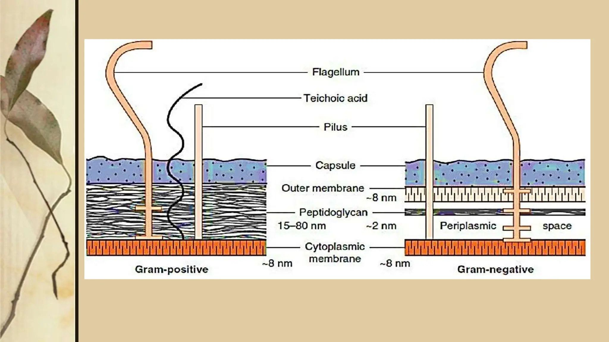
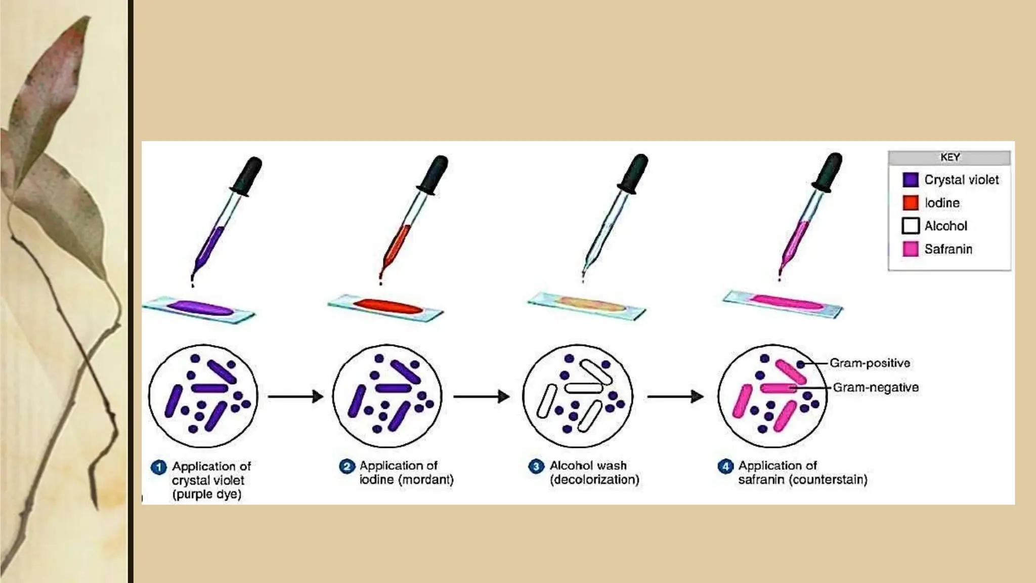
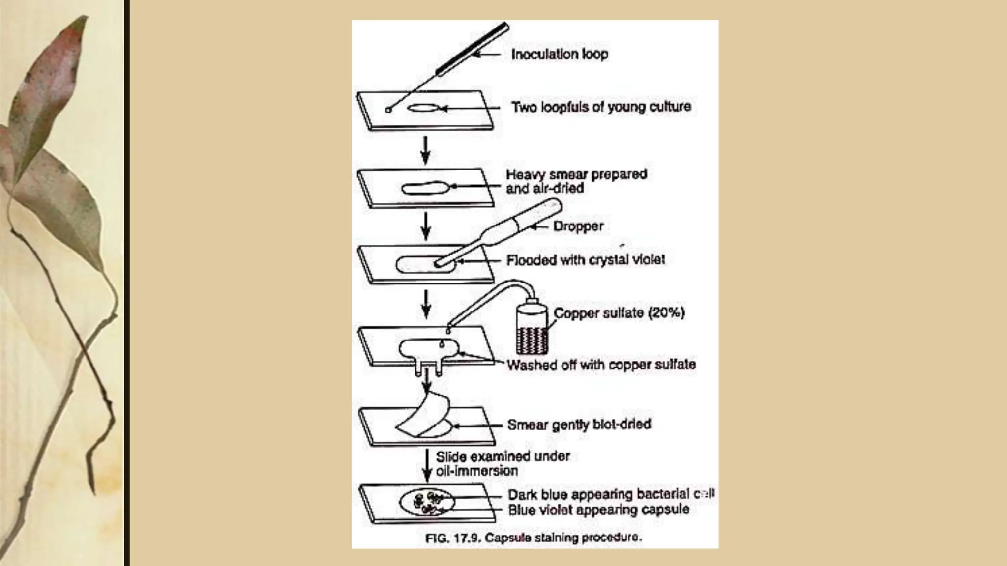
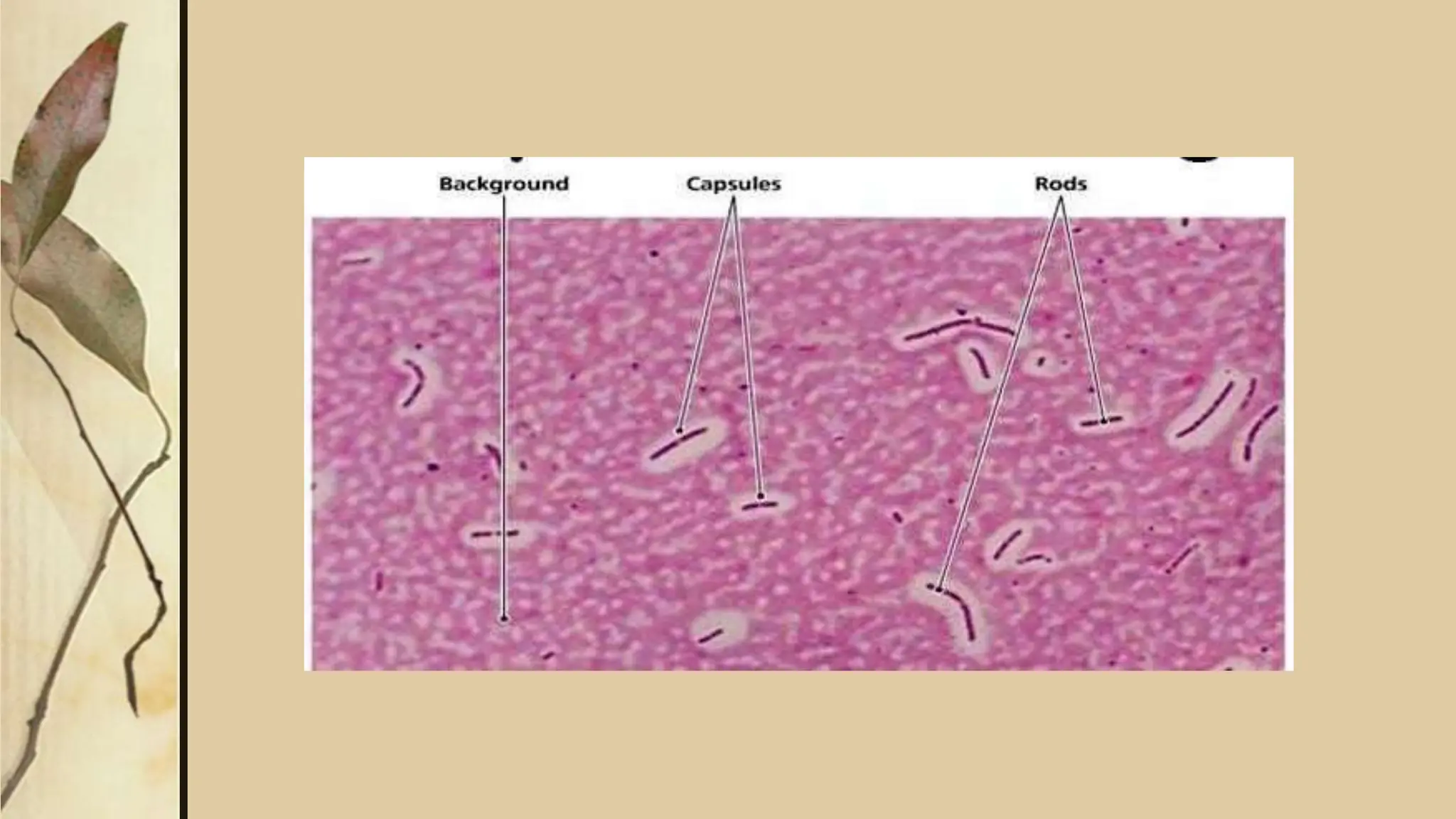
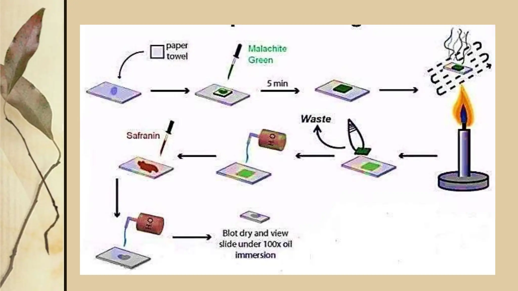
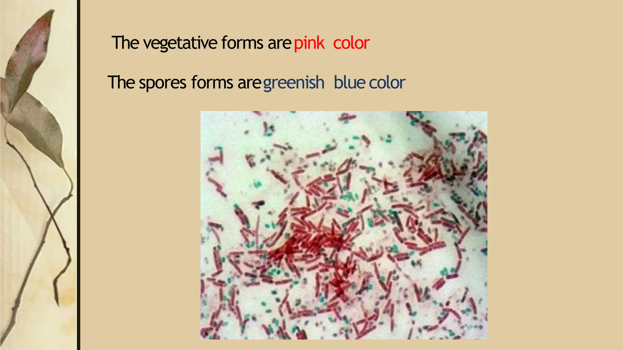
SlideShare a Scribd company logo
Title Layout
Subtitle
Staining
 Micro organism cannot beseenbynakedeyes,that why we must magnifythe
sizeof micro-organism.
 Forthat reason we needa magnifier which ismicroscopewhich enlargesthe
organismsalmost 1000more than its actualsize.
 Thesizeof bacteria ismeasuresby micronmeter(µm)
 Thesizeof most bacteria ragesfrom 1to 3µm.
 Thesmallestbacteria isMycoplasma0.2µm, where asthe largestisBorrelia
10µm
• Most of the micro-organism are enableto seenevenby microscope,we
need to stain theorganism
• According to pHall stainsare acidic,alkali or neutral.
• Generallyacidic materials are stainswith alkali or in contrast alkali materials
are stainswith acidic.
• In general we classify the staining methodsasfollow.
1. Simplestaining
2. Differentialstaining ( Gramstaining)
3. Acidfast staining
4. Negativestaining
5. Flagellastaining
6. Capsulestaining
7. Sporestaining
1.Simple staining
In simple staining we useasingledye(methylene blue, carbol
fuchsin, Safranin).
Here just observe the shapeof organismor bacteria either its cocci,
rods orspirochetes
• Procedure;
1. Makeathin smearon aglassslide of agivensample
2. Air dry it and cover with Methylene blue.
3. Washwith tap water and againair dryit
4. Observeunder the microscope ,
5. if the given sampleisbacteria then we will seeBacilli or Coccishapesof
bacteria
Safranin stainedbacilli
Methylene Blue stained
cocci
2.Differential staining(Gram Staining)
 Gramstain isthe most important staining procedure which differentiate
all the bacteria in two main categories either gramnegativeor gram
positive bacteria.
 Thismethod wasdiscoveredby HansChristianGramin Germanyin 1884
 In this staining method we usemore then one type ofdyewhich include
 Crystalviolet
 Safranin
 Themechanismof grampositive & gramnegative bacteria isbasedon
bacterialcellwall which gramnegative bacteria haveathin peptidoglycan
coveredby anouterlipid-containingmembrane, whereas gram-positive
bacteria haveathickpeptidoglycanand noouter membrane.
• Gramstaining Procedure
1. Makeasmearof bacterial specimenon aglassslide.
2. Air dry it ancoverwith crystal violet for 2-3 minutes.
3. Washwith tap water and cover withgram’siodine for 2 minutes.
4. Washwith tap water and cover with gram’salcohol (95%)for 30seconds
to1minute.
5. Washwith tap water and cover with Safraninfor2 minutes.
6. Washwith tap water and observed under the microscope which will
results either purple or pink, red coloredbacteria.
7. ThePurplecolored bacteria isnamed grampositivebacteria & the Pinkor
redcolored bacteria named gramnegative bacteria.
Acidfaststaining (Ziehl–Neelsenstain)
 Ziehl-Neelsen stain was discovered by the bacteriologist Franz Ziehl
(1859–1926)andthe pathologist FriedrichNeelsen(1854 – 1898).
 It is a special bacteriological stain used to identify acid fast
organisms.
 The mycobacterium tuberculosis is deferent from other bacteria in the
composition of cell wall which in addition to peptidoglycan, the outer
membrane or envelope of the acid-fast cell wall of contains large amounts of
glycolipids,especially mycolicacidsthat in the genusMycobacterium, makeup
approximately 60%of the acid-fast cellwall.
Structure of Mycobacteriumtuberculosis
• Procedure
1. Makeasmearof sputum on glassslide andfixe with ethanol &air
dry it
2. Coverslide with Carbolfuchsinandheated for 5minutes
3. Washwith tap water & cover withacidalcoholfor 1-2 minutes
4. Washwith tap water & coverthe slide with Methylene bluefor 2
minutes
5. Washthe slide and observed the redrodsunder microscope which
will be mycobacterium tuberculosis
TBslideafterAFB stain
Negativestaining (IndiaInk)
Negativestainingisarecognizedmethod, often usedin diagnosticmicroscopy, for contrasting a
thin specimenwith anoptically opaquefluid.
In this technique, the backgroundisstained,leavingthe actualspecimen untouched, andthus
visible
• Procedure
1. Makeasmearfrom agivensample& mix with Negrosinor Congreddye
2. Air dry itandobservedunder Bright field microscope
• Result
Thebackground will takethe colorbut the bacteriawill not , which the bacteria will appear
bright
Note:Mostly usedfor Spirillaor for electron microscopy
Flagellastain
• Theflagellastainallows observation of bacterial flagellaunder
the light microscope.
• Bacterial flagellaare normally too thin to beseen
• Therefore the flagellated bacteria are stained with specialdyeis
commercially available.
Capsulestaining
• Acapsuleisagelatinous outer layer secreted by bacterial cell and
that surrounds and adheresto the cellwall
• Themain purpose of capsulestain isto distinguish capsularmaterial
from the bacterialcell.
• Most capsulesare composedof polysaccharides, but someare composed
of polypeptides.
• Thecapsulestain employsanacidic stain and abasicstain to detect
capsule production
• Procedure;
1. Placeasmall drop of anegativestain(India Ink, CongoRed,
Nigrosin, or Eosin)on the slide.
2. Add aloopful of bacterial culture to slide, smearingit in the dye.
3. Usethe other slideto dragthe ink-cell mixture into athin film
along the first slide and let stand for 5-7minutes.
4. Allow to air dry (do not heat fix).
5. Flood the smear with crystalviolet stain(this will stain the
cells but not the capsules)for about 1minutes.
6. Coverthe slide with copper sulfate(20%).
7. Drain the crystal violet by tilting the slideat a45 degreeangleand
let stain run off until itair dries .
8. Examinethe smear microscopically (100X)for the presenceof
encapsulatedcells asindicated by clear zonessurrounding the
cells.
Sporestaining
 Theendosporestainisadifferential stainusedto visualizebacterial
endospores.
 Endosporesare formed by afew generaof bacteria, suchasBacillus
 Byforming spores,bacteria cansurvive in hostileconditions.
• Sporesare resistant to heat, dryness,chemicals,andradiation
• Procedure;
1. Preparesmearsof organisms to be tested for presenceofendospores on aclean
microscope slide and air dry it.
2. Heat fix the smear.
3. Placeasmall piece of blotting paper (absorbent paper) over the smear and place the
slide (smear side up) on awire gauzeon aring stand.
4. Heat the slide gently till it starts to evaporate (either by putting the slide on a
staining rack that hasbeen placed over aboiling water bath or via bunsenburner).
5. Removethe heat and reheat the slide asneeded to keepthe slidesteamingfor about 3-
5 minutes.Asthe paper begins to dry add adrop or two of malachitegreento keep it
moist, but don’t add somuch at one time that the temperatureis appreciably
reduced.
6. Remove the blotting paper and allow the slide to cool to room temperature for 2
minutes.
7. Washthe slide with tap water (to wash the malachite green from both sidesof the
microscope slide).
8. Stain the smear with safraninfor 2minutes.
9. Washboth side of the slide to remove the secondarystain and blot the slide/ air dry.
The vegetative forms arepink color
The spores forms aregreenish bluecolor

More Related Content

Similar to bacterial staining.pptx

403149684 stainingtechniquesgp-150203151421-conversion-gate02-pdf
403149684 stainingtechniquesgp-150203151421-conversion-gate02-pdf403149684 stainingtechniquesgp-150203151421-conversion-gate02-pdf
403149684 stainingtechniquesgp-150203151421-conversion-gate02-pdf
ssuserd72fc5
 
(2)MORPHOLOGY OF BACTERIA.pptx
(2)MORPHOLOGY OF BACTERIA.pptx(2)MORPHOLOGY OF BACTERIA.pptx
(2)MORPHOLOGY OF BACTERIA.pptx
anilapj
 
diagnosticmicrobiology-140523042250-phpapp01.pdf
diagnosticmicrobiology-140523042250-phpapp01.pdfdiagnosticmicrobiology-140523042250-phpapp01.pdf
diagnosticmicrobiology-140523042250-phpapp01.pdf
Fatima Fasih
 
Diagnostic microbiology.
Diagnostic microbiology.Diagnostic microbiology.
Diagnostic microbiology.DCROWN
 
Identification of bacteria by staining methods
Identification of bacteria by staining methodsIdentification of bacteria by staining methods
Identification of bacteria by staining methods
NAGALAKSHMI R
 
Staining
StainingStaining
Staining
Madhuri Bobburi
 
Observation Of Water In Sycamore Creek
Observation Of Water In Sycamore CreekObservation Of Water In Sycamore Creek
Observation Of Water In Sycamore Creek
Brianna Johnson
 
Lec 01. introduction to microbiology
Lec 01. introduction to microbiologyLec 01. introduction to microbiology
Lec 01. introduction to microbiology
Sebghatullah Mansoor
 
Microbiology
MicrobiologyMicrobiology
Microbiology
Suvarna Wagh
 
bacterial different staining technique
bacterial different staining techniquebacterial different staining technique
bacterial different staining techniqueKoi Bdk Pahang
 
Staining.pptx
Staining.pptxStaining.pptx
Staining.pptx
sneha_pharmacist
 
Afb stain by manoj
Afb stain by manojAfb stain by manoj
Afb stain by manoj
Manoj Mahato
 
Identification of bacteria
Identification of bacteriaIdentification of bacteria
Identification of bacteria
Dhanashri Mali
 
STAINING TECHNIQUES AND TYPES PROCEDURE.
STAINING TECHNIQUES AND TYPES PROCEDURE.STAINING TECHNIQUES AND TYPES PROCEDURE.
STAINING TECHNIQUES AND TYPES PROCEDURE.
Shylesh M
 
identificationof bacteria.pptx
identificationof bacteria.pptxidentificationof bacteria.pptx
identificationof bacteria.pptx
obedcudjoe1
 
Lab Report: Isolation of Pure Culture, Gram-staining, and Microscopic Observa...
Lab Report: Isolation of Pure Culture, Gram-staining, and Microscopic Observa...Lab Report: Isolation of Pure Culture, Gram-staining, and Microscopic Observa...
Lab Report: Isolation of Pure Culture, Gram-staining, and Microscopic Observa...
Annisa Hayatunnufus
 
Common Laboratory investigations in dermatology
Common Laboratory investigations in dermatologyCommon Laboratory investigations in dermatology
Common Laboratory investigations in dermatology
Kezha Zutso
 
Mycobacterium tuberculosis (Practical Medical Microbiology, 14)
Mycobacterium tuberculosis (Practical Medical Microbiology, 14)Mycobacterium tuberculosis (Practical Medical Microbiology, 14)
Mycobacterium tuberculosis (Practical Medical Microbiology, 14)
Hussein Al-tameemi
 

Similar to bacterial staining.pptx (20)

403149684 stainingtechniquesgp-150203151421-conversion-gate02-pdf
403149684 stainingtechniquesgp-150203151421-conversion-gate02-pdf403149684 stainingtechniquesgp-150203151421-conversion-gate02-pdf
403149684 stainingtechniquesgp-150203151421-conversion-gate02-pdf
 
(2)MORPHOLOGY OF BACTERIA.pptx
(2)MORPHOLOGY OF BACTERIA.pptx(2)MORPHOLOGY OF BACTERIA.pptx
(2)MORPHOLOGY OF BACTERIA.pptx
 
diagnosticmicrobiology-140523042250-phpapp01.pdf
diagnosticmicrobiology-140523042250-phpapp01.pdfdiagnosticmicrobiology-140523042250-phpapp01.pdf
diagnosticmicrobiology-140523042250-phpapp01.pdf
 
Diagnostic microbiology.
Diagnostic microbiology.Diagnostic microbiology.
Diagnostic microbiology.
 
Identification of bacteria by staining methods
Identification of bacteria by staining methodsIdentification of bacteria by staining methods
Identification of bacteria by staining methods
 
Staining
StainingStaining
Staining
 
Observation Of Water In Sycamore Creek
Observation Of Water In Sycamore CreekObservation Of Water In Sycamore Creek
Observation Of Water In Sycamore Creek
 
Lec 01. introduction to microbiology
Lec 01. introduction to microbiologyLec 01. introduction to microbiology
Lec 01. introduction to microbiology
 
Microbiology
MicrobiologyMicrobiology
Microbiology
 
3 cytology
3 cytology3 cytology
3 cytology
 
bacterial different staining technique
bacterial different staining techniquebacterial different staining technique
bacterial different staining technique
 
Staining.pptx
Staining.pptxStaining.pptx
Staining.pptx
 
Afb stain by manoj
Afb stain by manojAfb stain by manoj
Afb stain by manoj
 
Identification of bacteria
Identification of bacteriaIdentification of bacteria
Identification of bacteria
 
STAINING TECHNIQUES AND TYPES PROCEDURE.
STAINING TECHNIQUES AND TYPES PROCEDURE.STAINING TECHNIQUES AND TYPES PROCEDURE.
STAINING TECHNIQUES AND TYPES PROCEDURE.
 
identificationof bacteria.pptx
identificationof bacteria.pptxidentificationof bacteria.pptx
identificationof bacteria.pptx
 
Lab Report: Isolation of Pure Culture, Gram-staining, and Microscopic Observa...
Lab Report: Isolation of Pure Culture, Gram-staining, and Microscopic Observa...Lab Report: Isolation of Pure Culture, Gram-staining, and Microscopic Observa...
Lab Report: Isolation of Pure Culture, Gram-staining, and Microscopic Observa...
 
Lab 4
Lab 4Lab 4
Lab 4
 
Common Laboratory investigations in dermatology
Common Laboratory investigations in dermatologyCommon Laboratory investigations in dermatology
Common Laboratory investigations in dermatology
 
Mycobacterium tuberculosis (Practical Medical Microbiology, 14)
Mycobacterium tuberculosis (Practical Medical Microbiology, 14)Mycobacterium tuberculosis (Practical Medical Microbiology, 14)
Mycobacterium tuberculosis (Practical Medical Microbiology, 14)
 

Recently uploaded

How STIs Influence the Development of Pelvic Inflammatory Disease.pptx
How STIs Influence the Development of Pelvic Inflammatory Disease.pptxHow STIs Influence the Development of Pelvic Inflammatory Disease.pptx
How STIs Influence the Development of Pelvic Inflammatory Disease.pptx
FFragrant
 
Vision-1.pptx, Eye structure, basics of optics
Vision-1.pptx, Eye structure, basics of opticsVision-1.pptx, Eye structure, basics of optics
Vision-1.pptx, Eye structure, basics of optics
Sai Sailesh Kumar Goothy
 
Pharma Pcd Franchise in Jharkhand - Yodley Lifesciences
Pharma Pcd Franchise in Jharkhand - Yodley LifesciencesPharma Pcd Franchise in Jharkhand - Yodley Lifesciences
Pharma Pcd Franchise in Jharkhand - Yodley Lifesciences
Yodley Lifesciences
 
ANATOMY AND PHYSIOLOGY OF URINARY SYSTEM.pptx
ANATOMY AND PHYSIOLOGY OF URINARY SYSTEM.pptxANATOMY AND PHYSIOLOGY OF URINARY SYSTEM.pptx
ANATOMY AND PHYSIOLOGY OF URINARY SYSTEM.pptx
Swetaba Besh
 
CDSCO and Phamacovigilance {Regulatory body in India}
CDSCO and Phamacovigilance {Regulatory body in India}CDSCO and Phamacovigilance {Regulatory body in India}
CDSCO and Phamacovigilance {Regulatory body in India}
NEHA GUPTA
 
Colonic and anorectal physiology with surgical implications
Colonic and anorectal physiology with surgical implicationsColonic and anorectal physiology with surgical implications
Colonic and anorectal physiology with surgical implications
Dr Maria Tamanna
 
Aortic Association CBL Pilot April 19 – 20 Bern
Aortic Association CBL Pilot April 19 – 20 BernAortic Association CBL Pilot April 19 – 20 Bern
Aortic Association CBL Pilot April 19 – 20 Bern
suvadeepdas911
 
Gram Stain introduction, principle, Procedure
Gram Stain introduction, principle, ProcedureGram Stain introduction, principle, Procedure
Gram Stain introduction, principle, Procedure
Suraj Goswami
 
263778731218 Abortion Clinic /Pills In Harare ,
263778731218 Abortion Clinic /Pills In Harare ,263778731218 Abortion Clinic /Pills In Harare ,
263778731218 Abortion Clinic /Pills In Harare ,
sisternakatoto
 
Maxilla, Mandible & Hyoid Bone & Clinical Correlations by Dr. RIG.pptx
Maxilla, Mandible & Hyoid Bone & Clinical Correlations by Dr. RIG.pptxMaxilla, Mandible & Hyoid Bone & Clinical Correlations by Dr. RIG.pptx
Maxilla, Mandible & Hyoid Bone & Clinical Correlations by Dr. RIG.pptx
Dr. Rabia Inam Gandapore
 
micro teaching on communication m.sc nursing.pdf
micro teaching on communication m.sc nursing.pdfmicro teaching on communication m.sc nursing.pdf
micro teaching on communication m.sc nursing.pdf
Anurag Sharma
 
Physiology of Special Chemical Sensation of Taste
Physiology of Special Chemical Sensation of TastePhysiology of Special Chemical Sensation of Taste
Physiology of Special Chemical Sensation of Taste
MedicoseAcademics
 
Ophthalmology Clinical Tests for OSCE exam
Ophthalmology Clinical Tests for OSCE examOphthalmology Clinical Tests for OSCE exam
Ophthalmology Clinical Tests for OSCE exam
KafrELShiekh University
 
Thyroid Gland- Gross Anatomy by Dr. Rabia Inam Gandapore.pptx
Thyroid Gland- Gross Anatomy by Dr. Rabia Inam Gandapore.pptxThyroid Gland- Gross Anatomy by Dr. Rabia Inam Gandapore.pptx
Thyroid Gland- Gross Anatomy by Dr. Rabia Inam Gandapore.pptx
Dr. Rabia Inam Gandapore
 
BRACHYTHERAPY OVERVIEW AND APPLICATORS
BRACHYTHERAPY OVERVIEW  AND  APPLICATORSBRACHYTHERAPY OVERVIEW  AND  APPLICATORS
BRACHYTHERAPY OVERVIEW AND APPLICATORS
Krishan Murari
 
Local Advanced Lung Cancer: Artificial Intelligence, Synergetics, Complex Sys...
Local Advanced Lung Cancer: Artificial Intelligence, Synergetics, Complex Sys...Local Advanced Lung Cancer: Artificial Intelligence, Synergetics, Complex Sys...
Local Advanced Lung Cancer: Artificial Intelligence, Synergetics, Complex Sys...
Oleg Kshivets
 
Best Ayurvedic medicine for Gas and Indigestion
Best Ayurvedic medicine for Gas and IndigestionBest Ayurvedic medicine for Gas and Indigestion
Best Ayurvedic medicine for Gas and Indigestion
SwastikAyurveda
 
Dehradun #ℂall #gIRLS Oyo Hotel 9719300533 #ℂall #gIRL in Dehradun
Dehradun #ℂall #gIRLS Oyo Hotel 9719300533 #ℂall #gIRL in DehradunDehradun #ℂall #gIRLS Oyo Hotel 9719300533 #ℂall #gIRL in Dehradun
Dehradun #ℂall #gIRLS Oyo Hotel 9719300533 #ℂall #gIRL in Dehradun
chandankumarsmartiso
 
Pharynx and Clinical Correlations BY Dr.Rabia Inam Gandapore.pptx
Pharynx and Clinical Correlations BY Dr.Rabia Inam Gandapore.pptxPharynx and Clinical Correlations BY Dr.Rabia Inam Gandapore.pptx
Pharynx and Clinical Correlations BY Dr.Rabia Inam Gandapore.pptx
Dr. Rabia Inam Gandapore
 
Hemodialysis: Chapter 4, Dialysate Circuit - Dr.Gawad
Hemodialysis: Chapter 4, Dialysate Circuit - Dr.GawadHemodialysis: Chapter 4, Dialysate Circuit - Dr.Gawad
Hemodialysis: Chapter 4, Dialysate Circuit - Dr.Gawad
NephroTube - Dr.Gawad
 

Recently uploaded (20)

How STIs Influence the Development of Pelvic Inflammatory Disease.pptx
How STIs Influence the Development of Pelvic Inflammatory Disease.pptxHow STIs Influence the Development of Pelvic Inflammatory Disease.pptx
How STIs Influence the Development of Pelvic Inflammatory Disease.pptx
 
Vision-1.pptx, Eye structure, basics of optics
Vision-1.pptx, Eye structure, basics of opticsVision-1.pptx, Eye structure, basics of optics
Vision-1.pptx, Eye structure, basics of optics
 
Pharma Pcd Franchise in Jharkhand - Yodley Lifesciences
Pharma Pcd Franchise in Jharkhand - Yodley LifesciencesPharma Pcd Franchise in Jharkhand - Yodley Lifesciences
Pharma Pcd Franchise in Jharkhand - Yodley Lifesciences
 
ANATOMY AND PHYSIOLOGY OF URINARY SYSTEM.pptx
ANATOMY AND PHYSIOLOGY OF URINARY SYSTEM.pptxANATOMY AND PHYSIOLOGY OF URINARY SYSTEM.pptx
ANATOMY AND PHYSIOLOGY OF URINARY SYSTEM.pptx
 
CDSCO and Phamacovigilance {Regulatory body in India}
CDSCO and Phamacovigilance {Regulatory body in India}CDSCO and Phamacovigilance {Regulatory body in India}
CDSCO and Phamacovigilance {Regulatory body in India}
 
Colonic and anorectal physiology with surgical implications
Colonic and anorectal physiology with surgical implicationsColonic and anorectal physiology with surgical implications
Colonic and anorectal physiology with surgical implications
 
Aortic Association CBL Pilot April 19 – 20 Bern
Aortic Association CBL Pilot April 19 – 20 BernAortic Association CBL Pilot April 19 – 20 Bern
Aortic Association CBL Pilot April 19 – 20 Bern
 
Gram Stain introduction, principle, Procedure
Gram Stain introduction, principle, ProcedureGram Stain introduction, principle, Procedure
Gram Stain introduction, principle, Procedure
 
263778731218 Abortion Clinic /Pills In Harare ,
263778731218 Abortion Clinic /Pills In Harare ,263778731218 Abortion Clinic /Pills In Harare ,
263778731218 Abortion Clinic /Pills In Harare ,
 
Maxilla, Mandible & Hyoid Bone & Clinical Correlations by Dr. RIG.pptx
Maxilla, Mandible & Hyoid Bone & Clinical Correlations by Dr. RIG.pptxMaxilla, Mandible & Hyoid Bone & Clinical Correlations by Dr. RIG.pptx
Maxilla, Mandible & Hyoid Bone & Clinical Correlations by Dr. RIG.pptx
 
micro teaching on communication m.sc nursing.pdf
micro teaching on communication m.sc nursing.pdfmicro teaching on communication m.sc nursing.pdf
micro teaching on communication m.sc nursing.pdf
 
Physiology of Special Chemical Sensation of Taste
Physiology of Special Chemical Sensation of TastePhysiology of Special Chemical Sensation of Taste
Physiology of Special Chemical Sensation of Taste
 
Ophthalmology Clinical Tests for OSCE exam
Ophthalmology Clinical Tests for OSCE examOphthalmology Clinical Tests for OSCE exam
Ophthalmology Clinical Tests for OSCE exam
 
Thyroid Gland- Gross Anatomy by Dr. Rabia Inam Gandapore.pptx
Thyroid Gland- Gross Anatomy by Dr. Rabia Inam Gandapore.pptxThyroid Gland- Gross Anatomy by Dr. Rabia Inam Gandapore.pptx
Thyroid Gland- Gross Anatomy by Dr. Rabia Inam Gandapore.pptx
 
BRACHYTHERAPY OVERVIEW AND APPLICATORS
BRACHYTHERAPY OVERVIEW  AND  APPLICATORSBRACHYTHERAPY OVERVIEW  AND  APPLICATORS
BRACHYTHERAPY OVERVIEW AND APPLICATORS
 
Local Advanced Lung Cancer: Artificial Intelligence, Synergetics, Complex Sys...
Local Advanced Lung Cancer: Artificial Intelligence, Synergetics, Complex Sys...Local Advanced Lung Cancer: Artificial Intelligence, Synergetics, Complex Sys...
Local Advanced Lung Cancer: Artificial Intelligence, Synergetics, Complex Sys...
 
Best Ayurvedic medicine for Gas and Indigestion
Best Ayurvedic medicine for Gas and IndigestionBest Ayurvedic medicine for Gas and Indigestion
Best Ayurvedic medicine for Gas and Indigestion
 
Dehradun #ℂall #gIRLS Oyo Hotel 9719300533 #ℂall #gIRL in Dehradun
Dehradun #ℂall #gIRLS Oyo Hotel 9719300533 #ℂall #gIRL in DehradunDehradun #ℂall #gIRLS Oyo Hotel 9719300533 #ℂall #gIRL in Dehradun
Dehradun #ℂall #gIRLS Oyo Hotel 9719300533 #ℂall #gIRL in Dehradun
 
Pharynx and Clinical Correlations BY Dr.Rabia Inam Gandapore.pptx
Pharynx and Clinical Correlations BY Dr.Rabia Inam Gandapore.pptxPharynx and Clinical Correlations BY Dr.Rabia Inam Gandapore.pptx
Pharynx and Clinical Correlations BY Dr.Rabia Inam Gandapore.pptx
 
Hemodialysis: Chapter 4, Dialysate Circuit - Dr.Gawad
Hemodialysis: Chapter 4, Dialysate Circuit - Dr.GawadHemodialysis: Chapter 4, Dialysate Circuit - Dr.Gawad
Hemodialysis: Chapter 4, Dialysate Circuit - Dr.Gawad
 

bacterial staining.pptx

  • 2. Staining  Micro organism cannot beseenbynakedeyes,that why we must magnifythe sizeof micro-organism.  Forthat reason we needa magnifier which ismicroscopewhich enlargesthe organismsalmost 1000more than its actualsize.  Thesizeof bacteria ismeasuresby micronmeter(µm)  Thesizeof most bacteria ragesfrom 1to 3µm.  Thesmallestbacteria isMycoplasma0.2µm, where asthe largestisBorrelia 10µm
  • 3. • Most of the micro-organism are enableto seenevenby microscope,we need to stain theorganism • According to pHall stainsare acidic,alkali or neutral. • Generallyacidic materials are stainswith alkali or in contrast alkali materials are stainswith acidic. • In general we classify the staining methodsasfollow. 1. Simplestaining 2. Differentialstaining ( Gramstaining) 3. Acidfast staining 4. Negativestaining 5. Flagellastaining 6. Capsulestaining 7. Sporestaining
  • 4. 1.Simple staining In simple staining we useasingledye(methylene blue, carbol fuchsin, Safranin). Here just observe the shapeof organismor bacteria either its cocci, rods orspirochetes • Procedure; 1. Makeathin smearon aglassslide of agivensample 2. Air dry it and cover with Methylene blue. 3. Washwith tap water and againair dryit 4. Observeunder the microscope , 5. if the given sampleisbacteria then we will seeBacilli or Coccishapesof bacteria
  • 6. 2.Differential staining(Gram Staining)  Gramstain isthe most important staining procedure which differentiate all the bacteria in two main categories either gramnegativeor gram positive bacteria.  Thismethod wasdiscoveredby HansChristianGramin Germanyin 1884  In this staining method we usemore then one type ofdyewhich include  Crystalviolet  Safranin  Themechanismof grampositive & gramnegative bacteria isbasedon bacterialcellwall which gramnegative bacteria haveathin peptidoglycan coveredby anouterlipid-containingmembrane, whereas gram-positive bacteria haveathickpeptidoglycanand noouter membrane.
  • 7.
  • 8.
  • 9.
  • 10. • Gramstaining Procedure 1. Makeasmearof bacterial specimenon aglassslide. 2. Air dry it ancoverwith crystal violet for 2-3 minutes. 3. Washwith tap water and cover withgram’siodine for 2 minutes. 4. Washwith tap water and cover with gram’salcohol (95%)for 30seconds to1minute. 5. Washwith tap water and cover with Safraninfor2 minutes. 6. Washwith tap water and observed under the microscope which will results either purple or pink, red coloredbacteria. 7. ThePurplecolored bacteria isnamed grampositivebacteria & the Pinkor redcolored bacteria named gramnegative bacteria.
  • 11.
  • 12.
  • 13. Acidfaststaining (Ziehl–Neelsenstain)  Ziehl-Neelsen stain was discovered by the bacteriologist Franz Ziehl (1859–1926)andthe pathologist FriedrichNeelsen(1854 – 1898).  It is a special bacteriological stain used to identify acid fast organisms.  The mycobacterium tuberculosis is deferent from other bacteria in the composition of cell wall which in addition to peptidoglycan, the outer membrane or envelope of the acid-fast cell wall of contains large amounts of glycolipids,especially mycolicacidsthat in the genusMycobacterium, makeup approximately 60%of the acid-fast cellwall.
  • 15. • Procedure 1. Makeasmearof sputum on glassslide andfixe with ethanol &air dry it 2. Coverslide with Carbolfuchsinandheated for 5minutes 3. Washwith tap water & cover withacidalcoholfor 1-2 minutes 4. Washwith tap water & coverthe slide with Methylene bluefor 2 minutes 5. Washthe slide and observed the redrodsunder microscope which will be mycobacterium tuberculosis
  • 16.
  • 18. Negativestaining (IndiaInk) Negativestainingisarecognizedmethod, often usedin diagnosticmicroscopy, for contrasting a thin specimenwith anoptically opaquefluid. In this technique, the backgroundisstained,leavingthe actualspecimen untouched, andthus visible • Procedure 1. Makeasmearfrom agivensample& mix with Negrosinor Congreddye 2. Air dry itandobservedunder Bright field microscope • Result Thebackground will takethe colorbut the bacteriawill not , which the bacteria will appear bright Note:Mostly usedfor Spirillaor for electron microscopy
  • 19.
  • 20. Flagellastain • Theflagellastainallows observation of bacterial flagellaunder the light microscope. • Bacterial flagellaare normally too thin to beseen • Therefore the flagellated bacteria are stained with specialdyeis commercially available.
  • 21. Capsulestaining • Acapsuleisagelatinous outer layer secreted by bacterial cell and that surrounds and adheresto the cellwall • Themain purpose of capsulestain isto distinguish capsularmaterial from the bacterialcell. • Most capsulesare composedof polysaccharides, but someare composed of polypeptides. • Thecapsulestain employsanacidic stain and abasicstain to detect capsule production
  • 22. • Procedure; 1. Placeasmall drop of anegativestain(India Ink, CongoRed, Nigrosin, or Eosin)on the slide. 2. Add aloopful of bacterial culture to slide, smearingit in the dye. 3. Usethe other slideto dragthe ink-cell mixture into athin film along the first slide and let stand for 5-7minutes. 4. Allow to air dry (do not heat fix). 5. Flood the smear with crystalviolet stain(this will stain the cells but not the capsules)for about 1minutes. 6. Coverthe slide with copper sulfate(20%). 7. Drain the crystal violet by tilting the slideat a45 degreeangleand let stain run off until itair dries . 8. Examinethe smear microscopically (100X)for the presenceof encapsulatedcells asindicated by clear zonessurrounding the cells.
  • 23.
  • 24.
  • 25. Sporestaining  Theendosporestainisadifferential stainusedto visualizebacterial endospores.  Endosporesare formed by afew generaof bacteria, suchasBacillus  Byforming spores,bacteria cansurvive in hostileconditions. • Sporesare resistant to heat, dryness,chemicals,andradiation
  • 26. • Procedure; 1. Preparesmearsof organisms to be tested for presenceofendospores on aclean microscope slide and air dry it. 2. Heat fix the smear. 3. Placeasmall piece of blotting paper (absorbent paper) over the smear and place the slide (smear side up) on awire gauzeon aring stand. 4. Heat the slide gently till it starts to evaporate (either by putting the slide on a staining rack that hasbeen placed over aboiling water bath or via bunsenburner). 5. Removethe heat and reheat the slide asneeded to keepthe slidesteamingfor about 3- 5 minutes.Asthe paper begins to dry add adrop or two of malachitegreento keep it moist, but don’t add somuch at one time that the temperatureis appreciably reduced. 6. Remove the blotting paper and allow the slide to cool to room temperature for 2 minutes. 7. Washthe slide with tap water (to wash the malachite green from both sidesof the microscope slide). 8. Stain the smear with safraninfor 2minutes. 9. Washboth side of the slide to remove the secondarystain and blot the slide/ air dry.
  • 27.
  • 28. The vegetative forms arepink color The spores forms aregreenish bluecolor