Το χαρακτηριστικό τωνΤοχαρακτηριστικό των
εκπτυσσόμενων κυλίνδρων είναι ηεκπτυσσόμενων κυλίνδρων είναι η
δυνατότηταδυνατότητα in situin situ ΔιάτασηςΔιάτασης
α) Επιτυγχάνοντας ισχυρό οστικόα) Επιτυγχάνοντας ισχυρό οστικό
αγκυροβόλιο αυξάνοντας τη σταθερότητααγκυροβόλιο αυξάνοντας τη σταθερότητα
β) Είναι εύκολη η εφαρμογή λόγω τωνβ) Είναι εύκολη η εφαρμογή λόγω των
μικρών διαστάσεων των πριν τη διάτασημικρών διαστάσεων των πριν τη διάταση
γ) Αποκαθίσταται το ύψος τουγ) Αποκαθίσταται το ύψος του
μεσοσπονδυλίου διαστήματος καιμεσοσπονδυλίου διαστήματος και
διευρύνονται τα σπονδυλικά τρήματαδιευρύνονται τα σπονδυλικά τρήματα
δ) Αποκαθίσταται η λόρδωση δίνοντας 3δ) Αποκαθίσταται η λόρδωση δίνοντας 3οο
––
66οο
πρόσθια κλίσηπρόσθια κλίση
Επιπλέον :
4.
Disc – O– TechDisc – O – Tech
The B-TwinThe B-Twin
ExpandableExpandable SpinalSpinal SystemSystem
Medical Technologies
L.T.D.
7.
EXPANDABLE TWIN SPINALSPACEREXPANDABLE TWIN SPINAL SPACER
FOR POSTERIOR LUMBARFOR POSTERIOR LUMBAR
INTERBODY STABILIZATION:INTERBODY STABILIZATION:
MECHANICAL TESTINGMECHANICAL TESTING
Folman YFolman Y.. & Gepstein R& Gepstein R..
– Department of Orthopaedic Surgery , Hillel-Yaffe MC, Hadera,Department of Orthopaedic Surgery , Hillel-Yaffe MC, Hadera,
IsraelIsrael
– Spinal Care Unit, Meir MC, Kefar Save, IsraelSpinal Care Unit, Meir MC, Kefar Save, Israel
– Orthopaedic Bioengineering Research Laboratory,Orthopaedic Bioengineering Research Laboratory,
University of UTAH, U.S.A.University of UTAH, U.S.A.
8.
Έλεγχος Αξονικής Συμπίεσης– Στατική φόρτισηΈλεγχος Αξονικής Συμπίεσης – Στατική φόρτιση
((Axial compression test – static loadingAxial compression test – static loading))
Compression test static loading configuration
(Tensometric M 350 – 10 kN. Tensometric, Rochdale, USA)
• Μέση Δύναμη Υποχώρησης του
εμφυτεύματος ήταν 2660.0 ± 483.0 Ν.
• Μέση τιμή της Τελικής Δύναμης ήταν
4131.6 ± 420.7 Ν.
• Τα πτερύγια δεν έσπασαν κατά τη
διάρκεια του τεστ αλλά λύγισαν στη
βάση τους ακολουθώντας την πορεία
λόρδωσης του εμφυτεύματος.
9.
Έλεγχος αξονικής συμπίεσης-Έλεγχος αξονικής συμπίεσης -
αντοχήαντοχή
(Axial compression test - fatigue loading)(Axial compression test - fatigue loading)
• Η αντοχή του εμφυτεύματος
υπολογίστηκε στους 5.000.000 κύκλους
φόρτισης.
• Το όριο αντοχής βρέθηκε ίσο με 931 Ν
Compression test Cyclic (fatigue) loading configuration
(Instron 8871, Canton, Ma, USA)
10.
Έλεγχος απαγκίστρωσης –στατικήΈλεγχος απαγκίστρωσης – στατική
φόρτισηφόρτιση
(Shear Pull Out (Expulsion) – Static loading)(Shear Pull Out (Expulsion) – Static loading)
• Το μέσο φορτίο που
οδήγησε σε υποχώρηση
ήταν 1645.0 ± 148.0 Ν.
Pull out test (Expulsion) - Static loading
(Tensometric M 350 – 10 kN. Tensometric, Rochdale, USA)
11.
Έλεγχος απαγκίστρωσης –αντοχήΈλεγχος απαγκίστρωσης – αντοχή
(Pull Out (Expulsion) – Fatigue loading)(Pull Out (Expulsion) – Fatigue loading)
• Κάθε εμφύτευμα υποβλήθηκε σε
εναλλασσόμενα φορτία εύρους 49 Ν έως
και 249 Ν με συνεχή ρυθμό 10 Hz.
• Τα εμφυτεύματα παρατηρήθηκαν μάκρο
και μικροσκοπικά για αστοχία υλικού κατά
τόπους.
• Δεν παρατηρήθηκαν σημεία αστοχίας.
Pull out test (Expulsion) - Fatigue loading
(Tensometric M 350 – 10 kN. Tensometric, Rochdale, USA)
12.
Έλεγχος έκπτυξης υπόαξονική φόρτισηΈλεγχος έκπτυξης υπό αξονική φόρτιση
(λειτουργία γρύλλου)(λειτουργία γρύλλου)
Jacking test (Expansion under axial loading)Jacking test (Expansion under axial loading)
• Έκπτυξη εμφυτευμάτων υπό αξονικό
φορτίο 245 Ν.
• Τα εμφυτεύματα εκπτύχθηκαν
χρησιμοποιώντας τη συσκευή
τοποθέτησης έως ότου εκπτυχθούν
πλήρως.
• Η μέση μεταβολή του ύψους ( πρίν και
μετά την έκπτυξη) ήταν 1.16 ± 0.52 mm.
• Σε όλες τις περιπτώσεις η συσκευή
εκπτύχθηκε χωρίς περαιτέρω
παραμόρφωση
Κριτήρια επιλογήςΚριτήρια επιλογής
Επίμονος,οσφυαλγία,Επίμονος, οσφυαλγία,
προκαλούσα λειτουργικήπροκαλούσα λειτουργική
ανικανότητα του ασθενούς γιαανικανότητα του ασθενούς για
περισσότερο από 6 μήνεςπερισσότερο από 6 μήνες
Περιορισμένη ή ανεπαρκήςΠεριορισμένη ή ανεπαρκής
ανταπόκριση στη συντηρητικήανταπόκριση στη συντηρητική
θεραπείαθεραπεία
Βέβαιη διάγνωση ότι ταΒέβαιη διάγνωση ότι τα
συμπτώματα απορρέουν απόσυμπτώματα απορρέουν από
εκφύλιση μεσοσπονδυλίουεκφύλιση μεσοσπονδυλίου
δίσκουδίσκου
Ακτινολογική επιβεβαίωσηΑκτινολογική επιβεβαίωση
(απλές ακτινογραφίες,(απλές ακτινογραφίες, M.R.I.M.R.I.
κ.λ.π.)κ.λ.π.)
ΣπονδυλολίσθησηΣπονδυλολίσθηση ≤ 1≤ 1ουου
βαθμούβαθμού
16.
Κριτήρια αποκλεισμούΚριτήρια αποκλεισμού
Παθήσειςπου επηρεάζουνΠαθήσεις που επηρεάζουν
αρνητικά την οστικήαρνητικά την οστική
πυκνότηταπυκνότητα
1.1. Φλεγμονή της Σ.Σ.Φλεγμονή της Σ.Σ.
2.2. ΝεοπλασίαΝεοπλασία
3.3. Μεταβολική οστική νόσοςΜεταβολική οστική νόσος
4.4. Κατάχρηση οινοπνεύματοςΚατάχρηση οινοπνεύματος
5.5. Διαταραχές συμπεριφοράςΔιαταραχές συμπεριφοράς
ΤοΤο Expandable TwinSpinal SpacerExpandable Twin Spinal Spacer
μπορεί να εφαρμοστεί με ασφάλειαμπορεί να εφαρμοστεί με ασφάλεια
λόγω:λόγω:
α) του ειδικού σχεδιασμούα) του ειδικού σχεδιασμού
β) των διαστάσεων του εμφυτεύματοςβ) των διαστάσεων του εμφυτεύματος
γ) των μηχανικών ιδιοτήτωνγ) των μηχανικών ιδιοτήτων
19.
Οι κύλινδροι διάτασηςΟικύλινδροι διάτασης B – TwinB – Twin
Expandable Spinal Spacers (Disc-O-Expandable Spinal Spacers (Disc-O-
Tech)Tech) έδωσαν ικανοποιητικάέδωσαν ικανοποιητικά
λειτουργικά και ακτινολογικάλειτουργικά και ακτινολογικά
αποτελέσματα στους 12-36 μήνεςαποτελέσματα στους 12-36 μήνες
μετεγχειρητικής παρακολούθησης μεμετεγχειρητικής παρακολούθησης με
ποσοστό επιτυχίας 95%ποσοστό επιτυχίας 95%
20.
Εκ των μέχριτώραΕκ των μέχρι τώρα
αποτελεσμάτωναποτελεσμάτων
η Ο.Ο.Μ.Σ.η Ο.Ο.Μ.Σ.
με το σύστημα Β-με το σύστημα Β-TwinTwin
Expandable Cages (Disc-Expandable Cages (Disc-
O-Tech)O-Tech)
δεν κατέστησε φανερή τηνδεν κατέστησε φανερή την
ανάγκη εφαρμογήςανάγκη εφαρμογής
επιπλέον συστήματοςεπιπλέον συστήματος
εσωτερικήςεσωτερικής
σταθεροποίησηςσταθεροποίησης
(διαυχενικές βίδες(διαυχενικές βίδες --
ράβδοι)ράβδοι)
21.
Minimally InvasiveMinimally Invasive
LumbarFusion &Lumbar Fusion &
Fixation with the SextantFixation with the Sextant
Percutaneous PediclePercutaneous Pedicle
Screw-Rod SystemScrew-Rod SystemSS
Minimally InvasiveMinimally Invasive
LumbarFusionLumbar Fusion
Achieve the same goals as openAchieve the same goals as open
fusion while minimizing approach-fusion while minimizing approach-
related morbidity (“fusionrelated morbidity (“fusion
disease”)disease”)
Sextant Rod InsertionSextantRod Insertion
SystemSystem
• MaterialsMaterials
• Cannulated,Cannulated,
Multi- AxialMulti- Axial
ScrewsScrews
• Rod ExtensionRod Extension
SleevesSleeves
• Rod InserterRod Inserter
• Pre-contouredPre-contoured
26.
Clinical ApplicationClinical Application
Maybe used anytime pedicle fixationMay be used anytime pedicle fixation
is felt to be necessary and/oris felt to be necessary and/or
desirabledesirable
Posterior supplementation for ALIF,Posterior supplementation for ALIF,
minimally invasive PLIF, or minimallyminimally invasive PLIF, or minimally
invasive TLIFinvasive TLIF
Supplement to minimally invasiveSupplement to minimally invasive
posterolateral fusionposterolateral fusion
Limitations of theLimitationsof the
Standard TreatmentsStandard Treatments
Discectomy: relieves pain, but does notDiscectomy: relieves pain, but does not
stabilize the vertebrae.stabilize the vertebrae.
Fusion/pedicle screw fixation: providesFusion/pedicle screw fixation: provides
stability, but eliminates mobility andstability, but eliminates mobility and
natural cushioning.natural cushioning.
Total disc replacement: provides stabilityTotal disc replacement: provides stability
and mobility, but eliminates cushioningand mobility, but eliminates cushioning
and procedure isand procedure is moremore invasive.invasive.
63.
The Alternative:The Alternative:
APDN DeviceA PDN Device
Preserves range of motion
Provides cushioning
Maintains disc height
Retards the degenerative
cascade
Relieves pain associated
with degenerative disc
disease
Disc Blood SupplyDiscBlood Supply
AvascularAvascular
– Intradisc pressureIntradisc pressure
higher than arterialhigher than arterial
pressurepressure
Nutrient ExchangeNutrient Exchange
– External diffusion fromExternal diffusion from
peripheral capillariesperipheral capillaries
– Internal diffusionInternal diffusion
through cartilaginousthrough cartilaginous
endplatesendplates
66.
Internal Fluid CycleInternalFluid Cycle
Nocturnal CycleNocturnal Cycle
– Horizontal postureHorizontal posture
– Water and nutrients moveWater and nutrients move
into discinto disc
– Thickness increasesThickness increases
Diurnal CycleDiurnal Cycle
– Vertical postureVertical posture
– Increased pressure forcesIncreased pressure forces
water and waste out of disc.water and waste out of disc.
– Disc thickness decreasesDisc thickness decreases
67.
Disc CompressionDisc Compression
VerticalLoadingVertical Loading
– Nucleus getsNucleus gets
compressed andcompressed and
radiates outward.radiates outward.
– Nucleus pushes onNucleus pushes on
anulus from within.anulus from within.
– Anulus fibers are inAnulus fibers are in
tension.tension.
71.
PDN GoalsPDN Goals
RELIEVEPAINRELIEVE PAIN
Stabilize disc heightStabilize disc height
Prevent furtherPrevent further
anulus tearinganulus tearing
Restore vertebralRestore vertebral
biomechanicsbiomechanics
Stop theStop the
degenerativedegenerative
cascadecascade
PDN DEVICE BACKGROUND
•Multi-block copolymer hydrogel
(HYPAN®
)
• Absorbs 80% w/w water at full
hydration
• Can absorb energy and increase in
height when loaded in the hydrated
state
Hydrogel Core
74.
PDN DEVICE BACKGROUND
•Oriented high-molecular weight
polyethylene fiber
• Woven circular tube
construction
• Constrains hydrogel core
• Allows device to function
independent of the anulus
Woven Jacket
75.
PDN DEVICE BACKGROUND
•90% platinum / 10%
iridium
• Aid device positioning
under fluoroscopy
• Minimal scatter under CT
Wire Markers
76.
Mimics Natural FluidMimicsNatural Fluid
Fluctuation Within the DiscFluctuation Within the Disc
Hydrophilic nature of the PDN
device allows it to absorb fluid
like the nucleus pulposus.
77.
Mechanical Testing ofMechanicalTesting of
PDN DevicePDN Device
Device was tested for:Device was tested for:
- Fatigue- Fatigue
- Burst strength- Burst strength
- Maintenance of disc- Maintenance of disc
heightheight
78.
Mechanical Testing ProtocolsMechanicalTesting Protocols
Fatigue Test
– Single hydrated device sinusoidally loaded between 200 and 800N at 4Hz
up to 50 million cycles (approximately 20-50 yrs in body)
– After every 10 million cycles devices are retested in load/deflection to
evaluate continued long-term maintenance of disc height and structural
integrity of the device
Burst Test
– Single hydrated device loaded to 2000, 4000, and 6000N and evaluated
for structural integrity of the weave, stitch, and pellet
Maintenance of Disc Height
– Load/deflection testing to determine device height at loads of 200N
(supine), 800N (standing), and 1600N (holding a 20kg weight)
In all tests the device continued to function as designedIn all tests the device continued to function as designed
Patient Inclusion CriteriaPatientInclusion Criteria
Age of at least 18 yearsAge of at least 18 years
Symptomatic DDD from L2-S1Symptomatic DDD from L2-S1
Symptoms have not responded toSymptoms have not responded to
nonsurgical treatment for at least 6nonsurgical treatment for at least 6
monthsmonths
Patient is experiencing low back pain withPatient is experiencing low back pain with
or without leg painor without leg pain
A radiographic study correlates withA radiographic study correlates with
symptoms and signs of discogenic originsymptoms and signs of discogenic origin
82.
Patient Exclusion CriteriaPatientExclusion Criteria
Disc height of affected level is less thanDisc height of affected level is less than
5mm5mm
Severe degeneration, listhesis, Schmorl’sSevere degeneration, listhesis, Schmorl’s
nodules, or other significant defects atnodules, or other significant defects at
affected disc levelaffected disc level
A body mass index equal/greater than 3A body mass index equal/greater than 355.0.0
Pre & postop,Pre & post op,
L4/L5 mild to moderate DDD, disc reduction to 7mmL4/L5 mild to moderate DDD, disc reduction to 7mm
L5/S1 severe DDD with endplate changes, height <L5/S1 severe DDD with endplate changes, height <
5mm5mm
7mm
89.
L4/5 = 2PDNsL4/5 = 2 PDNs
L5/S1 = Total DiscL5/S1 = Total Disc
Post Operative CarePostOperative Care
24 hours bedrest24 hours bedrest
Ambulate with a brace or corsetAmbulate with a brace or corset
Wear brace or corset for 6 weeksWear brace or corset for 6 weeks
Begin gentle physio after 2 weeksBegin gentle physio after 2 weeks
No strenuous exercise for 3 monthsNo strenuous exercise for 3 months
93.
6-Month Post PDN-SOLOImplant6-Month Post PDN-SOLO Implant
PDN-SOLO
Patient # 1 Patient # 2
94.
41y man afterseveral disc operations L5/S1 and41y man after several disc operations L5/S1 and
total disc replacement L5/S1, developedtotal disc replacement L5/S1, developed
acute discogenic pain L4/5 with segmental instabilityacute discogenic pain L4/5 with segmental instability
7 mm SOLO
X-ays by Dr C. Brinkmann, Germany
Oswestry Low BackPain and
Disability Index
0
10
20
30
40
50
60
PainScore%
P
re
-O
p
3-m
o
s
6-m
o
s
12
-m
o
s
24
-m
o
s
48
-m
o
s
Severe Pain
Moderate Pain
Minimal Pain
N = 152 at 12-months, 45 at 24-months, 7 at 48-months
99.
Prolo Functional andEconomic
Index
0
1
2
3
4
5
6
7
8
P
re
-O
p
3-m
o
s
6-m
o
s
12
-m
o
s
24
-m
o
s
Severe
Moderate
Minimal
N = 133 at 12-months, 40 at 24-months
Mild
PainScore
100.
Visual Analog ScalePain Scores
0
1
2
3
4
5
6
7
8
P
re
-O
p
3-m
o
s
6-m
o
s
12
-m
o
s
24
-m
o
s
None
Minimal
Severe
N = 119 at 12-months, 27 at 24-months
Moderate
PainScore
Surgical Field
Make a3-4 cm incision parallelMake a 3-4 cm incision parallel
to the spine.to the spine.
Separate paraspinal musclesSeparate paraspinal muscles
from spinous processes andfrom spinous processes and
laminae.laminae.
Remove ligamentum flavum.Remove ligamentum flavum.
Perform partial laminotomyPerform partial laminotomy
– Only large enough to accommodateOnly large enough to accommodate
devicedevice
Retract nerve root and duraRetract nerve root and dura..
Make a small incision inMake a small incision in
annulus using a scalpel.annulus using a scalpel.
108.
Annulotomy
Dilators are usedtoDilators are used to
expand the annulotomy.expand the annulotomy.
The small dilator enlargesThe small dilator enlarges
the access laterally tothe access laterally to
approximately 9 mm.approximately 9 mm.
The large dilator enlargesThe large dilator enlarges
the access laterally tothe access laterally to
approximately 12 mmapproximately 12 mm..
109.
Removal of NucleusMaterial
The nucleus pulposus isThe nucleus pulposus is
removed using pituitaryremoved using pituitary
rongeurs of varying sizes,rongeurs of varying sizes,
shapes, and angles.shapes, and angles.
It is important to clean theIt is important to clean the
disc space completely.disc space completely.
Confirm with fluoroscopyConfirm with fluoroscopy
and contrast agent that alland contrast agent that all
nucleus material has beennucleus material has been
removed.removed.
110.
Final Placement of
thePDN Device
Use the footed impactor toUse the footed impactor to
situate the device in itssituate the device in its
final position.final position.
Confirm with fluoroscopy.Confirm with fluoroscopy.
Remove the insertion guideRemove the insertion guide
while holding the devicewhile holding the device
with the footed impactor towith the footed impactor to
minimize movement.minimize movement.
111.
Wound Closure Procedure
Irrigatedisc space with minimumIrrigate disc space with minimum
10 ml of normal saline to begin10 ml of normal saline to begin
device-hydration process.device-hydration process.
Remove retractors and allowRemove retractors and allow
paraspinal muscles to repositionparaspinal muscles to reposition
over laminae.over laminae.
Suture lumbodorsal fascia andSuture lumbodorsal fascia and
subcutaneous tissue.subcutaneous tissue.
Close skin.Close skin.
Apply dressing.Apply dressing.
6. 5 cm
112.
Intended Results
Disc heightis maintainedDisc height is maintained
or increased.or increased.
Disc regains cushioningDisc regains cushioning
and load transmittingand load transmitting
functions.functions.
Vertebral segments remainVertebral segments remain
mobile.mobile.
Reduction of the painReduction of the pain
associated withassociated with
degenerative disc disease.degenerative disc disease.
WHAT ISWHAT IS
THEDIAM ?THE DIAM ?
• Interspinous « shock absorber »
• Silicone made device with polyethylene jacket
• 2 laces in polyethylene
• Radiotransparent
• Positioned in between lumbar interspinous process
• L1-L5, sometimes L5-S1 possible
• 4 sizes: 8/ 10/ 12/ 14
• Compact easy instrumentation
• No vigilance reports after more than 7000 cases
• Conceptor :
Dr Jean Taylor
(ortho, France)
SIMPLESIMPLE
AND SAFEAND SAFE
115.
DIAMDIAM
EFFECTSEFFECTS
• Reduce loadingof the disc
• Restore the posterior tension band
• Realign the joint-lines restoring the facets’ congruence
• The neural arch is distracted.
• The root is moved away from the bony encroachment
DYNAMICDYNAMIC
116.
DIAMDIAM
BENEFITSBENEFITS
• Dynamic stabilization:
–conserving the spinal motion
– preserving upper and lower levels functions
• Less risky/heavy surgery:
– no need to go in the pedicles (pedicular screws),
– no need to resect a lot of bone (except osteophytes)
– Quick surgery
• Minimal invasive technique
– not compromising future surgery when necessary
– Non-iatrogenic (not creating instability)
RESTORATIVERESTORATIVE
117.
DIAMDIAM
SURGICALSURGICAL
TECHNIQUETECHNIQUE
• 3 options:from invasive (traditional) to mini-invasive
– Median bilateral approach with posterior ligament resection
– Median bilateral approach keeping posterior ligament,
– Unilateral approach (in this case, the lace is removed),
• Possibility to preserve the stabilizing anatomy:
– Posterior ligaments and especially the supraspinous ligament
– Maximized preservation of posterior muscles
• Small incision, minimal blood loss
• General anesthesia is most common but spinal or
epidural anesthesia can be used successfully
MINIMAL INVASIVEMINIMAL INVASIVE
118.
SELLINGSELLING
POINTSPOINTS
Simple and Safe
–For the surgeon and the patient leading to good acceptability
Dynamic :
– The DIAM is a shock absorber to allow optimal mechanical
behavior in compression and flexion
Restorative :
– The DIAM will restore the normal balance to the posterior
spine providing stability and absorbing loads
Mini-invasive :
– The Diam can be placed through a posterior mini-invasive
approach
119.
INDICATIONINDICATION
• Indications
– Stenosis
–Disc Herniation with posterior instability
– Transitional disease adjacent to fused segment
• Indications that are usually addressed with:
– conservative therapy
– lesser surgical procedures (such as discectomy)
– in some cases screw insertion with bone grafting
120.
INDICATIONINDICATION
STENOSISSTENOSIS
• Narrowing ofthe spinal canal and/or lateral foramen through
which the nerves travel:
– Central, lateral or acquired stenosis
• Clinical symptom: back pain and leg pain, relief with flexing
forward
• Surgical treatment (after conservative treatment) for stenosis /
intractable pain candidates for surgery:
– DIAM with partial laminectomy, facetectomy and/or foraminotomy
121.
INDICATIONINDICATION
DISCDISC
HERNIATIONHERNIATION
WITH OVERWITH OVER
LOADOFLOAD OF
THE FACETSTHE FACETS
• Most often encountered at L4-L5
• Imaging findings:
– facets hypertrophy,
– slackness of the posterior
supra-spinous ligament,
– abnormal approximation of
the spinous process (kissing
spine)
• Clinical consequences:
– posterior transfer of loads
– with degenerative instability such as retrolisthesis, asymetric
disc height in the sagital plane (hyper-opening of the anterior
disc space on lateral radiographs)
• Surgical treatment: DIAM with/out Discectomy
122.
INDICATIONINDICATION
ADJACENTADJACENT
TO FUSEDTO FUSED
SEGMENTSEGMENT
•Consequences of newly created constraints of a fused
segment:
– Creation of additional loads to the adjacent segments (below
or above fusion)
– Result in accelerated degeneration of the adjacent segments
• Surgical treatment: topping off above or below the
fused segment:
– DIAM will assist the adjacent segment to support the newly
created loads
– Same approach as the segment being fused without requiring
larger incision
123.
SURGICALSURGICAL
TECHNIQUETECHNIQUE
STEPSSTEPS
• 3 possibletechniques:
– Median bilateral approach
with posterior ligament resection
– Median bilateral approach
keeping posterior ligament
– Unilateral approach
in this case, the lace is removed
Αφαίρεση τμήματος τουΑφαίρεσητμήματος του
πυρήνα με σκοπό τηπυρήνα με σκοπό τη
μείωση της ενδοδισκικήςμείωση της ενδοδισκικής
πίεσης και την υποχώρησηπίεσης και την υποχώρηση
της προβολής του δίσκουτης προβολής του δίσκου
Δημιουργία πεδίου ιονισμένωνΔημιουργίαπεδίου ιονισμένων
σωματιδίων με αποτέλεσμασωματιδίων με αποτέλεσμα
την αποδόμηση των πρωτεϊνώντην αποδόμηση των πρωτεϊνών
του πυρήνατου πυρήνα
Θερμοκρασία <70 βαθμούςΘερμοκρασία <70 βαθμούς
(προς ελαχιστοποίηση(προς ελαχιστοποίηση
της ιστικής βλάβης)της ιστικής βλάβης)
Τα προιόντα αποδομήσεωςΤα προιόντα αποδομήσεως
απομακρύνονται δια μέσουαπομακρύνονται δια μέσου
της βελόνηςτης βελόνης
132.
HH θερμική επεξεργασίαθερμικήεπεξεργασία
προκαλεί τη δημιουργίαπροκαλεί τη δημιουργία
μιας σειράς καναλιώνμιας σειράς καναλιών
μέσα στο δίσκομέσα στο δίσκο
με σκοπό τη μείωση τηςμε σκοπό τη μείωση της
ενδοδισκικής πίεσηςενδοδισκικής πίεσης
Πειραματική μελέτη τηςΠειραματικήμελέτη της
μεταβολής της ενδοδισκικήςμεταβολής της ενδοδισκικής
πίεσης μετάπίεσης μετά
πυρηνοπλαστική σεπυρηνοπλαστική σε
ανθρώπινα πτώματαανθρώπινα πτώματα
(Υ.(Υ.Chen, S.Lee, D.Chen, Spine 2003)Chen, S.Lee, D.Chen, Spine 2003)
141.
ΣΥΜΠΕΡΑΣΜΑ
Η πυρηνοπλαστικήΗ πυρηνοπλαστική
αποτυγχάνεινα μειώσειαποτυγχάνει να μειώσει
την Ενδοδισκική Πίεσητην Ενδοδισκική Πίεση
ανάλογαανάλογα
προς το βαθμό εκφύλισης τουπρος το βαθμό εκφύλισης του
μεσοσπονδύλιου δίσκουμεσοσπονδύλιου δίσκου
142.
ΠΛΕΟΝΕΚΤΗΜΑΤΑΠΛΕΟΝΕΚΤΗΜΑΤΑ
Ελάχιστη κάκωση ιστώνΕλάχιστηκάκωση ιστών
Όχι παραμονή στο νοσοκομείοΌχι παραμονή στο νοσοκομείο
Τοπική αναισθησίαΤοπική αναισθησία
Σύντομη ανάρρωσηΣύντομη ανάρρωση
Μικρότερο κόστοςΜικρότερο κόστος
143.
ΑΝΤΙΛΟΓΟΣΑΝΤΙΛΟΓΟΣ
Ένα φτωχό αποτέλεσμαή ένας τυχόνΈνα φτωχό αποτέλεσμα ή ένας τυχόν
τραυματισμός της ρίζας υπερβαίνουν τατραυματισμός της ρίζας υπερβαίνουν τα
πλεονεκτήματα έναντι μίας μικροδισκεκτομήςπλεονεκτήματα έναντι μίας μικροδισκεκτομής
MMία μικροδισκεκτομή απαιτεί επίσης μικρήία μικροδισκεκτομή απαιτεί επίσης μικρή
προσπέλαση και μονοήμερη παραμονή στοπροσπέλαση και μονοήμερη παραμονή στο
νοσοκομείονοσοκομείο
Πολλές από τις περιπτώσεις θα ανταποκρίνοντανΠολλές από τις περιπτώσεις θα ανταποκρίνονταν
το ίδιο καλά σε ένα επισκληρίδιοτο ίδιο καλά σε ένα επισκληρίδιο blockblock
PositioningPositioning
The Smith &NephewThe Smith & Nephew
DecompressionDecompression
Catheter is uniquelyCatheter is uniquely
designed to providedesigned to provide
focalfocal decompressiondecompression
of containedof contained
herniated discs.herniated discs.
147.
Decompression Catheter
Strong,braided polyimide shaftStrong, braided polyimide shaft
adds rigidity and durabilityadds rigidity and durability
1.5 cm heating coil provides1.5 cm heating coil provides focalfocal
thermal decompression at the sitethermal decompression at the site
of pathologyof pathology
Integrated thermocouple (TC) inIntegrated thermocouple (TC) in
probe closely monitors tissueprobe closely monitors tissue
temperaturetemperature
Stiffer catheter construction addsStiffer catheter construction adds
enhanced navigation andenhanced navigation and
steerabilitysteerability
Radio-opaque markers for easyRadio-opaque markers for easy
visualizationvisualization
148.
Thermal Map –In-VitroThermal Map – In-Vitro
7 Cadaver Discs7 Cadaver Discs
2 thermometry probes2 thermometry probes
each containing 12each containing 12
thermocouplesthermocouples
65°C to 95° C heating65°C to 95° C heating
rangerange
““Steady state”Steady state”
temperaturestemperatures
achievedachieved
MaximumMaximum
temperatures 0-10temperatures 0-10
mm distances frommm distances from
catheter measuredcatheter measured
Axial View of Catheter Placement and
Thermometry Probe Placement within a Lumbar Disc
Thermal
Probe 1
Thermal
Probe 2Catheter
Heating
Segment
Individual TCs
149.
Thermal Map –In-VitroThermal Map – In-Vitro
Temperatures of at least 60°C achieved in regions up
to 5 mm from catheter at 85°C setting
In-vitro temperatures may not be representative of in-
vivo values
Average Maximum Temperatures at Various
Decompression Catheter Temperature Settings
35
40
45
50
55
60
65
70
75
80
85
90
-15 -12 -9 -6 -3 0 3 6 9 12 15
Distance from Catheter (mm)
MaxTemp(C)
70C
75C
80C
85C
90C
95C
150.
The Decompression CatheterisThe Decompression Catheter is
intended to be used for theintended to be used for the
coagulation and decompressioncoagulation and decompression
of disc material to treatof disc material to treat
symptomatic patients withsymptomatic patients with
contained herniated discscontained herniated discs
510K Approved Indications
151.
InclusionInclusion
Contained discContained disc
herniationherniation
LegpainLeg pain ≥≥ back painback pain
OR Back and legOR Back and leg
painpain
Failure to respond toFailure to respond to
conservative therapyconservative therapy
ExclusionExclusion
Large extrusion orLarge extrusion or
sequestered discsequestered disc
Degenerative discDegenerative disc
(height(height ≤≤ 50%)50%)
Spinal stenosisSpinal stenosis
Spinal fracture,Spinal fracture,
tumor, infectiontumor, infection
Objective findingsObjective findings
of segmentalof segmental
instabilityinstability
Inclusion/Exclusion
152.
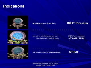
Journal of Arthroscopy,Vol. 12, No.4,Journal of Arthroscopy, Vol. 12, No.4,
August 1996 Hayashi et alAugust 1996 Hayashi et al
Indications
Herniation with back and leg pain IDET/Decompression
Herniation with radiculopathy DECOMPRESSION
Axial Discogenic Back Pain IDET™ Procedure
Large extrusion or sequestration OTHER
153.
The Decompression CatheterisThe Decompression Catheter is
NOT intended to replaceNOT intended to replace
discectomy or microdiscectomydiscectomy or microdiscectomy
for the treatment of largefor the treatment of large
extrusions or free nuclearextrusions or free nuclear
fragmentsfragments
Indications
154.
DiagnosticsDiagnostics
Positive MRI orCT showing contained focal discPositive MRI or CT showing contained focal disc
herniationherniation
– Protrusion no greater than 6mm beyond disc marginProtrusion no greater than 6mm beyond disc margin
– Above 6mm is likely an extrusionAbove 6mm is likely an extrusion
Positive Straight Leg Raise (SLR)Positive Straight Leg Raise (SLR)
Leg pain greater than back painLeg pain greater than back pain
Back and leg painBack and leg pain
Positive neurological findings on physicalPositive neurological findings on physical
examination, including weakness or numbnessexamination, including weakness or numbness
155.
Decompression vs. OtherProcedures
Patient Selection Radicular Axial and radicular Radicular
Heating Method Controlled, focal, broad Hot, localized
Hot, localized,
unpredictable
Catheter Placement Good access to pathology Nucleus Nucleus
Therapy Time ~ 12 minutes/disc ~ 8 minutes/disc ~ 30 minutes/disc
Post-Op
Management
Light activities Light activities Light activities
Ease of Use Highly steerable catheter Redirection is difficult Technically very difficult,
Complex OR set-up
Clinical Experience IDET
67 publications and
presentations
5 published clinical studies
with 2 year outcomes
Over 30,000 procedures
Limited Limited
Cost Decompression: $922 PercD: $900-$1000
Laser: ~ $100,000
LASE Kit: $1,445
(endoscope, guide needle,
dilator, cannula, tubing)
Topic Decompression
Catheter
Nucleoplasty LASER
Vertebroplasty - KyphoplastyVertebroplasty- Kyphoplasty
advantagesadvantages
May be performedMay be performed
under localunder local
anaesthesia as aanaesthesia as a
day caseday case
VERTEBROPLASTYVERTEBROPLASTY
It seems tobe moreIt seems to be more
favourable in recentfavourable in recent
vertebral fracturesvertebral fractures
(osteoporotic etc)(osteoporotic etc)
without majorwithout major
deformitydeformity
Balloon KyphoplastyBalloon Kyphoplasty
advantagesadvantages
Restoressufficiently theRestores sufficiently the
height of the collapsedheight of the collapsed
vertebravertebra
Is associated with inferiorIs associated with inferior
possibility of cementpossibility of cement
leakageleakage
184.
Balloon KyphoplastyBalloon Kyphoplasty
disadvantagesdisadvantages
Therisk of fracture in the adjacent levelsThe risk of fracture in the adjacent levels
is enhanced in the balloon kyphoplastyis enhanced in the balloon kyphoplasty
Increased operative time and radiationIncreased operative time and radiation
exposureexposure
185.
The minimal invasiveTheminimal invasive
procedureprocedure
((Vertebroplasty – Kyphoplasty)Vertebroplasty – Kyphoplasty)
is an effective and ais an effective and a
versatile methodversatile method
of treatment for:of treatment for:
Metastatic spinalMetastatic spinal
lesionslesions
(single or multiple)(single or multiple)