鎖骨下動脈盗血症候群
•
7 likes•13,484 views

The document describes a case of a 79-year-old female patient who fell down the stairs after suddenly losing consciousness. She has a history of diabetes and hypertension. Upon examination, her left radial pulse was weak and there was a 20 mmHg difference in blood pressure between the left and right arms, suggesting subclavian steal syndrome. Further tests such as CT angiography would be needed to confirm.
![Med Sci Monit, 2012: 18(5): RA57-63
Subclavian Steal Syndrome
Review Article Med Sci Monit, 2012: 18(5): RA57-63
HISTORICAL BACKGROUND
Subclavian steal phenomenon occurs when a subclavian ar-
tery stenosis proximal to the vertebral origin causes retro-
grade flow in the ipsilateral vertebral artery. Contorni [1]
鎖骨下動脈 起始部の閉塞, 狭窄により
was the first to recognize and describe this retrograde flow
in 1960 using angiography in a patient who had an absent ra-
dial pulse. A year later, Reivich [2] associated this phenome-
non with transient ischemic attack (TIA) and hence became
椎骨動脈の逆流を生じ,
the first scientist to correlate it with neurological symptoms.
The term “subclavian steal”, however, was coined by Fisher
[3] in 1961. This was after he reviewed Reivich’s article and
observed that the anomaly caused the ipsilateral subclavian
椎骨-脳底動脈系の虚血を生じる病態
artery to receive retrograde flow from the contralateral circu-
lation at the expense of the vertebro-basilar circulation [3].
Subclavian steal syndrome (SSS) has since been defined
as a group of symptoms that arise from this reversed blood
flow in the ipsilateral vertebral artery. It is often a differen-
一過性の脳虚血やめまい, 失神,
tial diagnosis in any patient who presents with a pulse def-
icit or a systolic blood pressure difference of greater than
20 mmHg between the arms [4]. The subclavian steal, in
上肢の間欠性跛行を生じる.
the absence of other anatomical anomalies, is usually as-
ymptomatic and often an incidental finding. Rarely, how-
ever, some patients may provoke the syndrome with exer-
cise and present with transient ipsilateral arm claudication,
ataxia and/or angina. The latter is prominent in those un-
dergoing coronary artery bypass graft (CABG) surgery with
橈骨動脈の脈の欠損や減弱,
the left internal mammary artery (LIMA) as the graft [5].
Angiography was the initial test used to screen for subcla-
血圧の左右差(20mmHg以上)は
vian steal, with only high probability patients being inves-
tigated [6]. This selection bias in testing gave the false im-
pression that SSS was not only rare, but also symptomatic
[7]. However, with the emergence of noninvasive techniques
所見として認められることがある
such as ultrasound in 1970 and magnetic resonance angiog-
raphy (MRA) in 1992, a greater number of asymptomatic
patients have been identified, reflecting the more benign
nature of the condition [8,9]. The prevalence and natural
history of SSS has, nevertheless, only recently been recent-
ly reported [10].](data:image/gif;base64,R0lGODlhAQABAIAAAAAAAP///yH5BAEAAAAALAAAAAABAAEAAAIBRAA7)
More Related Content
What's hot
What's hot (20)
Viewers also liked
Similar to 鎖骨下動脈盗血症候群
Similar to 鎖骨下動脈盗血症候群 (20)
More from Katsushige Takagishi
Recently uploaded
Recently uploaded (20)
鎖骨下動脈盗血症候群
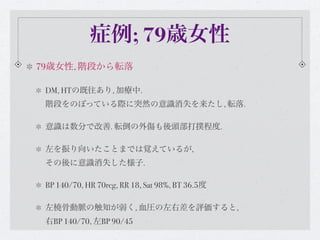
- 1. 症例; 79歳女性 79歳女性, 階段から転落 DM, HTの既往あり, 加療中. 階段をのぼっている際に突然の意識消失を来たし, 転落. 意識は数分で改善. 転倒の外傷も後頭部打撲程度. 左を振り向いたことまでは覚えているが, その後に意識消失した様子. BP 140/70, HR 70reg, RR 18, Sat 98%, BT 36.5度 左橈骨動脈の触知が弱く, 血圧の左右差を評価すると, 右BP 140/70, 左BP 90/45
- 2. Med Sci Monit, 2012: 18(5): RA57-63 Subclavian Steal Syndrome Review Article Med Sci Monit, 2012: 18(5): RA57-63 HISTORICAL BACKGROUND Subclavian steal phenomenon occurs when a subclavian ar- tery stenosis proximal to the vertebral origin causes retro- grade flow in the ipsilateral vertebral artery. Contorni [1] 鎖骨下動脈 起始部の閉塞, 狭窄により was the first to recognize and describe this retrograde flow in 1960 using angiography in a patient who had an absent ra- dial pulse. A year later, Reivich [2] associated this phenome- non with transient ischemic attack (TIA) and hence became 椎骨動脈の逆流を生じ, the first scientist to correlate it with neurological symptoms. The term “subclavian steal”, however, was coined by Fisher [3] in 1961. This was after he reviewed Reivich’s article and observed that the anomaly caused the ipsilateral subclavian 椎骨-脳底動脈系の虚血を生じる病態 artery to receive retrograde flow from the contralateral circu- lation at the expense of the vertebro-basilar circulation [3]. Subclavian steal syndrome (SSS) has since been defined as a group of symptoms that arise from this reversed blood flow in the ipsilateral vertebral artery. It is often a differen- 一過性の脳虚血やめまい, 失神, tial diagnosis in any patient who presents with a pulse def- icit or a systolic blood pressure difference of greater than 20 mmHg between the arms [4]. The subclavian steal, in 上肢の間欠性跛行を生じる. the absence of other anatomical anomalies, is usually as- ymptomatic and often an incidental finding. Rarely, how- ever, some patients may provoke the syndrome with exer- cise and present with transient ipsilateral arm claudication, ataxia and/or angina. The latter is prominent in those un- dergoing coronary artery bypass graft (CABG) surgery with 橈骨動脈の脈の欠損や減弱, the left internal mammary artery (LIMA) as the graft [5]. Angiography was the initial test used to screen for subcla- 血圧の左右差(20mmHg以上)は vian steal, with only high probability patients being inves- tigated [6]. This selection bias in testing gave the false im- pression that SSS was not only rare, but also symptomatic [7]. However, with the emergence of noninvasive techniques 所見として認められることがある such as ultrasound in 1970 and magnetic resonance angiog- raphy (MRA) in 1992, a greater number of asymptomatic patients have been identified, reflecting the more benign nature of the condition [8,9]. The prevalence and natural history of SSS has, nevertheless, only recently been recent- ly reported [10].
- 3. Med Sci Monit, 2012: 18(5): RA57-63 Southern Medical Journal 2001;94:445-447 Review Article Med Sci Monit, 2012: 18(5): RA57-63 盗血現象は患側の上肢を運動させたり, H B ISTORICAL ACKGROUND Subclavian steal phenomenon occurs when a subclavian ar- 首を曲げる, 左右を向くといった運動, tery stenosis proximal to the vertebral origin causes retro- grade flow in the ipsilateral vertebral artery. Contorni [1] was the first to recognize and describe this retrograde flow in 1960 using angiography in a patient who had an absent ra- 血圧が低下した際に増加. dial pulse. A year later, Reivich [2] associated this phenome- non with transient ischemic attack (TIA) and hence became the first scientist to correlate it with neurological symptoms. The term “subclavian steal”, however, was coined by Fisher >> 脳底動脈の循環不全を来す. [3] in 1961. This was after he reviewed Reivich’s article and observed that the anomaly caused the ipsilateral subclavian artery to receive retrograde flow from the contralateral circu- lation at the expense of the vertebro-basilar circulation [3]. Subclavian steal syndrome (SSS) has since been defined 椎骨動脈のみでは無く, as a group of symptoms that arise from this reversed blood flow in the ipsilateral vertebral artery. It is often a differen- tial diagnosis in any patient who presents with a pulse def- icit or a systolic blood pressure difference of greater than 20 mmHg between the arms [4]. The subclavian steal, in the absence of other anatomical anomalies, is usually as- 例えば内胸動脈からのCABG後の患者では ymptomatic and often an incidental finding. Rarely, how- ever, some patients may provoke the syndrome with exer- cise and present with transient ipsilateral arm claudication, 冠血流の盗血を来たし, 心筋虚血となる ataxia and/or angina. The latter is prominent in those un- dergoing coronary artery bypass graft (CABG) surgery with the left internal mammary artery (LIMA) as the graft [5]. Coronary subclavianAngiographysyndromeという subcla- stealwith only high probability patients being inves- vian steal, was the initial test used to screen for tigated [6]. This selection bias in testing gave the false im- 病態もある. pression that SSS was not only rare, but also symptomatic [7]. However, with the emergence of noninvasive techniques such as ultrasound in 1970 and magnetic resonance angiog- raphy (MRA) in 1992, a greater number of asymptomatic patients have been identified, reflecting the more benign nature of the condition [8,9]. The prevalence and natural history of SSS has, nevertheless, only recently been recent- ly reported [10].
- 4. Figure 2. Schematic diagram showing the retrograde from the left coronary artery through the mammary artery bypass grafts in a patient with left subclavian artery Stenosis. (Reproduced and modified with permission from Takach et Coronary subclavian steal syndrome al., 2006). Med Sci Monit, 2012: 18(5): RA57-63
- 5. SSSの疫学 頻度は報告によりバラツキがあるが, 0.6-6.4% PAD(+)群では11-18%と頻度も高くなる ただし, 症候性となると更に頻度は低下. 狭窄を認める患者のうち, 5.3%が神経症状を認める. また, 解剖学的に左側が殆どを占める. 後天性のSSSでは82.3%が左側. 右:左 = 1:4とする報告が多い. 若年性では高安病が主な原因. 30歳台で女性で多く, 50歳以上では動脈硬化によるものが多い. その場合は男性例. 先天性は動脈奇形, 右大動脈弓など, 解剖学的な奇形による. Med Sci Monit, 2012: 18(5): RA57-63
- 6. SSSの分類 重症度はGrade I-IIIに分類; Grade I; pre-subclavian steal - 椎骨動脈の逆流(-), 流速は遅い. Grade II; intermittent/partial/latent - 収縮期のみに逆流あり Review Article Grade III; permanent/advanced - 持続的に逆流を認める. Prevelance of grades subclavian steal 100 これらのGradeは 90 Absent Partial 80 Complete Presence of SSS patients 血圧の左右差に比例する 70 60 50 40 30 20 10 0 Med Sci Monit, 2012: 18(5): RA57-63 20–30 31–40 41–50 >50 Arm blood pressure differential (mmHg)
- 7. Prevelance of grades subclavian steal Prevelance of symptoms and intervention for SSS 100 40 90 Absent % of symptomatic Partial 35 % of requiring 80 Complete intervention 30 Presence of SSS patients Precent of SSS patients 症状の有無も 70 60 25 50 20 血圧の左右差に比例 40 15 30 10 20 10 5 血圧差20-30mmHg程度では, 0 0 20–30 31–40 41–50 >50 20–30 31–40 41–50 >50 症候性となる例は少ない(1.38%). Arm blood pressure differential (mmHg) Arm blood pressure differential (mmHg) Figure 4. Prevalence of grades of subclavian steal with increasing arm Figure 5. Prevalence of symptoms and interventions in patients with BP differential. Grade 1: BP differential 20–30 mmHg; Grade SSS with increasing arm Blood Pressure (BP) differential. 血圧差>50mmHgでは38.5%が症候性 (Reproduced and modified with permission from 2: BP differential 31–40 mmHg; Grade 3: BP differential 41–50 mmHg, Grade 4: BP differential >50. (Reproduced Labropoulos et al., 2010). and modified with permission from Labropoulos et al., 2010). pathway for the affected arm [31]. Significant ischemia of CLASSIFICATION the arm is therefore rare, even in patients who have com- plete occlusion of the proximal subclavian artery [16,32]. The subclavian steal phenomenon has been characterized Some authors have suggested that the patients who develop either by the territory from which the blood is “stolen” [28] symptoms from this phenomenon usually have additional or the severity of hemodynamic disturbances in the verte- vascular pathology involving either the intracranial or ex- bral artery [29]. Territories are classified as vertebral-verte- tra-cranial vessels [33]. However, while there is some log- bral, carotid-basilar, external carotid-basilar, or carotid-sub- ic to this assertion, cases of patients who had no significant clavian. A case has also been reported of a patient who had stenosis in any other cervical or intracranial artery yet still partial bilateral (carotid – carotid and carotid – vertebral) suffered from chronic posterior circulation ischemia as a subclavian steal syndrome, with blood supply to both arms result of SSS has also been reported [34]. somewhat maintained by collateral vessels [30]. Med Sci Monit, 2012: 18(5): RA57-63 Recent studies have shown a linear correlation between in-
- 8. SSSの診断 血圧の左右差はスクリーニングとして有用 感度は55-88%と幅があるが, 初期検査として血圧差と橈骨動脈の左右差の評価は重要. 画像評価は血管エコーが血管狭窄と血流評価に優れる. 鎖骨下動脈の狭窄のみではなく, 椎骨動脈の逆流, 流速も評価可能 その際, 血圧計のカフを患側に巻き, sBPより20mmHg高い圧で 数分保持し, その後減圧 ⇒ 上肢血流を増やし盗血を誘発する方法もある. Med Sci Monit, 2012: 18(5): RA57-63
- 9. CTやMRIによるAngiographyは確定的検査. USにて疑いが強い場合に選択すべき検査と言える. Med Sci Monit, 2012: 18(5): RA57-63
- 10. SSSの治療 症状や程度に応じて考慮するが, 基本的には保存治療が推奨される. 動脈硬化によるものならば, 血圧コントロール, 高脂血症, DMへの対応, 禁煙など. 上記を行いながら, USでフォローしてゆくことが多い. 重症例ではバイパス術や, 血管内治療が選択される.
- 11. Carotid-subclavian bypass surgery; 10年間で95%の開存率を見込める有用な治療. 成功率も良好(98%)とされる. Endovascular therapy; 開存率は3年間で93%(狭窄例), 65%(閉塞例) 長期的にフォローしたstudyは乏しく, 32 長期的か依存率はバイパス術に劣る可能性が高い. Vascular Medicine 16(1) Table 1. Restenosis rates from major series of subclavian stenting Vasc Med 2011:16;29-34 Author Year Subjects Approximate primary restenosis (%) Mean follow-up (months) Sullivan34 1998 83 4.5 14 Al-Mubarak35 1999 38 6 20 Bates36 2004 91 14.6 36 De Vries37 2005 110 7.8 34 Patel14 2008 170 14.6 36
- 12. J Endovasc Ther. 2012;19:44–51 1989年∼2010年に治療された252例のSSS患者 Balloon-expandable stentsでの治療が148例, Extrathoracic surgical bypassesでの治療が104例. 手技自体の成功率はステントで97.3%, THER J ENDOVASC 2012;19:44–51 バイパス術99.0%と同等 手技による合併症も有意差無し. 血管開存率; ステント バイパス @1y 91% 99% @3y 78% 97% @5y 67% 95% @10y 49% 89% 生存率は両者変わらず.