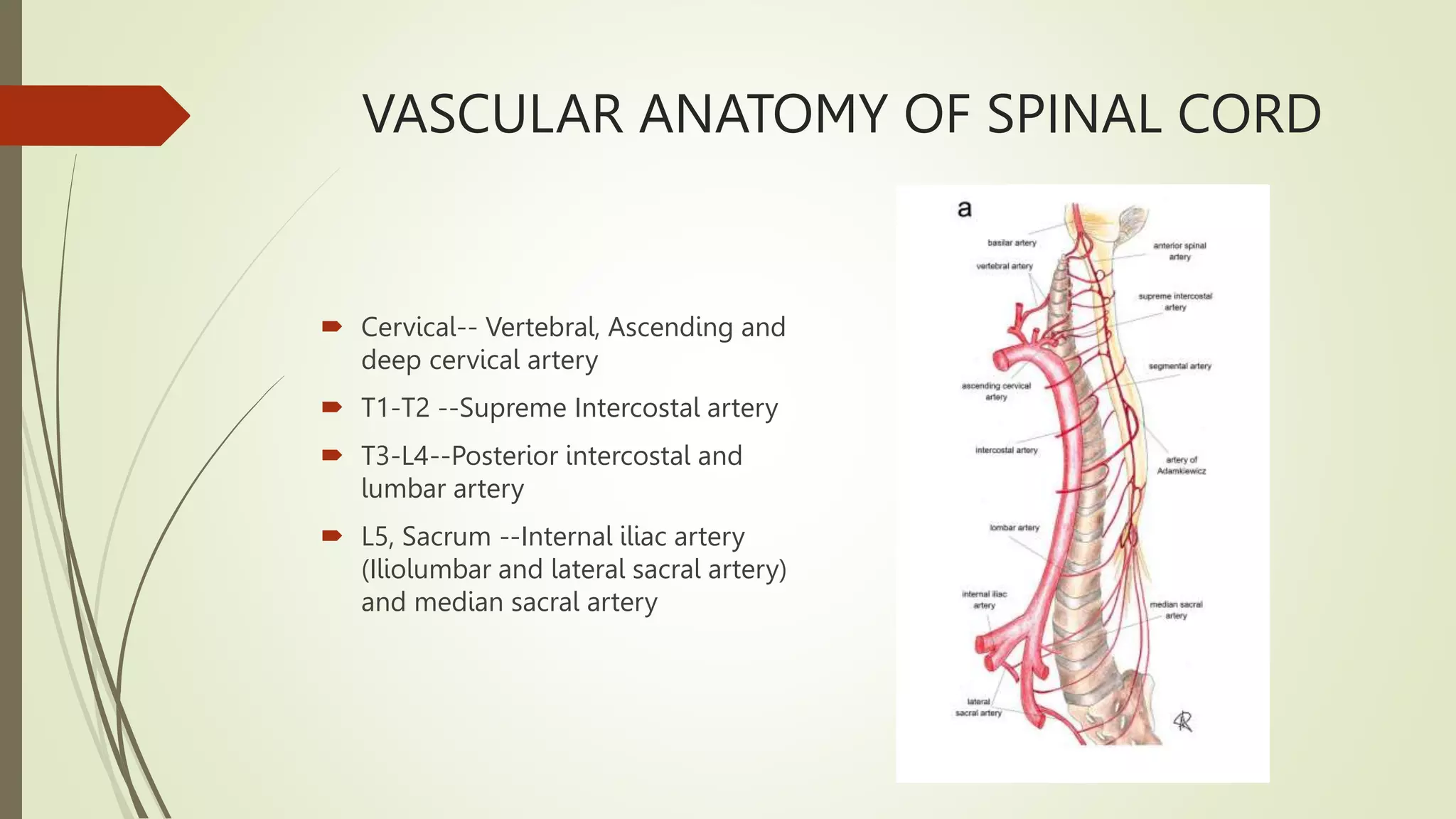
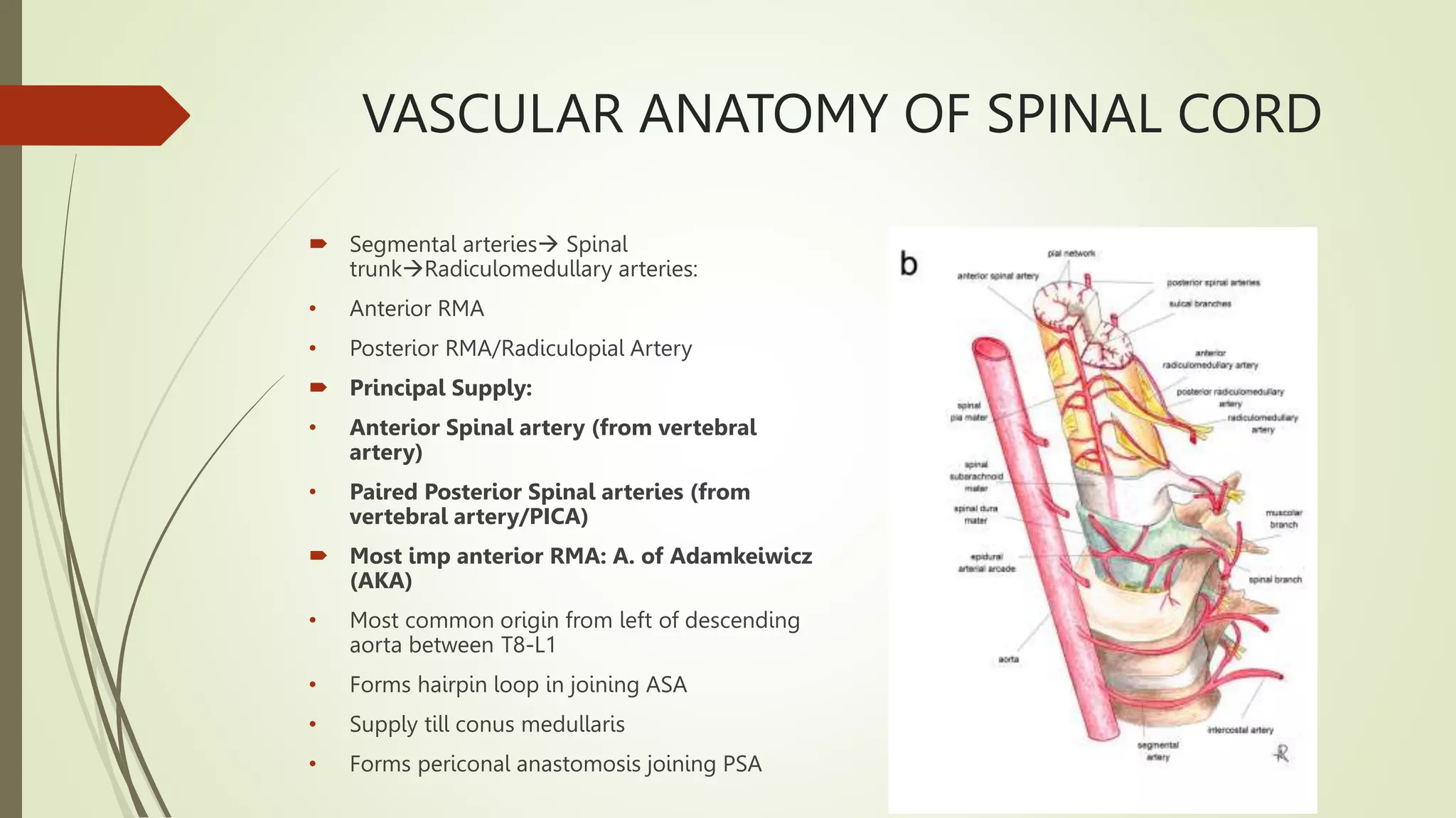
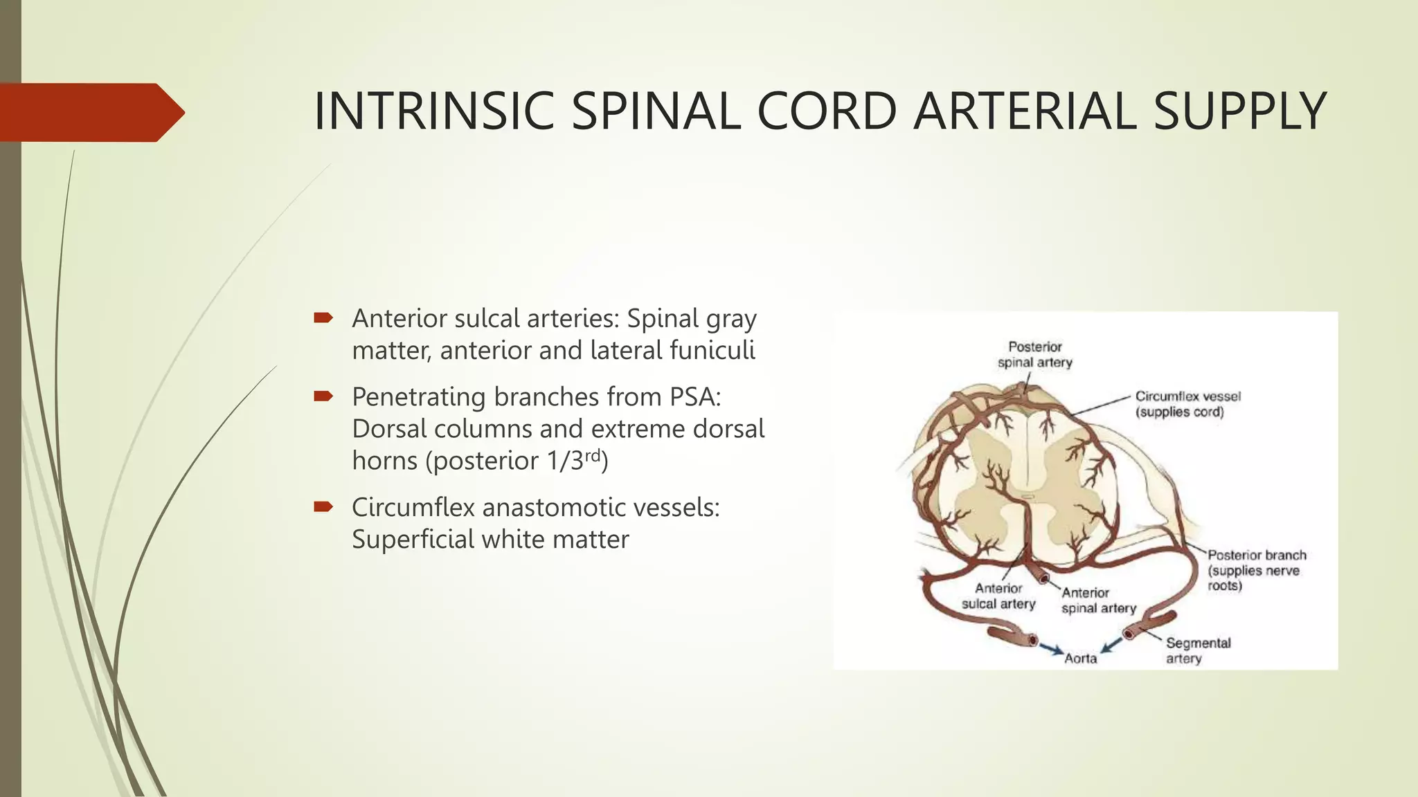
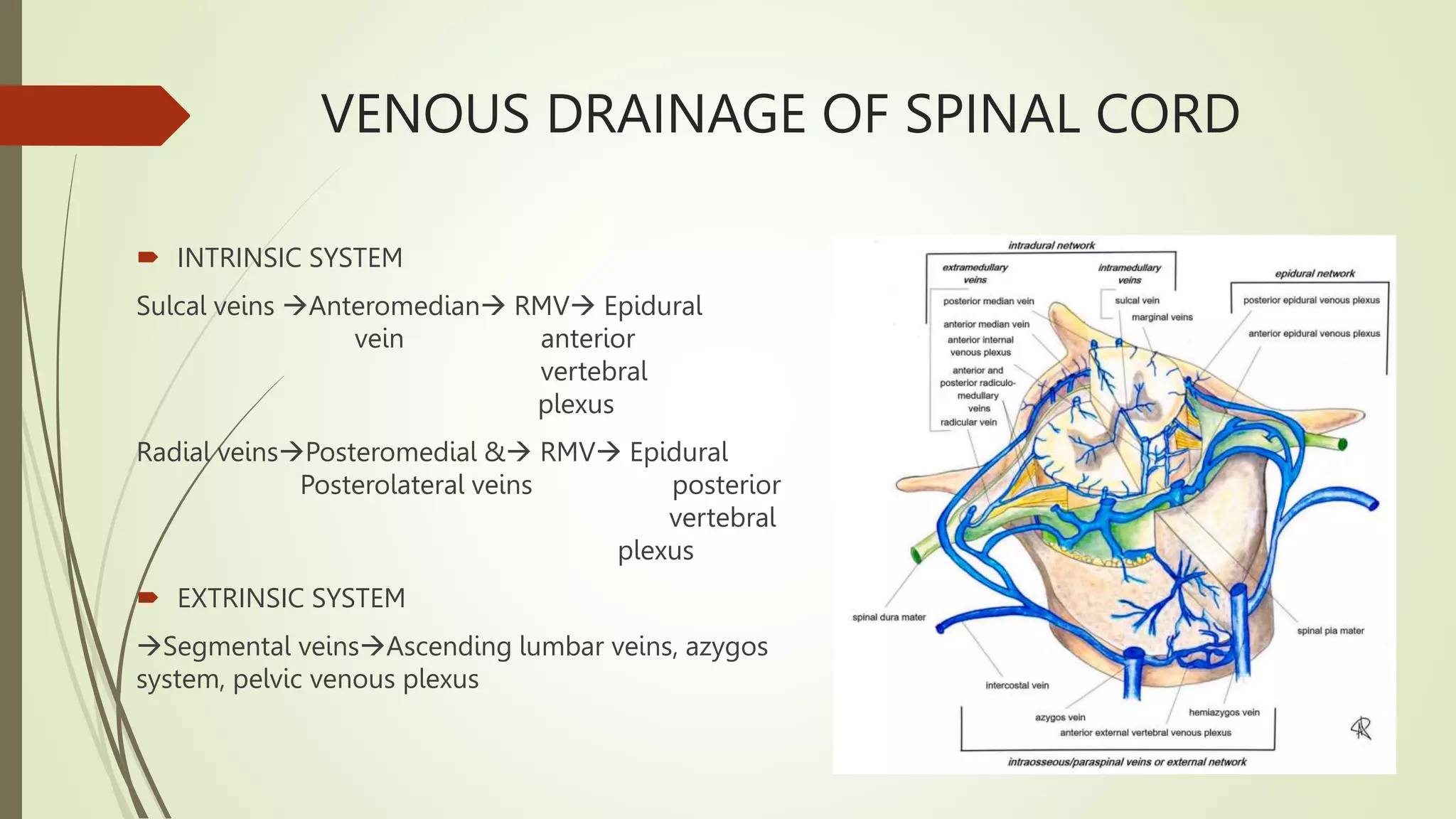
SPINAL CORD VASCULAR
SYNDROME- DIAGNOSIS AND
MANAGEMENT
DR. DEVASHISH GUPTA
SR 1 NEUROLOGY
VASCULAR ANATOMY OF SPINAL CORD
 Cervical-- Vertebral, Ascending and
deep cervical artery
 T1-T2 --Supreme Intercostal artery
 T3-L4--Posterior intercostal and
lumbar artery
 L5, Sacrum --Internal iliac artery
(Iliolumbar and lateral sacral artery)
and median sacral artery
VASCULAR ANATOMY OF SPINAL CORD
 Segmental arteries Spinal
trunkRadiculomedullary arteries:
• Anterior RMA
• Posterior RMA/Radiculopial Artery
 Principal Supply:
• Anterior Spinal artery (from vertebral
artery)
• Paired Posterior Spinal arteries (from
vertebral artery/PICA)
 Most imp anterior RMA: A. of Adamkeiwicz
(AKA)
• Most common origin from left of descending
aorta between T8-L1
• Forms hairpin loop in joining ASA
• Supply till conus medullaris
• Forms periconal anastomosis joining PSA
INTRINSIC SPINAL CORD ARTERIAL SUPPLY
 Anterior sulcal arteries: Spinal gray
matter, anterior and lateral funiculi
 Penetrating branches from PSA:
Dorsal columns and extreme dorsal
horns (posterior 1/3rd)
 Circumflex anastomotic vessels:
Superficial white matter
VENOUS DRAINAGE OF SPINAL CORD
 INTRINSIC SYSTEM
Sulcal veins Anteromedian RMV Epidural
vein anterior
vertebral
plexus
Radial veinsPosteromedial & RMV Epidural
Posterolateral veins posterior
vertebral
plexus
 EXTRINSIC SYSTEM
Segmental veinsAscending lumbar veins, azygos
system, pelvic venous plexus
SPINAL CORD ANATOMY
SPINAL CORD VASCULAR SYNDROME
 Abnormality in physiological spinal vascularity either due to impaired arterial supply or
venous drainage Spinal cord injury
 PATHOPHYSIOLOGY
• MC Cause: Iatrogenic injury to thoracoabdominal aorta during surgery
• Mechanical compression of radicular artery
• Diseases of aorta: Atherosclerotic, Aortic dissection
• Systemic hypoperfusion
• Radiotherapy
• Vascular malformations
• Venous infarcts
• Fibrocartilaginous emboli from ruptured disc
• Vasculitic and thrombotic causes: Meningovascular syphilis, Crohn’s, PAN, Giant cell arteritis
ARTERIAL SPINAL CORD ISCHEMIA
 ANTERIOR SPINAL ARTERY SYNDROME
• Bilateral loss of motor function and pain/temperature sensation
• Relative sparing of proprioception and vibratory senses
• Acute: Flaccidity and loss of deep tendon reflexes
• Later: Spasticity and hyperreflexia.
• Autonomic dysfunction may be present
 INCOMPLETE SYNDROME OF THE SPINAL ARTERY SYNDROME
• Localized at the level of the anterior horns
1) Acute paraplegia (pseudopoliomyelitic form) without sensory abnormalities and sphincter
dysfunction
2) Painful bilateral brachial diplegia in the case of a cervical lesion (the man-in-the-barrel syndrome)
3) Progressive distal amyotrophy due to chronic lesions of the anterior horns; misdiagnosed as lateral
amyotrophic sclerosis.
ARTERIAL SPINAL CORD ISCHEMIA
 POSTERIOR SPINAL ARTERY SYNDROME.
• Loss of proprioception and vibratory senses below the level of the injury and total anesthesia at the level of the
injury.
• Mild and transient weakness.
• Unilateral > bilateral
 SULCOCOMMISSURAL SYNDROME Presents as a partial Brown-Sequard syndrome clinical picture with sparing
of postural sensibility.
• Hemiparesis with a contralateral spinothalamic sensory deficit.
 INFARCTION AT THE LEVEL OF CONUS MEDULLARIS May be misdiagnosed as a cauda equina syndrome.
 CENTRAL SPINAL INFARCT
• Bilateral spinothalamic sensory deficit with sparing of the posterior columns.
• Motor deficit and sphincter dysfunction are usually absent.
 TRANSVERSE MEDULLARY INFARCTION Paraplegia/paresis or, in cases of higher cord lesions,
tetraplegia/paresis.
• May be complete sensory loss
• Sphincter dysfunction
INVESTIGATIONS IN ARTERIAL SPINAL
CORD ISCHEMIA
 MRI
• Most common pattern “Owl-eyes sign/Double dot pattern” on axial scan bilateral anterior
horns
• “H shaped pattern” on axial scan  Entire central gray matter
• “Pencil-like appearance” in the sagittal plane.
• Hyperacute and acute phases: Diffusion restriction, consistent with cytotoxic oedema,
hyperintense T2 signal with slight spinal cord swelling affecting a long segment.
• Subacute phase: Variable contrast enhancement, may persist upto 3 weeks.
• Chronic phase : atrophy may be observed
 MYELOGRAPHY: Usually normal
 CTA/MRA: Vertebral or aortic dissection
 MRI BRAIN: Inflammatory etiology
 CSF: Inflammatory/Infectious process
TREATMENT OF ARTERIAL SPINAL CORD
ISCHEMIA
 Infarction due to aortic surgery  Increase MAP with vasopressors and place
lumbar drain, Thrombolytics
 Inflammatory myelopathy  Corticosteroids
 Bed rest, Reversal of proximate causes (Arrythmia, hypovolemia)
 Monitor for development of autonomic dysreflexia or neurogenic shock
 Prophylaxis to prevent DVT, GI ulceration
VENOUS SPINAL CORD ISCHEMIA
 SPINAL AV MALFORMATIONS
Developmental/ acquired abnormal direct communication of normal to enlarged
radiculomedullary arteries with enlarged radiculomedullary veins without intervening
capillary network
 ANSON AND SPETZLER CLASSIFICATION
Type I: Dural AVF, IA (single feeding artery) IB (multiple feeding arteries)
Type II: Intramedullary glomus type AVM
Type III: Intramedullary juvenile type AVM, frequent extramedullary and sometimes
extradural component
Type IV: Intradural extramedullary AVF – subtypes IVA, IVB , IVC with progressively
increasing AV shunting
SPINAL DURAL ARTERIOVENOUS FISTULAS
(SDAVFS)
• Vein arterialization impaired venous drainage and venous congestion
intramedullary oedema, decreased arterial perfusion and chronic hypoxia  later
may affect additional spinal levels in an ascending fashion
• MC symptom: Sensorimotor disturbances , usually worsen with exercise,
prolonged standing, and Valsalva.
• Sensory symptoms (numbness or hypoesthesia) may localize into the perianal area,
while a sensory level occurs up to one-third of patients.
• Legs weakness includes upper and lower motor neuron features.
• Dysautonomia (bowel, bladder, and sexual dysfunction) is more tardive
• Spinal bruit: Highly specific
INVESTIGATIONS IN SDAVFS
 MRI +_MRA: Initial diagnosis of choice
 Cord edema and prominent perimedullary vessels posterior to cord
 T2: Swelling and increased cord signal, Conglomerate perimedullary/
intramedullary blood vessels seen as flow voids
 Post contrast variable enhancement
 DSA : Gold standard  “T-shaped” anastomosis between the injected RMA and the
draining vein, as well as a network of dilated perimedullary veins
SPINAL ARTERIOVENOUS
MALFORMATIONS (SAVMS)
 Intramedullary glomus-type AVMs : Intramedullary nidus of shunting vessels
usually located in the anterior half of the spinal cord fed by one or more spinal
arteries, draining into spinal veins.
 Mostly in thoracic spine
 MRI: Hematomyelia  Severe back, neck or radicular pain along with
sensorimotor/autonomic deficits
 Non-hemorrhagic spinal AVMs: Gradual myelopathic symptoms
TREATMENT OF SPINAL AV
MALFORMATION
 Surgical resection and/or angiographically directed embolization
 Surgical approach  Better rate of obliteration of fistula
 Endovascular approach  Though less invasive, has the potential to cause
additional infarction in case of migration of embolic material
 Sequential approach of Embolization followed by surgery is common
 Radioablation : Being explored for some lesions
SPINAL CAVERNOUS MALFORMATIONS
 Intramedullary Slow flow vascular malformations without AV shunting
 Typical presentation: Direct episodic neurological deficit with variable recovery
between episodes
 MRI: Heterogenous T1/T2 signal with hypointense hemosiderin rim on T2
“Popcorn appearance”
 GRE: Profound hypointensity due to blood products
 Enhancement is not typical
TREATMENT OF SPINAL CAVERNOUS
MALFORMATIONS
 Asymptomatic  No treatment
 Symptomatic  Surgical exploration and resection
SPINAL HEMORRHAGE
 Subarachnoid hemorrhage
 Intramedullary hemorrhage
 Subdural hemorrhage
 Epidural hemorrhage
SPINAL SAH
 MC Cause : Spinal angioma
 Trauma, coarctation of aorta, rupture of spinal artery aneurysmal rupture, PAN, spinal tumors,
iatrogenic (LP), blood dyscrasia, therapeutic thrombolytics and anticoagulants
 Correct diagnosis requires strong clinical suspicion
 C/F: Sudden onset severe back pain near level of hemorrhage  Diffuse pain and signs of
meningeal irritation
 Multiple radiculopathies and myelopathy
 Headache, cranial neuropathy, decreased level of consciousness
 Grossly bloody CSF
 Elevated ICP, Papilledema
 Treatment : Treat the underlying cause
HEMATOMYELIA/ INTRAMEDULLARY
SPINAL HEMORRHAGE
 MC Cause: Direct trauma to spinal column/ hyperextension injury of spine
 Bleeding from spinal vascular malformation, hemorrhage into spinal tumor or syrinx, bleeding diathesis,
anticoagulant use, venous infarction, radiation therapy
 Disruption of gray matter > white matter
 Spinal shock associated with sudden onset severe backpain, often radicular
 Later spasticity below level of lesion with LMN signs
 Diagnosis:
• MRI : Acute  Intramedullary T1 hyper, T2 hypointensity with cord swelling  Signal depends on stage of
blood
• CSF: consistent with SAH
 Treatment
• Supportive
• Laminectomy and drainage of hematoma followed by resection of tumor / vascular malformation
SPINAL EDH AND SDH
SPINAL EDH
 More common
 MC in men, Bimodal distribution
(childhood, 5th-6th decade)
 Childhood  Cervical lesions
Adults  Thoracic and lumbar
 Cause : Spontaneous, post exertion/
trauma, post LP and epidural
anesthesia
 More likely in anticoagulated patients
SPINAL SDH
 Less common
 Women, any age (Mostly in 6th
decade)
 MC in thoracic and lumbar regions
 Causes: Hemorrhagic diathesis (MC),
trauma, LP, vascular malformation,
spinal surgery
SPINAL EDH AND SDH
 C/F: Indistinguishable
 Severe back pain at level of bleed
 Myelopathy or cauda equina syndrome with motor and sensory deficits
 Rapidly decreasing platelet count or <40,000 platelets/µl are at risk of developing SEH or SSH as
complication of LP  Should receive platelet transfusion prior to procedure
 DIAGNOSIS
• CSF: Normal/ Xanthochromia
• MRI: Delineate size and location
• CEMRI, MRA: Underlying vascular malformation
• CT Myelography: Partial filling defect or complete blockage to flow of contrast below level of lesion
 TREATMENT: Laminectomy with evacuation of clot as soon as possible
SPINAL SDH SPINAL EDH
REFERENCES
 Bradley and Daroff’s Neurology in clinical practice 2, 8th edition 2021
 Vargas MI, Gariani J, Sztajzel R, Barnaure-Nachbar I, Delattre BM, Lovblad KO,
Dietemann JL. Spinal cord ischemia: practical imaging tips, pearls, and pitfalls.
American Journal of Neuroradiology. 2015 May 1;36(5):825-30.
 Da Ros V, Picchi E, Ferrazzoli V, Schirinzi T, Sabuzi F, Grillo P, Muto M, Garaci F, Muto
M, Di Giuliano F. Spinal vascular lesions: anatomy, imaging techniques and
treatment. European Journal of Radiology Open. 2021 Jan 1;8:100369.

SPINAL CORD VASCULAR SYNDROME (1).pptx

  • 1.
    SPINAL CORD VASCULAR SYNDROME-DIAGNOSIS AND MANAGEMENT DR. DEVASHISH GUPTA SR 1 NEUROLOGY
  • 2.
    VASCULAR ANATOMY OFSPINAL CORD  Cervical-- Vertebral, Ascending and deep cervical artery  T1-T2 --Supreme Intercostal artery  T3-L4--Posterior intercostal and lumbar artery  L5, Sacrum --Internal iliac artery (Iliolumbar and lateral sacral artery) and median sacral artery
  • 3.
    VASCULAR ANATOMY OFSPINAL CORD  Segmental arteries Spinal trunkRadiculomedullary arteries: • Anterior RMA • Posterior RMA/Radiculopial Artery  Principal Supply: • Anterior Spinal artery (from vertebral artery) • Paired Posterior Spinal arteries (from vertebral artery/PICA)  Most imp anterior RMA: A. of Adamkeiwicz (AKA) • Most common origin from left of descending aorta between T8-L1 • Forms hairpin loop in joining ASA • Supply till conus medullaris • Forms periconal anastomosis joining PSA
  • 4.
    INTRINSIC SPINAL CORDARTERIAL SUPPLY  Anterior sulcal arteries: Spinal gray matter, anterior and lateral funiculi  Penetrating branches from PSA: Dorsal columns and extreme dorsal horns (posterior 1/3rd)  Circumflex anastomotic vessels: Superficial white matter
  • 5.
    VENOUS DRAINAGE OFSPINAL CORD  INTRINSIC SYSTEM Sulcal veins Anteromedian RMV Epidural vein anterior vertebral plexus Radial veinsPosteromedial & RMV Epidural Posterolateral veins posterior vertebral plexus  EXTRINSIC SYSTEM Segmental veinsAscending lumbar veins, azygos system, pelvic venous plexus
  • 6.
  • 7.
    SPINAL CORD VASCULARSYNDROME  Abnormality in physiological spinal vascularity either due to impaired arterial supply or venous drainage Spinal cord injury  PATHOPHYSIOLOGY • MC Cause: Iatrogenic injury to thoracoabdominal aorta during surgery • Mechanical compression of radicular artery • Diseases of aorta: Atherosclerotic, Aortic dissection • Systemic hypoperfusion • Radiotherapy • Vascular malformations • Venous infarcts • Fibrocartilaginous emboli from ruptured disc • Vasculitic and thrombotic causes: Meningovascular syphilis, Crohn’s, PAN, Giant cell arteritis
  • 8.
    ARTERIAL SPINAL CORDISCHEMIA  ANTERIOR SPINAL ARTERY SYNDROME • Bilateral loss of motor function and pain/temperature sensation • Relative sparing of proprioception and vibratory senses • Acute: Flaccidity and loss of deep tendon reflexes • Later: Spasticity and hyperreflexia. • Autonomic dysfunction may be present  INCOMPLETE SYNDROME OF THE SPINAL ARTERY SYNDROME • Localized at the level of the anterior horns 1) Acute paraplegia (pseudopoliomyelitic form) without sensory abnormalities and sphincter dysfunction 2) Painful bilateral brachial diplegia in the case of a cervical lesion (the man-in-the-barrel syndrome) 3) Progressive distal amyotrophy due to chronic lesions of the anterior horns; misdiagnosed as lateral amyotrophic sclerosis.
  • 9.
    ARTERIAL SPINAL CORDISCHEMIA  POSTERIOR SPINAL ARTERY SYNDROME. • Loss of proprioception and vibratory senses below the level of the injury and total anesthesia at the level of the injury. • Mild and transient weakness. • Unilateral > bilateral  SULCOCOMMISSURAL SYNDROME Presents as a partial Brown-Sequard syndrome clinical picture with sparing of postural sensibility. • Hemiparesis with a contralateral spinothalamic sensory deficit.  INFARCTION AT THE LEVEL OF CONUS MEDULLARIS May be misdiagnosed as a cauda equina syndrome.  CENTRAL SPINAL INFARCT • Bilateral spinothalamic sensory deficit with sparing of the posterior columns. • Motor deficit and sphincter dysfunction are usually absent.  TRANSVERSE MEDULLARY INFARCTION Paraplegia/paresis or, in cases of higher cord lesions, tetraplegia/paresis. • May be complete sensory loss • Sphincter dysfunction
  • 10.
    INVESTIGATIONS IN ARTERIALSPINAL CORD ISCHEMIA  MRI • Most common pattern “Owl-eyes sign/Double dot pattern” on axial scan bilateral anterior horns • “H shaped pattern” on axial scan  Entire central gray matter • “Pencil-like appearance” in the sagittal plane. • Hyperacute and acute phases: Diffusion restriction, consistent with cytotoxic oedema, hyperintense T2 signal with slight spinal cord swelling affecting a long segment. • Subacute phase: Variable contrast enhancement, may persist upto 3 weeks. • Chronic phase : atrophy may be observed  MYELOGRAPHY: Usually normal  CTA/MRA: Vertebral or aortic dissection  MRI BRAIN: Inflammatory etiology  CSF: Inflammatory/Infectious process
  • 12.
    TREATMENT OF ARTERIALSPINAL CORD ISCHEMIA  Infarction due to aortic surgery  Increase MAP with vasopressors and place lumbar drain, Thrombolytics  Inflammatory myelopathy  Corticosteroids  Bed rest, Reversal of proximate causes (Arrythmia, hypovolemia)  Monitor for development of autonomic dysreflexia or neurogenic shock  Prophylaxis to prevent DVT, GI ulceration
  • 13.
    VENOUS SPINAL CORDISCHEMIA  SPINAL AV MALFORMATIONS Developmental/ acquired abnormal direct communication of normal to enlarged radiculomedullary arteries with enlarged radiculomedullary veins without intervening capillary network  ANSON AND SPETZLER CLASSIFICATION Type I: Dural AVF, IA (single feeding artery) IB (multiple feeding arteries) Type II: Intramedullary glomus type AVM Type III: Intramedullary juvenile type AVM, frequent extramedullary and sometimes extradural component Type IV: Intradural extramedullary AVF – subtypes IVA, IVB , IVC with progressively increasing AV shunting
  • 14.
    SPINAL DURAL ARTERIOVENOUSFISTULAS (SDAVFS) • Vein arterialization impaired venous drainage and venous congestion intramedullary oedema, decreased arterial perfusion and chronic hypoxia  later may affect additional spinal levels in an ascending fashion • MC symptom: Sensorimotor disturbances , usually worsen with exercise, prolonged standing, and Valsalva. • Sensory symptoms (numbness or hypoesthesia) may localize into the perianal area, while a sensory level occurs up to one-third of patients. • Legs weakness includes upper and lower motor neuron features. • Dysautonomia (bowel, bladder, and sexual dysfunction) is more tardive • Spinal bruit: Highly specific
  • 16.
    INVESTIGATIONS IN SDAVFS MRI +_MRA: Initial diagnosis of choice  Cord edema and prominent perimedullary vessels posterior to cord  T2: Swelling and increased cord signal, Conglomerate perimedullary/ intramedullary blood vessels seen as flow voids  Post contrast variable enhancement  DSA : Gold standard  “T-shaped” anastomosis between the injected RMA and the draining vein, as well as a network of dilated perimedullary veins
  • 19.
    SPINAL ARTERIOVENOUS MALFORMATIONS (SAVMS) Intramedullary glomus-type AVMs : Intramedullary nidus of shunting vessels usually located in the anterior half of the spinal cord fed by one or more spinal arteries, draining into spinal veins.  Mostly in thoracic spine  MRI: Hematomyelia  Severe back, neck or radicular pain along with sensorimotor/autonomic deficits  Non-hemorrhagic spinal AVMs: Gradual myelopathic symptoms
  • 22.
    TREATMENT OF SPINALAV MALFORMATION  Surgical resection and/or angiographically directed embolization  Surgical approach  Better rate of obliteration of fistula  Endovascular approach  Though less invasive, has the potential to cause additional infarction in case of migration of embolic material  Sequential approach of Embolization followed by surgery is common  Radioablation : Being explored for some lesions
  • 23.
    SPINAL CAVERNOUS MALFORMATIONS Intramedullary Slow flow vascular malformations without AV shunting  Typical presentation: Direct episodic neurological deficit with variable recovery between episodes  MRI: Heterogenous T1/T2 signal with hypointense hemosiderin rim on T2 “Popcorn appearance”  GRE: Profound hypointensity due to blood products  Enhancement is not typical
  • 25.
    TREATMENT OF SPINALCAVERNOUS MALFORMATIONS  Asymptomatic  No treatment  Symptomatic  Surgical exploration and resection
  • 26.
    SPINAL HEMORRHAGE  Subarachnoidhemorrhage  Intramedullary hemorrhage  Subdural hemorrhage  Epidural hemorrhage
  • 27.
    SPINAL SAH  MCCause : Spinal angioma  Trauma, coarctation of aorta, rupture of spinal artery aneurysmal rupture, PAN, spinal tumors, iatrogenic (LP), blood dyscrasia, therapeutic thrombolytics and anticoagulants  Correct diagnosis requires strong clinical suspicion  C/F: Sudden onset severe back pain near level of hemorrhage  Diffuse pain and signs of meningeal irritation  Multiple radiculopathies and myelopathy  Headache, cranial neuropathy, decreased level of consciousness  Grossly bloody CSF  Elevated ICP, Papilledema  Treatment : Treat the underlying cause
  • 29.
    HEMATOMYELIA/ INTRAMEDULLARY SPINAL HEMORRHAGE MC Cause: Direct trauma to spinal column/ hyperextension injury of spine  Bleeding from spinal vascular malformation, hemorrhage into spinal tumor or syrinx, bleeding diathesis, anticoagulant use, venous infarction, radiation therapy  Disruption of gray matter > white matter  Spinal shock associated with sudden onset severe backpain, often radicular  Later spasticity below level of lesion with LMN signs  Diagnosis: • MRI : Acute  Intramedullary T1 hyper, T2 hypointensity with cord swelling  Signal depends on stage of blood • CSF: consistent with SAH  Treatment • Supportive • Laminectomy and drainage of hematoma followed by resection of tumor / vascular malformation
  • 31.
    SPINAL EDH ANDSDH SPINAL EDH  More common  MC in men, Bimodal distribution (childhood, 5th-6th decade)  Childhood  Cervical lesions Adults  Thoracic and lumbar  Cause : Spontaneous, post exertion/ trauma, post LP and epidural anesthesia  More likely in anticoagulated patients SPINAL SDH  Less common  Women, any age (Mostly in 6th decade)  MC in thoracic and lumbar regions  Causes: Hemorrhagic diathesis (MC), trauma, LP, vascular malformation, spinal surgery
  • 32.
    SPINAL EDH ANDSDH  C/F: Indistinguishable  Severe back pain at level of bleed  Myelopathy or cauda equina syndrome with motor and sensory deficits  Rapidly decreasing platelet count or <40,000 platelets/µl are at risk of developing SEH or SSH as complication of LP  Should receive platelet transfusion prior to procedure  DIAGNOSIS • CSF: Normal/ Xanthochromia • MRI: Delineate size and location • CEMRI, MRA: Underlying vascular malformation • CT Myelography: Partial filling defect or complete blockage to flow of contrast below level of lesion  TREATMENT: Laminectomy with evacuation of clot as soon as possible
  • 33.
  • 34.
    REFERENCES  Bradley andDaroff’s Neurology in clinical practice 2, 8th edition 2021  Vargas MI, Gariani J, Sztajzel R, Barnaure-Nachbar I, Delattre BM, Lovblad KO, Dietemann JL. Spinal cord ischemia: practical imaging tips, pearls, and pitfalls. American Journal of Neuroradiology. 2015 May 1;36(5):825-30.  Da Ros V, Picchi E, Ferrazzoli V, Schirinzi T, Sabuzi F, Grillo P, Muto M, Garaci F, Muto M, Di Giuliano F. Spinal vascular lesions: anatomy, imaging techniques and treatment. European Journal of Radiology Open. 2021 Jan 1;8:100369.

Editor's Notes

  • #7 Cross sectional MRI anatomy of the spinal cord in cervical segment. The grey matter appears hyperintense on T2 sequences and its H-shaped borders the three white matter columns: the anterior column (yellow dotted line), the lateral column (green dotted line) and the dorsal column (red dotted line)
  • #12 Evolution of ischemia. The first MR image shows the subtle signal anomaly on T2 and diffusion sequences (arrows, A–C). Follow-up 48 hours later shows an important tumefaction and high signal on T2WI associated with a restriction of diffusion of the cervical spinal cord at the C4 –C7 levels (arrows, D–G)
  • #16 Spinal dural arteriovenous fistula (SDAVF): a low-flow shunt between a radiculomeningeal artery and a medullary vein inside the dura mater results in vein arterialization and retrograde blood flow into a congested coronal venous plexus.
  • #18 Sagittal T2-TSE weighted (a) and T1-TSE weighted (b) images; axial T2-TSE weighted images at D7 (c) and D11 (d). T2 sequences show abnormal hyperintensity and swollen appearance of the dorsal spinal cord with cross-sectional involvement (bracket in a, white arrows in c and d).
  • #19 Spinal dural arteriovenous fistula (SDAVF). Microcatheterism (A) and 3D reconstruction (B) following injection of the right L3 segmental artery show an anomalous arteriovenous shunt between a radiculomeningeal artery and a medullary vein, located underneath the vertebral pedicle.
  • #21 . Intramedullary glomus-type arteriovenous malformations (AVMs): a compact intramedullary nidus, with or without superficial nidus compartments, supplied by multiple feeding vessels from spinal arteries and draining into spinal veins.
  • #22 T2 sag, T2 ax, Myelogram Intramedullary cluster of vessels at C7-D1 level with increased T2 signal in cord.
  • #25 T2 sag, T2 ax, SWI Right anterior intramedullary lesion with peripheral hemosiderin rim on T2 giving popcorn appearance and prominent blooming on SWI.
  • #29 A) Lumbar spine magnetic resonance imaging shows fusiform lesions with low signal intensity in the T2-sagittal image (arrow) and (B) iso-signal intensity in the T1-sagittal image (arrow) (C) within the dorsal aspect of the spinal canal at the L5 and S1 level. T2-axial image shows low signal intensity lesion within the thecal sac (arrow) which displaces the cauda equina laterally (arrow head).
  • #31 T1 SAG, T2 SAG, T2 AX: Intramedullary hyperintense lesionlate subacute hematoma
  • #34 T2 sag, T1 sag linear hyperintensity in subdural space in cervical spine (Late subacute SDH) T1 ax, T1 sag intermediate signal intensity lesion in posterior epidural space compressing the dorsal thecal sac (Acute EDH)