Acute rheumatic fever is an immune-mediated disease that can develop weeks after a Group A streptococcal throat infection. It commonly affects the heart, joints, skin, and brain in children ages 5-15. The disease is caused by an abnormal immune response to the bacterial infection. Symptoms may include migratory arthritis, heart inflammation (carditis), abnormal movements (chorea), and skin nodules or rashes. Diagnosis is based on the modified Jones criteria and treatment involves antibiotics, anti-inflammatories, and long-term prevention of recurrent episodes through antibiotics. Untreated, it can lead to permanent heart valve damage known as rheumatic heart disease.
Ventricular septal defect (VSD) is a congenital heart defect where there is an abnormal opening in the wall separating the two lower chambers of the heart, the left and right ventricles. Henri Roger first described VSD in 1879. VSDs occur in 2-3 per 1000 live births and account for 20% of congenital heart defects. The size of the defect and pulmonary vascular resistance determine the amount of blood shunting between the ventricles and the resulting pathophysiology. Large VSDs can lead to heart failure in infants, while small VSDs often close spontaneously. If left untreated, some large VSDs can progress to pulmonary hypertension and Eisenmenger's complex.
The interventricular septum separates the left and right ventricles of the heart. A ventricular septal defect (VSD) is an opening in the septum that allows blood to shunt between the ventricles. VSDs can occur anywhere in the septum and range in size from small to large. Larger VSDs often present in infants with symptoms like breathing difficulties. Echocardiography is the primary imaging method used to diagnose VSDs, and larger defects may require surgical closure to prevent complications like heart failure.
Natural history of common congenital heart diseasesRamachandra Barik
Most infants with ASDs are asymptomatic
They may present at 6 to 8 weeks of age with a soft systolic ejection murmur and possibly a fixed and widely split S2
CHF rare in the first decades of life but it can become common once the patient is older than 40 yrs
Ventricular septal defect DR NIKUNJ .R .SHRKHADA (MBBS,MS GEN SURG DNB CTS SR)DR NIKUNJ SHEKHADA
Ventricular septal defects (VSDs) are holes between the left and right ventricles. They can be classified based on their location as conoventricular (membranous), conal (outlet), inlet (AV canal), or muscular. Large VSDs with significant left-to-right shunting can cause pulmonary overcirculation and failure to thrive in infants, while small VSDs often close spontaneously. Echocardiography is used to diagnose VSDs, while cardiac catheterization is needed when measuring pulmonary vascular resistance and pressures to determine if surgery is required.
This document provides information on ventricular septal defects (VSDs), including their history, embryology, classification, pathophysiology, clinical features, and natural history. Some key points:
- VSDs are one of the most common congenital heart defects, occurring in around 2 per 1000 live births. They involve an abnormal opening in the wall separating the left and right ventricles.
- Their formation occurs during the first 8 weeks of fetal development. Errors in the formation and fusion of the endocardial cushions and bulbar ridges can result in VSDs.
- VSDs are classified based on their location, with the main types being perimembranous, muscular,
Natural history of right to left shuntsdinanathkumar
This document discusses different types of congenital heart disease with cyanosis. It describes three main types of cyanotic lesions:
1. Tetralogy of Fallot physiology - Presence of a non-restrictive VSD with decreased pulmonary blood flow, leading to right-to-left shunting. This includes conditions like TOF, DORV with VSD and PS.
2. Transposition of the great arteries physiology - Oxygenated blood flows to the lungs and deoxygenated blood to the body, creating two parallel circuits. This includes complete TGA and DORV with subpulmonic VSD.
3. Admixture physiology - Complete mixing of pulmonary and systemic venous return before it
This document discusses the embryology, anatomy, classifications, and associated anomalies of atrioventricular septal defects (AVSDs). It describes the embryologic development of endocardial cushions and septum that can lead to partial or complete AVSD. Partial AVSD involves a cleft in the anterior mitral valve leaflet while complete AVSD lacks fusion of endocardial cushions, forming a common atrioventricular valve. Complete AVSD is further classified and associated anomalies are discussed, including conotruncal defects, coronary anomalies, and ventricular disproportion. Valve abnormalities like double orifice, papillary muscle anomalies, and their implications are also summarized.
Tetralogy of Fallot is a congenital heart defect characterized by four abnormalities: pulmonary stenosis, ventricular septal defect, overriding aorta, and right ventricular hypertrophy. It was first described in detail by Etienne-Louis Arthur Fallot in 1888. Treatment options include medical management of symptoms as well as surgical repair to improve pulmonary blood flow and correct the defects. The document provides extensive details on the anatomical features, clinical presentation, diagnostic evaluation, and surgical/interventional management of Tetralogy of Fallot.
Ventricular septal defect (VSD) is the most common congenital heart defect, occurring when there is an abnormal opening in the dividing wall between the ventricles. VSDs range in size from a few millimeters to defects so large there is no interventricular septum. They are typically classified based on location, size, number, and associated conditions. While small VSDs may close spontaneously, moderate or large VSDs can cause heart failure in infants and children if left untreated. Surgical closure of the defect is usually recommended for larger VSDs with significant left-to-right shunting or those accompanied by other heart issues like pulmonary hypertension.
A 2-year-old child presented with recurrent chest infections, difficulty breathing, and failure to thrive. On examination, precordial bulging and a grade 3/6 pansystolic murmur were present. Investigation revealed pallor and echocardiography showed a ventricular septal defect.
Ventricular septal defects are one of the most common congenital heart defects. They involve an abnormal opening in the muscular or membranous septum separating the left and right ventricles. Most small defects close spontaneously, but larger defects require surgery to prevent pulmonary hypertension.
1. Ventricular septal defects (VSDs) are one of the most common congenital heart defects, accounting for 20-30% of cases in India.
2. The natural history and progression of a VSD depends on factors like its size, location, and the development of pulmonary hypertension.
3. Small VSDs have over a 50% chance of spontaneous closure by age 5, while larger defects often require surgical intervention. Without treatment, complications can include congestive heart failure, pulmonary vascular disease, bacterial endocarditis, and aortic regurgitation.
Atrioventricular septal defects (AVSDs) are congenital heart defects involving a defect in the atrioventricular septum and abnormal atrioventricular valves. They are broadly divided into partial and complete forms. Complete AVSD is associated with lack of fusion between the superior and inferior endocardial cushions and requires early surgical repair in infancy to prevent heart failure. Partial AVSD involves an incomplete fusion resulting in a cleft left anterior heart valve, and surgical repair is typically performed later in childhood. Early surgical repair is indicated for complete AVSD and partial AVSD with significant heart valve issues, while stable partial defects may be repaired later in childhood.
a cardiac surgery presentation about Atrioventricular septal defect,Definition, Prevalence,Anatomy,Classification,presentation ,diagnosis and management
Surgical management of ventricular septal defects (VSDs) involves evaluation using echocardiography and cardiac catheterization to determine the size, location, and hemodynamics of the defect. Indications for surgical intervention include symptoms, large defect size, and pulmonary vascular disease. Approaches include transatrial, transventricular, and transarterial depending on defect location. Complications include heart block, residual defects, and pulmonary hypertensive crisis. Long-term outcomes are generally good with surgical cure, though late complications like endocarditis can occur. Device closure is now an option for certain midmuscular and anterior muscular VSDs.
This document discusses ventricular septal defects (VSDs), including their anatomy, types, clinical presentation, diagnostic workup, and management. The key points are:
1. VSDs allow blood to pass abnormal from the left to the right ventricle. The patient presented has symptoms of a long-standing moderate VSD.
2. Echocardiography is the primary imaging modality used to characterize VSD location, size, complications like pulmonary hypertension.
3. Treatment indications for VSDs include the presence of heart failure symptoms or pulmonary hypertension. Surgical closure or catheter device closure are options.
The document discusses atrioventricular septal defects (AVSDs), which are characterized by the complete absence of the atrioventricular septum. It describes the anatomy, classification, epidemiology, presentation, investigations, and management of AVSDs. Key points include that AVSDs can be partial or complete, account for 4-5% of congenital heart disease, and require surgical repair in early infancy to prevent congestive heart failure and pulmonary hypertension. Left ventricular outflow tract obstruction is a potential postoperative complication.
External markers of congenital heart diseaseKurian Joseph
This document lists various congenital syndromes and their associated external physical features and cardiovascular system abnormalities. Some examples included are Down's syndrome which can cause short stature, brachydactyly, and defects like atrial septal defects; Ellis-Van Creveld syndrome with short limbs, polydactyly, and defects like atrial septal defects; and Turner's syndrome with short stature, webbed neck, and defects like coarctation of the aorta and bicuspid aortic valves. Many syndromes are associated with multiple external physical anomalies and cardiovascular system defects.
Atrial septal defect (ASD), ventricular septal defect (VSD), patent ductus arteriosus (PDA), and tetralogy of Fallot (TOF) are four common types of congenital heart disease. ASD is a hole in the atrial septum that allows blood to flow from the left to the right atrium. VSD is a hole in the ventricular septum that allows blood to flow between the ventricles. PDA is a persistent opening between the aorta and pulmonary artery that normally closes after birth. TOF involves four abnormalities that reduce pulmonary blood flow.
Double outlet right ventricle (DORV) is a heart defect where both the aorta and pulmonary artery arise completely or primarily from the right ventricle. It can cause varying degrees of cyanosis and congestive heart failure depending on pulmonary pressures and associated defects. Echocardiography is important for assessing the relationship of the great vessels to the ventricles, presence of a ventricular septal defect, and other structural issues to determine appropriate surgical repair. Management may involve biventricular repair in the neonatal period or staged palliation depending on the specific anatomy and physiology in each case.
This document discusses the embryology, anatomy, types, clinical features, investigations, and management of ventricular septal defects (VSDs). The key points are:
1. A VSD is a congenital heart defect where there is an abnormal opening in the wall separating the left and right ventricles of the heart.
2. There are four main types of VSDs classified by their location. The most common is type II or conoventricular VSD located near the membranous septum.
3. Clinical features vary depending on the size of the defect but may include a heart murmur, congestive heart failure, or pulmonary hypertension. Investigations include echocardiogram,
Ventricular septal defects (VSDs) are congenital holes in the wall separating the left and right ventricles of the heart. Henri Roger first described VSDs in 1879. VSDs are classified based on their location, with the main types being membranous, muscular, inlet, and outlet. The size of the defect determines the severity, with smaller defects having little impact and larger defects causing significant left-to-right shunting of blood. Symptoms range from none with small defects to heart failure in infants with large defects. Treatment may involve monitoring for closure, surgery, or device closure depending on the size and impact of the defect.
This document summarizes information about ventricular septal defects (VSDs). It begins by defining a VSD as a hole between the lower chambers (ventricles) of the heart, allowing blood to shunt between the left and right ventricles. It then describes the different types of VSDs and their causes. The document discusses how VSDs appear on 2D and Doppler echocardiograms, including features used to assess size and shunting. It provides details on catheterization procedures to close VSDs using occlusion devices. Risks are noted to include potential heart rhythm problems, device complications, or vessel injury.
This document summarizes ventricular septal defects (VSDs), the most common congenital heart defect. It describes the embryological development of the ventricular septum and the process of septation. It provides details on the classification of VSDs based on anatomy (perimembranous, outlet, inlet, muscular) and physiology (size and pulmonary vascular resistance). The document also discusses associations with other heart defects and imaging views used to identify VSDs.
Ventricular septal defects a brief and easy understanding of embryogenesis, pathophysiology, clinical features, types, diagnosis and management of various types of Ventricular septal defects
This document discusses atrioventricular septal defects (AVSDs). It begins with epidemiology, noting a prevalence of 4-5% of congenital heart defects. It then covers embryology, anatomy, pathology, classification, clinical features, diagnosis and management. Key points include abnormal development of endocardial cushions leading to absence of AV septum and common atrioventricular valves. Clinical features include congestive heart failure in infancy. Diagnosis is made via echocardiogram showing absent AV septum. Surgical repair aims to close defects and preserve left AV valve competence.
This document discusses the use of echocardiography in evaluating congenital heart diseases in adults. It outlines the indications for echocardiography and describes how to perform the examination and interpret findings. Key abnormalities that can be identified include atrial septal defects, ventricular septal defects, atrioventricular septal defects, anomalies of venous inflow, and abnormalities of ventricular morphology. Echocardiography is well-suited for diagnosing and monitoring these congenital heart conditions in adulthood.
Dr. Sudip Dutta Baruah presented on various types of atrioventricular septal defects (AVSDs). The basic defect is absence of the AV septum. There are several classifications including partial AVSD where the left AV valve leaflets attach to the ventricular septum with no interventricular communication, and complete AVSD where the leaflets are separate with a large interventricular communication. The morphology involves deficiencies of the atrial and ventricular septum as well as abnormal AV valves and varying degrees of communications between the atria and ventricles.
This presentation talks about the ventricular septal defect definition, incidence rate, Genetics, morphology, physiology, classification, investigations and management
1. Ventricular septal defects (VSDs) are the most common congenital heart defect, accounting for up to 40% of cases. They can be classified based on their location as perimembranous, muscular, inlet, or outlet.
2. The natural history and clinical presentation depends on the size of the defect. Small restrictive VSDs may close spontaneously, while larger defects can lead to pulmonary hypertension.
3. Intervention is recommended for symptomatic patients or those with evidence of pulmonary hypertension, left heart volume overload, or aortic regurgitation in the case of an outlet VSD.
This document discusses congenital heart disease, specifically atrial septal defect (ASD), ventricular septal defect (VSD), and patent ductus arteriosus (PDA). It defines each condition, describes their signs and symptoms, risk factors, pathophysiology, diagnosis, and management including both medical and surgical treatment options. The prognosis for each condition with and without treatment is addressed. The document provides a detailed yet concise overview of these three common types of acyanotic heart disease.
This document discusses congenital heart disease, specifically atrial septal defects and ventricular septal defects. It defines what each condition is, describes the causes and types, and outlines the pathophysiology, clinical features, diagnosis, and management. Atrial septal defects are abnormalities where blood passes from the left atrium to the right atrium, while ventricular septal defects allow blood to pass from the left ventricle to the right ventricle through a hole in the ventricular septum. Treatment may involve medication, surgery to repair the defects, or in some small cases, simply monitoring for spontaneous closure of the hole.
Ventricular septal defects (VSDs) are the most common congenital heart defect. They can be classified based on their surgical features, morphology, size, location, and other characteristics. Presentation varies depending on the size of the defect and pulmonary vascular resistance. Small, restrictive defects often have no symptoms, while large defects can cause heart failure in infants due to volume overload. Over time, pulmonary pressures may rise in untreated patients. Complications include aortic valve prolapse, right ventricular outflow tract obstruction, infective endocarditis, and arrhythmias.
Tetralogy of Fallot (TOF) is a congenital heart defect characterized by four abnormalities: ventricular septal defect, pulmonary stenosis, overriding aorta, and right ventricular hypertrophy. It has been successfully repaired surgically since the 1950s. Current surgical repair in infancy has excellent outcomes, aiming to relieve right ventricular outflow tract obstruction. Long term complications can include pulmonary regurgitation and right heart dysfunction, but most TOF patients now survive well into adulthood thanks to advances in diagnosis and treatment.
A ventricular septal defect (VSD) is a hole in the septum separating the left and right ventricles of the heart. VSDs are the most common type of congenital heart defect, occurring in about 2 out of every 1000 live births. They can range from small to large in size. Echocardiography is the primary way to diagnose a VSD and determine its location and size. Small VSDs may close on their own, but larger defects often require surgery to repair.
The lecture is for medical student. It is from Dr RUSINGIZA Emmanuel, MD, senior lecture at UR( UNIVERSITY OF RWANDA) .
It will help to understand heart diseases in newborn, infants and children.
The document discusses several congenital heart diseases including ventricular septal defects (VSD), atrial septal defects (ASD), patent ductus arteriosus (PDA), pulmonary stenosis, aortic stenosis, and coarctation of the aorta. It describes the pathophysiology, clinical presentation, investigations, and management of each condition. Cyanotic heart diseases are defined as those involving a right-to-left or left-to-right shunt leading to low oxygen saturation. The document provides classification, epidemiology, etiology and detailed information about specific lesions causing cyanosis.
This document provides information on atrial septal defects (ASD), including:
- ASD is the second most common congenital heart defect, accounting for 15-20% of cases. It involves a deficiency in the atrial septum.
- Types of ASD include ostium secundum (70% of cases), ostium primum, and sinus venosus defects.
- ASD causes a left-to-right shunt that can lead to enlargement of the right atrium and ventricle over time if left unrepaired. Closure is generally recommended for symptomatic patients or those with evidence of pulmonary hypertension.
- Diagnosis involves echocardiogram,
This document discusses congenital heart disease in adults. It notes that 1 million adults in the US have congenital heart disease, with 20,000 more reaching adulthood each year due to increased survival of children with CHD. Common adult presentations of CHD include effort dyspnea, atrial fibrillation, and right heart failure. The document reviews the pathophysiology, clinical features, diagnostic evaluation, and management of various CHD lesions that may present in adulthood, such as atrial septal defects, ventricular septal defects, patent ductus arteriosus, tetralogy of Fallot, Ebstein's anomaly, and coarctation of the aorta. Surgical and percutaneous interventions are discussed
This document presents information from a presentation on acyanotic congenital heart disease. It begins with objectives that cover fetal circulation, defining CHD and risk factors, classifying CHD, explaining acyanotic heart disease and specific defects. It then provides detailed information on ventricular septal defect, atrial septal defect, patent ductus arteriosus, aortic stenosis, pulmonary stenosis, and coarctation of aorta. For each defect, it discusses clinical manifestation, diagnostic criteria, management, and complications. It also includes summaries of two research papers on neurodevelopmental outcomes after surgery for acyanotic CHD and a comparison of renal function between cyanotic and acyanotic CHD in children.
Absent pulmonary valve syndrome is a rare congenital heart defect where the pulmonary valve is either completely absent or has rudimentary tissue. This causes the pulmonary arteries to dilate massively and compress the trachea during fetal development. After birth, affected infants often experience respiratory distress or complications like pneumonia. Surgical repair is needed to close the ventricular septal defect and restore competence to the pulmonary valve to address both the cardiac issues and pulmonary complications.
- An atrial septal defect (ASD) is an opening in the wall separating the left and right atria of the heart that was not present at birth.
- The most common type is an ostium secundum defect, which accounts for 70-75% of ASDs.
- Small ASDs may close on their own, but larger defects require closure to prevent long term complications like heart failure and pulmonary hypertension.
- Echocardiography is the primary diagnostic test used to identify the size, location and type of ASD.
1. An atrial septal defect is an opening in the septum between the left and right atria, allowing blood to shunt from the left to the right side of the heart.
2. It is one of the most common congenital heart defects found in adults.
3. Symptoms range from none in small defects to fatigue and shortness of breath from right heart strain in large defects that cause significant shunting of blood from the left to the right atrium.
An atrioventricular canal defect, also known as an endocardial cushion defect, is characterized by a complete absence of the atrioventricular septum. It results from abnormal differentiation and remodeling of endocardial cushion mesenchyme that fails to form the septal tissue. It presents with a common atrioventricular ring, a five leaflet valve guarding the common AV orifice, and an unwedged left ventricular outflow tract. Surgical repair is usually done between 2 to 4 months of age to close the septal defects and reconstruct the valves. Techniques include single patch, double patch, and modified single patch closure.
This document discusses acyanotic congenital heart disease, which includes conditions with left-to-right shunts such as atrial septal defects (ASD), ventricular septal defects (VSD), and patent ductus arteriosus (PDA). It provides details on the pathophysiology, clinical presentation, investigations, and treatment options for each condition. ASDs can cause volume overloading of the right heart and pulmonary hypertension if large. VSDs similarly cause left ventricular volume overloading. PDAs shunt blood from the aorta to the pulmonary artery. All conditions are typically addressed through either surgical or catheter-based closure.
This document provides information on congenital heart disease. It discusses various types of acyanotic heart defects including atrial septal defects (ASD), ventricular septal defects (VSD), patent ductus arteriosus (PDA), and coarctation of the aorta (CoA). For each condition, it describes the pathophysiology, clinical presentation, diagnostic evaluation, and treatment options including surgical repair. Common anatomical variations are defined for different subtypes of each condition.
- There are two main types of fibers in the heart - myocardial contractile fibers and pacemaker/conducting fibers.
- The conducting system includes the sinoatrial node, atrioventricular node, bundle of His, and Purkinje fibers which generate and conduct electrical impulses.
- The sinoatrial node has the highest automaticity and initiates each heartbeat, while the other nodes and fibers conduct the impulse through the atria and ventricles.
This document discusses various types of supraventricular arrhythmias including sinus arrhythmia, premature atrial contractions, atrial flutter, atrial fibrillation, AV nodal reentry SVT, and AV reentry SVT. It provides details on the characteristics, mechanisms, and features seen on ECG for each type. Common arrhythmias in neonates such as premature atrial contractions, atrial flutter, and different forms of supraventricular tachycardia are also mentioned. The classification of tachycardias based on site of origin and rhythm is summarized.
Kawasaki disease is an autoimmune vasculitis that primarily affects children under 5 years old. It is characterized by a high persistent fever and changes in the mouth, hands, feet, skin and eyes including redness of the eyes, red cracked lips, strawberry tongue, and a body rash. If left untreated, it can lead to coronary artery aneurysms in 20-25% of cases which increases the risk of heart attack and death. Treatment involves intravenous immunoglobulin and aspirin to reduce fever and risk of aneurysms developing. Long term risks include coronary artery stenosis and aortic root dilation.
This document discusses approach to inborn errors of metabolism. It begins with objectives of understanding normal metabolism, metabolic diseases, frequency and causes of inborn errors of metabolism (IEM). It describes how to recognize IEM in neonates with non-specific signs and symptoms, and how to use simple lab tests in diagnosis. It also covers initial management of life-threatening IEM conditions. The document defines IEM and discusses pathophysiology. It describes clinical presentations of IEM including acute life-threatening illness and pointers to specific IEM based on symptoms. Laboratory evaluation for IEM is also outlined.
This document discusses newborn screening in India. It notes that while individually rare metabolic disorders are not uncommon collectively, occurring in approximately 1 in 1000 newborns. However, India currently lacks a nationwide newborn screening program. The document outlines the components and goals of newborn screening programs, including early detection to prevent morbidity and mortality. It provides guidance on disease selection, sample collection from heel pricks onto filter paper, and conditions in India that could be included in basic and expanded newborn screening programs.
This document provides information on arterial blood gas (ABG) analysis for neonates, including indications, sample collection and processing, components analyzed, and interpretation. Key points discussed include:
1) ABG analysis is an important tool for assessing cardio-respiratory status in neonates and interpreting diagnosis, treatment, and prognosis.
2) Indications for ABG analysis include respiratory or metabolic disorders, signs of hypoxia or hypercarbia, shock, sepsis, and decreased cardiac output.
3) Components analyzed in an ABG include pH, pCO2, HCO3, pO2, oxygen saturation, and electrolytes. Precise collection and rapid processing are important for accurate results.
Dysplastic tricuspid valve and Ebstein's anomaly are congenital heart defects that involve malformations of the tricuspid valve. Dysplastic tricuspid valve involves a narrowed tricuspid valve that decreases blood flow from the right atrium to the right ventricle. Ebstein's anomaly is a rare defect where the tricuspid valve sits lower than normal in the right ventricle, allowing backflow of blood. Diagnosis involves echocardiography and cardiac catheterization. Treatment may involve tricuspid valve repair or replacement surgery. Lifelong follow-up is needed due to risks of arrhythmias and heart failure.
This document discusses shock in neonates. It defines shock and describes the unique pathophysiology of shock in newborns, including their immature cardiovascular systems. It outlines various types of shock seen in neonates such as cardiogenic, hypovolumic, obstructive, and distributive shock. Clinical scenarios that can cause neonatal shock are described. The use of echocardiography to evaluate shock is discussed. Parameters to assess shock are provided. Treatment approaches for different shock types are summarized, including fluid resuscitation, inotropic support, and other interventions.
This document describes techniques for point of care cranial ultrasound, echocardiography, and abdominal ultrasound imaging in neonates. It discusses various scan planes and views used to image the brain, heart, and abdomen and identifies normal anatomy as well as some common pathologies seen in neonates. Key topics include coronal, sagittal, and posterior coronal views of the brain; different echocardiography views and how they are used to assess cardiac structure and function; and using measurements of superior vena cava flow to estimate systemic blood flow and cerebral blood flow in neonates.
This document provides information on cardiovascular examination, specifically examining the arterial pulse. It defines an arterial pulse as the pressure wave felt along peripheral arteries with each left ventricular contraction. Key points discussed include the rate, rhythm, volume, and character of the pulse. Specific pulse abnormalities are defined, such as pulsus paradoxus, dicrotic pulse, and pulsus alternans. Methods for examining different peripheral pulses like the radial, brachial, femoral, and carotid are outlined.
1) The arterial pulse is caused by the pressure wave generated from left ventricular contraction and ejection of blood into the aorta. This pressure wave travels faster than blood flow through the arteries.
2) The characteristics of a normal pulse include a rate of 60-100 bpm, regular rhythm, and features of the pressure wave such as the anacrotic limb, dicrotic notch, and peak pressure before aortic valve closure.
3) Various abnormalities in pulse rate, rhythm, volume, and wave characteristics provide clues to underlying cardiovascular conditions such as aortic stenosis, which causes delayed upstroke, anacrotic pulse, and reduced volume; or aortic regurgitation, seen as a coll
This document provides an overview of shock in neonates. It discusses the pathophysiology of various types of shock seen in newborns, including cardiogenic shock, pulmonary hypertension, right heart hypoplasia/single ventricle lesions, left heart obstructive lesions, and distributive shock. It describes the role of echocardiography in evaluating neonatal shock and outlines the management principles, including aggressive fluid resuscitation, antibiotics for suspected sepsis, respiratory support, metabolic support with glucose and calcium monitoring, nutrition, and inotropic support when needed. The document emphasizes the importance of early recognition and intervention in shock for improved outcomes in neonates.
assessing neonatal systolic and diastolic cardiac function by echo. also assessing how PDA influences cardiac and systemic flow in neonates.
a new unique modility in NICU
8 Surprising Reasons To Meditate 40 Minutes A Day That Can Change Your Life.pptxHolistified Wellness
We’re talking about Vedic Meditation, a form of meditation that has been around for at least 5,000 years. Back then, the people who lived in the Indus Valley, now known as India and Pakistan, practised meditation as a fundamental part of daily life. This knowledge that has given us yoga and Ayurveda, was known as Veda, hence the name Vedic. And though there are some written records, the practice has been passed down verbally from generation to generation.
Cell Therapy Expansion and Challenges in Autoimmune DiseaseHealth Advances
There is increasing confidence that cell therapies will soon play a role in the treatment of autoimmune disorders, but the extent of this impact remains to be seen. Early readouts on autologous CAR-Ts in lupus are encouraging, but manufacturing and cost limitations are likely to restrict access to highly refractory patients. Allogeneic CAR-Ts have the potential to broaden access to earlier lines of treatment due to their inherent cost benefits, however they will need to demonstrate comparable or improved efficacy to established modalities.
In addition to infrastructure and capacity constraints, CAR-Ts face a very different risk-benefit dynamic in autoimmune compared to oncology, highlighting the need for tolerable therapies with low adverse event risk. CAR-NK and Treg-based therapies are also being developed in certain autoimmune disorders and may demonstrate favorable safety profiles. Several novel non-cell therapies such as bispecific antibodies, nanobodies, and RNAi drugs, may also offer future alternative competitive solutions with variable value propositions.
Widespread adoption of cell therapies will not only require strong efficacy and safety data, but also adapted pricing and access strategies. At oncology-based price points, CAR-Ts are unlikely to achieve broad market access in autoimmune disorders, with eligible patient populations that are potentially orders of magnitude greater than the number of currently addressable cancer patients. Developers have made strides towards reducing cell therapy COGS while improving manufacturing efficiency, but payors will inevitably restrict access until more sustainable pricing is achieved.
Despite these headwinds, industry leaders and investors remain confident that cell therapies are poised to address significant unmet need in patients suffering from autoimmune disorders. However, the extent of this impact on the treatment landscape remains to be seen, as the industry rapidly approaches an inflection point.
One health condition that is becoming more common day by day is diabetes.
According to research conducted by the National Family Health Survey of India, diabetic cases show a projection which might increase to 10.4% by 2030.
Osteoporosis - Definition , Evaluation and Management .pdfJim Jacob Roy
Osteoporosis is an increasing cause of morbidity among the elderly.
In this document , a brief outline of osteoporosis is given , including the risk factors of osteoporosis fractures , the indications for testing bone mineral density and the management of osteoporosis
Clinic ^%[+27633867063*Abortion Pills For Sale In Tembisa Central19various
Clinic ^%[+27633867063*Abortion Pills For Sale In Tembisa Central Clinic ^%[+27633867063*Abortion Pills For Sale In Tembisa CentralClinic ^%[+27633867063*Abortion Pills For Sale In Tembisa CentralClinic ^%[+27633867063*Abortion Pills For Sale In Tembisa CentralClinic ^%[+27633867063*Abortion Pills For Sale In Tembisa Central
These lecture slides, by Dr Sidra Arshad, offer a quick overview of the physiological basis of a normal electrocardiogram.
Learning objectives:
1. Define an electrocardiogram (ECG) and electrocardiography
2. Describe how dipoles generated by the heart produce the waveforms of the ECG
3. Describe the components of a normal electrocardiogram of a typical bipolar lead (limb II)
4. Differentiate between intervals and segments
5. Enlist some common indications for obtaining an ECG
6. Describe the flow of current around the heart during the cardiac cycle
7. Discuss the placement and polarity of the leads of electrocardiograph
8. Describe the normal electrocardiograms recorded from the limb leads and explain the physiological basis of the different records that are obtained
9. Define mean electrical vector (axis) of the heart and give the normal range
10. Define the mean QRS vector
11. Describe the axes of leads (hexagonal reference system)
12. Comprehend the vectorial analysis of the normal ECG
13. Determine the mean electrical axis of the ventricular QRS and appreciate the mean axis deviation
14. Explain the concepts of current of injury, J point, and their significance
Study Resources:
1. Chapter 11, Guyton and Hall Textbook of Medical Physiology, 14th edition
2. Chapter 9, Human Physiology - From Cells to Systems, Lauralee Sherwood, 9th edition
3. Chapter 29, Ganong’s Review of Medical Physiology, 26th edition
4. Electrocardiogram, StatPearls - https://www.ncbi.nlm.nih.gov/books/NBK549803/
5. ECG in Medical Practice by ABM Abdullah, 4th edition
6. Chapter 3, Cardiology Explained, https://www.ncbi.nlm.nih.gov/books/NBK2214/
7. ECG Basics, http://www.nataliescasebook.com/tag/e-c-g-basics
share - Lions, tigers, AI and health misinformation, oh my!.pptxTina Purnat
• Pitfalls and pivots needed to use AI effectively in public health
• Evidence-based strategies to address health misinformation effectively
• Building trust with communities online and offline
• Equipping health professionals to address questions, concerns and health misinformation
• Assessing risk and mitigating harm from adverse health narratives in communities, health workforce and health system
3. Acyanotic
CHD
Without shunt(normal
or decreased flow)
Right side of heart
PULMONARY
STENOSIS
Left side of heart
AORTIC STENOSIS
COARCTATIONOF
AORTA
L-> R SHUNT ↑ PBF
ASD
VSD
P.D.A.
Aorto-pulmonary
Window
4. Ventricular septal defect (VSD) is the most common congenital heart defect (excluding
bicuspid aortic valve [BAV]) and constitutes 20%–30% of all congenital heart defects.
The prevalence varies from 3 to 5/1000 live births. However, a much higher prevalence
(50/1000 live births) is reported due to ease of detection of small muscular VSDs by
echocardiography.
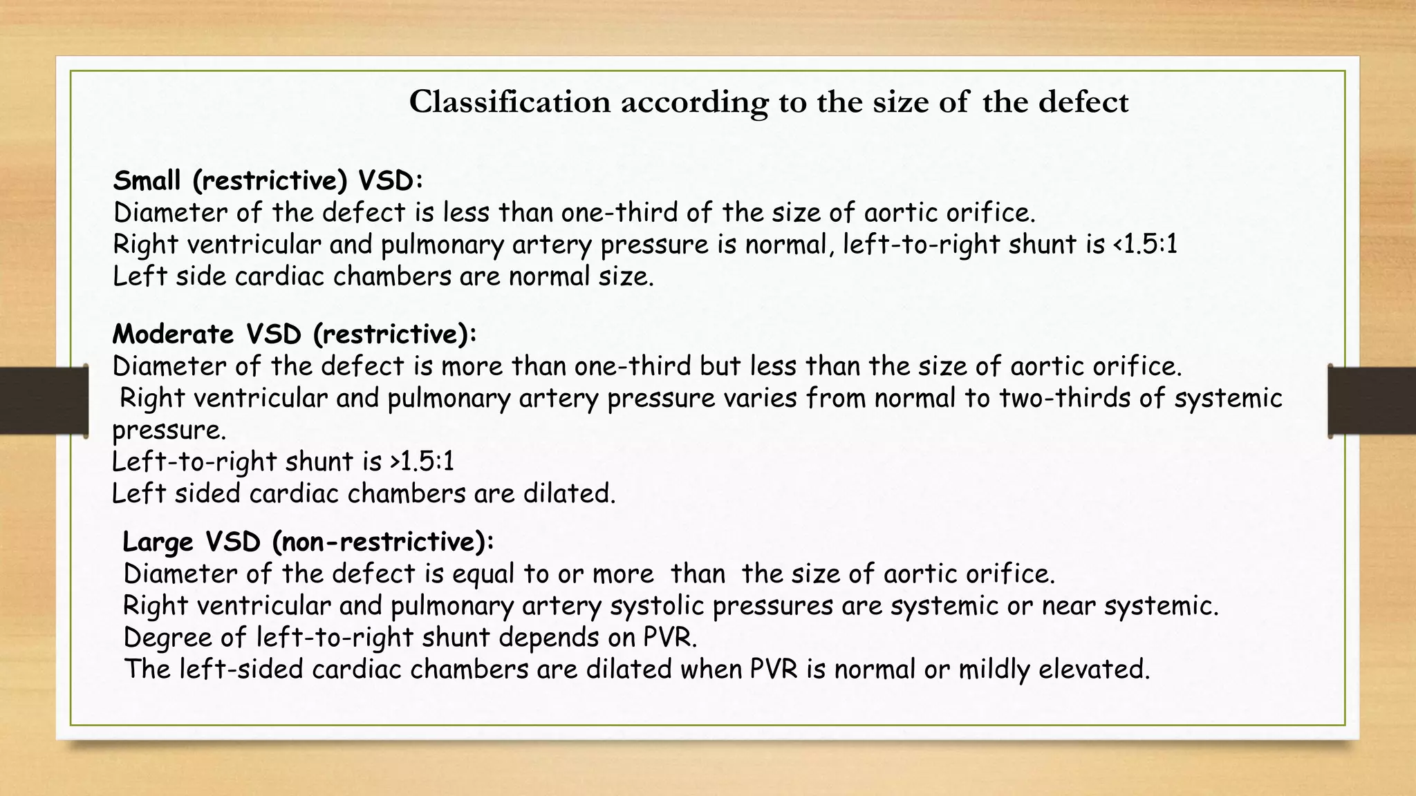
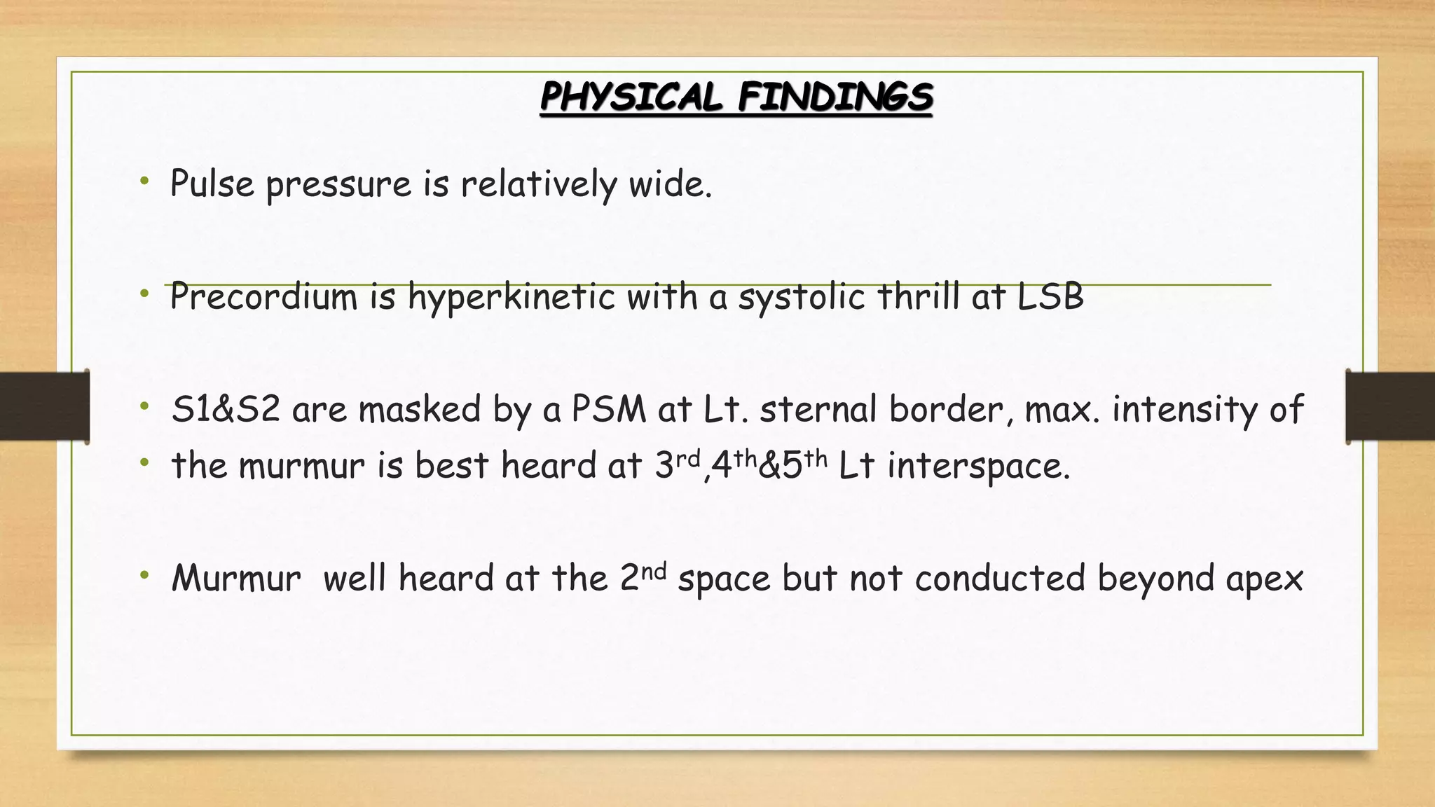
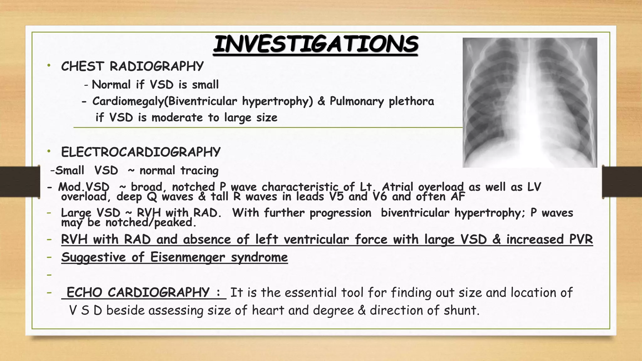
Clinical manifestations depend on the size of the defect and the pulmonary and systemic
vascular resistances. About 10% of patients with large VSDs die in 1st year, primarily due to
congestive heart failure.
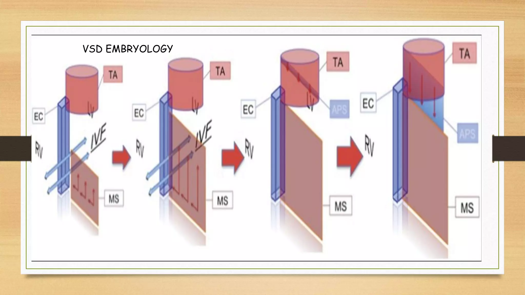
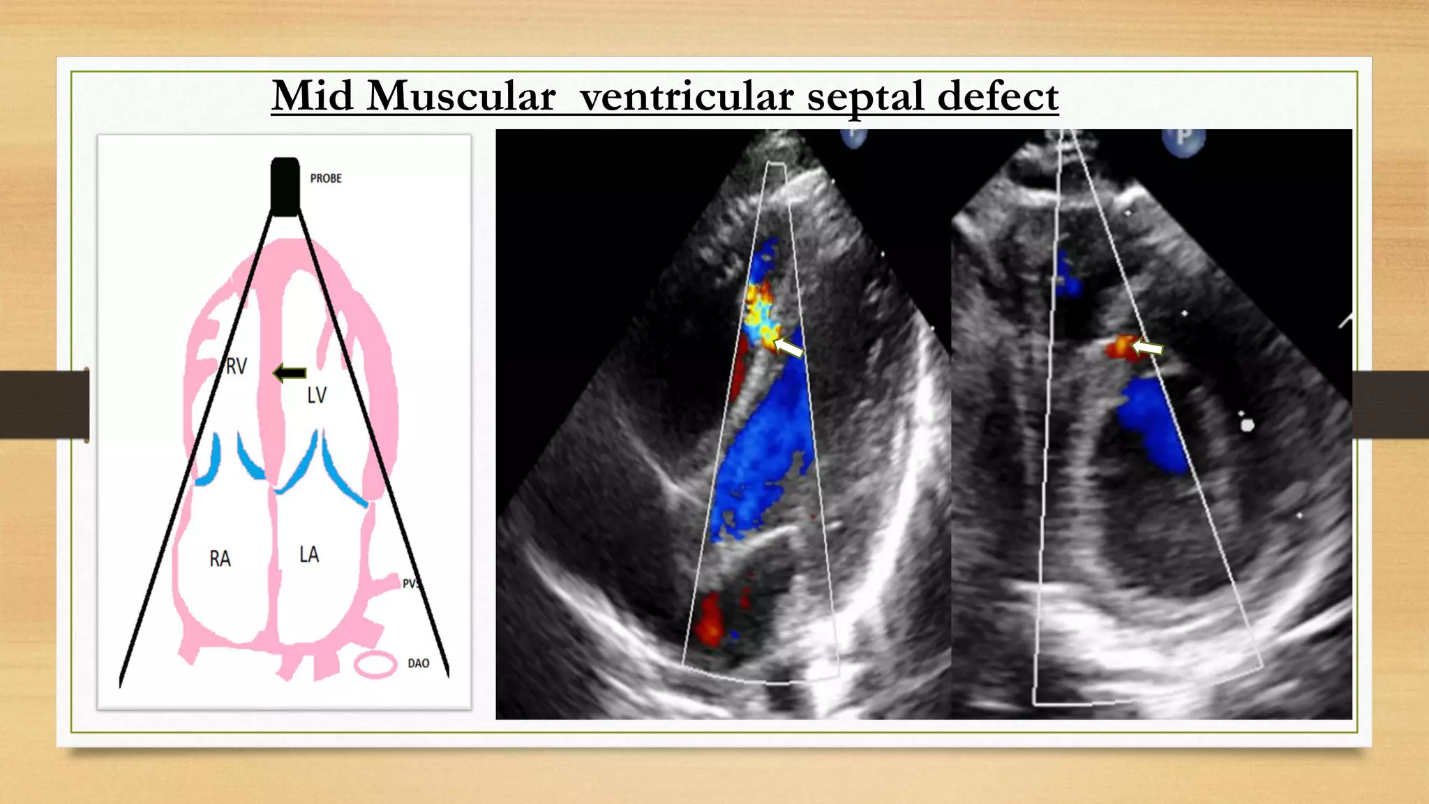
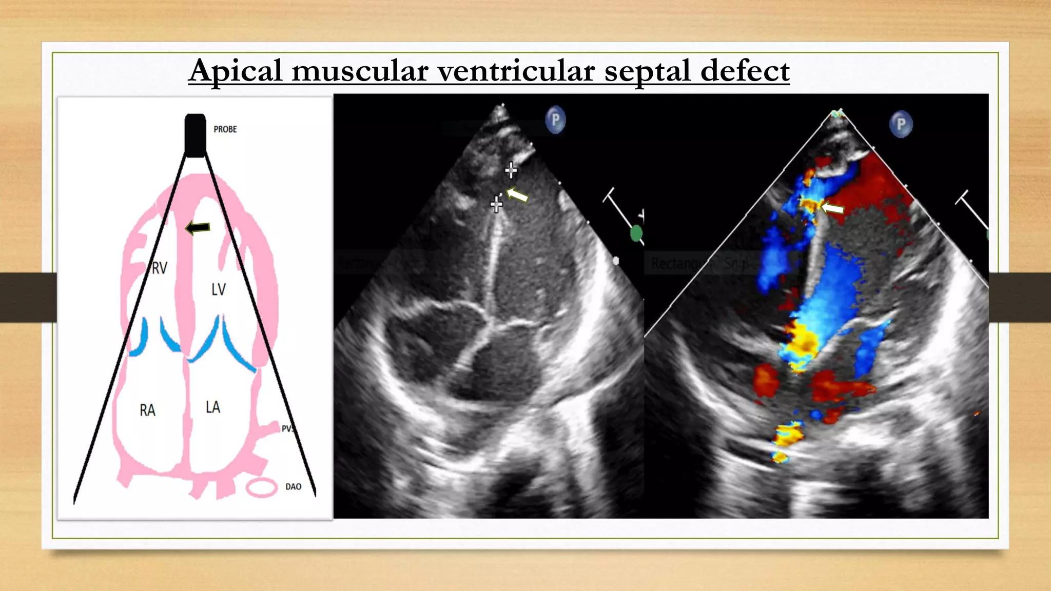
Rate of spontaneous closure depends on the size and location of the defect. Spontaneous
closure is uncommon in large VSDs. Inlet and malaligned VSDs almost never close spontaneously.
5. Muscular VSDs are more likely to close spontaneously, especially if they are not large. Decrease
in size of VSD is seen in 25% of patients.
Small VSDs have a >50% chance of spontaneous closure by 5 years of age and a >80% chance by
adolescence.
Progressive right ventricular outflow tract obstruction (Gasul phenomenon) may develop in 13%
and aortic regurgitation (AR) in 6% of patients.
In the historic series of Dr. Paul Wood, 52% of patients with large VSD developed irreversible
pulmonary vascular disease with the onset in infancy in four-fifths of them.
The incidence of IE in patients with small VSD is 1.3 per 1000 patient-years.
7. Diagrammatic representation of normal development of the IVS. The IVS is formed from 3 separate septa:
muscular, outlet, and inlet septa.
Early in embryologic development, the muscular septum (MS) grows upward from the floor of the ventricles
toward the already fused endocardial cushions (EC). The gap between the edge of the muscular IVS and EC is
called the interventricular foramen (IVF).
Meanwhile, 2 spiral ridges of tissue, the conotruncal ridges or truncoconal swellings, appear on the sides of the
truncus arteriosus (TA). The conotruncal ridges grow toward each other and fuse, forming a spiral-shaped
septum termed the aortopulmonary septum (APS). The APS divides the TA into the pulmonary trunk and aorta.
The conotruncal ridges also grow downward into the ventricles, meeting with the already fused endocardial
cushions and the muscular portion of the IVS. By the seventh to eighth week of gestation, the membranous
septum is formed when the APS, endocardial cushions, and muscular septum completely fuse, closing off the
IVS.
8. Diagrammatic representation of normal common developmental anomalies of the IVS. Defects in the fusion of the
muscular septum (MS) and the endocardial cushions (EC) result in membranous VSDs. Openings in the trabecular
portion of the IVS lead to muscular VSDs. Incomplete fusion of the aortopulmonary septum (APS) with the EC-MS
septum results in supracristal VSDs (SC-VSD).
10. Ventricular Septal Defects: Embryology and Imaging Findings
Rojas, Carlos Andres MD*; Jaimes, Camilo MD†; Abbara, Suhny
MD‡Author Information
Journal of Thoracic Imaging: March 2013 - Volume 28 - Issue 2 -
p W28-W34
doi: 10.1097/RTI.0b013e31824b5b95
Smaller defects, also called restrictive defects,
provide intrinsic resistance to flow and limit the amount of
shunted blood, maintaining a gradient between the 2
ventricles.
In contrast, large defects allow for unrestricted flow through
the defect and equalization of interventricular chamber
pressures.
11. Classification of ventricular septal defect
i. Peri-membranous: 80%
ii Outlet or sub-pulmonary (doubly committed): 5%–7%
iii. Inlet: 5%–8%
iv. Muscular: 5%–20%, these could be central (mid-muscular), apical, marginal (anterior,
septal-free wall area),
or multiple, “Swiss cheese” type.
12. Classification according to the size of the defect
Small (restrictive) VSD:
Diameter of the defect is less than one-third of the size of aortic orifice.
Right ventricular and pulmonary artery pressure is normal, left-to-right shunt is <1.5:1
Left side cardiac chambers are normal size.
Moderate VSD (restrictive):
Diameter of the defect is more than one-third but less than the size of aortic orifice.
Right ventricular and pulmonary artery pressure varies from normal to two-thirds of systemic
pressure.
Left-to-right shunt is >1.5:1
Left sided cardiac chambers are dilated.
Large VSD (non-restrictive):
Diameter of the defect is equal to or more than the size of aortic orifice.
Right ventricular and pulmonary artery systolic pressures are systemic or near systemic.
Degree of left-to-right shunt depends on PVR.
The left-sided cardiac chambers are dilated when PVR is normal or mildly elevated.
13.
14. CLINICAL FEATURES
• Race : no particular racial predilection
• Sex : no particular sex preference
• Age : of presentation -
Neonate and infants– difficult postnatal period, although ccf during first 6mths
is frequent, x ray chest cardiomegaly ECG LVH.
Children—after first year have variable clinical picture, depending upon the size
and location of the V S D.
small VSD – asymptomatic
large VSD – common symptoms, palpitation, feeding difficulties - suck & rest cycle
in neonate and infants, breathing difficulty on exertion, poor growth , frequent
cough and fever requiring hospitalization –suggestive of chest infections.
15. PHYSICAL FINDINGS
• Pulse pressure is relatively wide.
• Precordium is hyperkinetic with a systolic thrill at LSB
• S1&S2 are masked by a PSM at Lt. sternal border, max. intensity of
• the murmur is best heard at 3rd,4th&5th Lt interspace.
• Murmur well heard at the 2nd space but not conducted beyond apex
16. PHYSICAL FINDINGS
• Lt. 2nd space –widely and variable split second sound & accentuated P2
• Delayed diastolic murmur at the apex & S3
• Presence of mid-diastolic ,low pitched rumble at the apex is caused by increased
flow across the mitral valve & indicates Qp:Qs=2:1/greater
• Maladie de Roger – small VSD presenting in older children as a loud PSM without
other significant hemodynamic changes. S1 and S2 are distinct. Murmur ends well
before S2.
17. Natural History of VSD
Spontaneous diminution in size or closure : I 50-75% of restrictive perimembranous and
muscular VSD after birth
This occur in within first year, 60% before 3 years and 90% before 8 years.
Infant may develop -
Acquired RVOT or LVOT obstruction,
Develop AR due to AV prolapse
Infective endocarditis and
patient with VSD > 2/3 of aortic size and pulmonary arterial systolic pressure > 50% of
systemic arterial pressure is at risk of developing CHF initially and
pulmonary arterial obstruction later on in life if not treated appropriately.
18. VSD size & Haemodynamic
Size PA pressure PVR CHF
Restrictive <1/3 of aortic size Normal Normal NO
Moderately
restrictive
1/3-2/3 of aortic size < 50 % or systemic pressure LOW May be
large > 2/3 of aortic size >50% systemic pressure High Yes
19. INVESTIGATIONS
• CHEST RADIOGRAPHY
- Normal if VSD is small
- Cardiomegaly(Biventricular hypertrophy) & Pulmonary plethora
if VSD is moderate to large size
• ELECTROCARDIOGRAPHY
-Small VSD ~ normal tracing
- Mod.VSD ~ broad, notched P wave characteristic of Lt. Atrial overload as well as LV
overload, deep Q waves & tall R waves in leads V5 and V6 and often AF
- Large VSD ~ RVH with RAD. With further progression biventricular hypertrophy; P waves
may be notched/peaked.
- RVH with RAD and absence of left ventricular force with large VSD & increased PVR
- Suggestive of Eisenmenger syndrome
-
- ECHO CARDIOGRAPHY : It is the essential tool for finding out size and location of
V S D beside assessing size of heart and degree & direction of shunt.
20. P L A X VIEW
PERI MEMBRANOUS VSD
SUB-AORTIC VSD
SUB PUL. VSD
- ECHO CARDIOGRAPHY : It is the essential tool for finding out size and location of V S D
beside assessing size of heart, degree & direction of shunt for the management of patient.
38. The Tricuspid regurgitation with membranous VSD mainly occurred because of
1. short tricuspid valve septa – Dysplastic TV
2. Interminable anterior tricuspid valve septa – Richoceting TR
High velocity jet by impinging on anterior tricuspid leaflet and richoceting as TR.The TR is produced by the VSD jet (
Venturi effect) pushing the tricuspid anterior leaflet forward to open the tricuspid valve orifice and richoceting jet enter
right atrium as TR. In these patients, a moderate paramembranous VSD extended slightly below the septal tricuspid leaflet
with only partial obstruction of the VSD jet. We believe that when this mechanism for TR is found in association with a
moderate VSD, surgical VSD closure is warranted
3. Abnormal attachment point of the chordae tendineae to muscular VSD
4. Irregular adhesion of STL to right ventricular septal defect - Sometimes tricuspid septal aneurysm (which is closing large
VSD) can distort the tricuspid valve and cause tricuspid regurgitation.
If pulmonary arterial pressure is estimated by the tricuspid regurgitation method, the pulmonary artery systolic pressure
would be overestimated. Due to this the differential pressure of the tricuspid valve was not the same as that of the right
ventricle and right atrium, but instead the same as the left ventricle and right atrium.
Investigation of membranous ventricular septal defect complicated with tricuspid regurgitation in ventricular septal defect
occlusion
shu-ping liu, li li, ke-chun yao, na wang and jian-chang wang experimental and therapeutic medicine 5: 865-869, 2013
39.
40.
41.
42.
43.
44. 5. Gerbode VSD
6. Cleft in Tricuspid valve with inlet VSD - TR
7. Large VSD with Pulmonary arterial hypertension I Eisenmenger syndrome
45. Cardiac catheterization
It is required in patients with pulmonary hypertension and suspected pulmonary vascular
disease.
Cardiac catheterization is performed for interventional purpose - device closure
47. Where is shunt ?
74
78
74
95
90
90
95
95
Answer : At ventricular level
Left to right from LV to RV
V S D
Qp = 1/PAO2- PVO2 X 100= 1/90-95 X 100 =20
Qs= 1/SAO2- MVO2 X 100 = 1/95-74 X 100= 5
Qp/Qs=
𝟐𝟏
𝟓
= 4.2 :1
QeS = 1/ PAO2-MVO2 X 100 = 1/90-74 X 100 = 6.6
LEFT TO RIGHT SHUNT = Qp-QeS = 20-6.6 = 14.6
48. Qp= 1/PAO2-PVO2 X 100 = 1/87-94 X 100 = 1/7 X 100 = 14.2L
Qs = 1/SAO2-MVO2 X 100 = 1/95-68 X100= 1/27 X 100 = 3.8
QeS =1/ PVO2- MVO2 X 100 = 1/ 94-68 = 1/26 X 100 = 3.8
Qp/QS = 14.21/3.8 = 3.7
LT TO RT = QP – QeS = 14.2 -3.8 = 10. 4 L
RT TO LEFT = Qs- QeS = 3.8 – 3.8 = 0
NET SHUNTING 10.41 FROM LEFT TO RIGHT AT VSD
50. 74
78
74
90
74
74
100
90
Where is shunt ?
What is the direction of shunt ?
What is diagnosis?
Ventricular
Right to left
VSD
Qp=1/PAO2-PVO2 X 100= 1/74-100 X 100 = 1/26 X 100 = 4 L
Qs = 1/SAO2-MVO2X 100 = 1/90-74 X 100 = 1/16 X 100 = 6.6L
QeS = 1/PVO2-MVO2 X 100 = 1/100-74 X 100 = 1/26 X100 = 4
Qp /Qs = 4/6 = 0.6.6
RT to LT shunt = Qs –QeS = 6-4 = 2
LT to Rtshunt = Qp-QeS = 4-4 = 0
52. What is spectrum of VSD ?
VSD – Peri-memberanous, Subaortic, Sub-pulmonary, Inlet, Muscular
Fetal echo revealed
1st Step : – 4 CV lateral and basal view will revealed echo dropout with bidirectional color on color
doppler.
53. V S D
Peri-membranous Sub- aortic
Muscular Inlet
Sub -
pulmonary
Pul. Stenosis
Pul. Atresia
Absent Pul valve
PS
MR,TR ( AV Regurgitation
Associated cardiac abnormality - Fetus with VSD
54. What are the Associated extra cardiac abnormality
in Fetus with VSD ?
V S D
Peri-membranous Sub- aortic
Muscular Inlet
Sub -
pulmonary
21 trisomy with AVSD 40%
Sub aortic VSD one out let
Vessel - 22q11.1 deletion
Pul stenosis with absent radius
And thumb – Holt Oram syn
13T - Omphalocele,
Holo-procencephaly
cleft lip & palate
Polydactyly cutis aplasia
Micro-ophthalmia
18 T- Abnormal
fisting of hand
Low birth weight, under developed finger nail
Microcephaly, Ptosis, blephrophymosis
55. What is the possibility of genetic Etiology ?
Fetus VSD without extra cardiac abnormality – no genetic cause
Fetus with VSD and Extra-cardiac abnormality –
AVSD - 21 trisomy
VSD with Omphalocele and holoprocencephaly – 13T
Fisting of hand 18 T
Sub aortic VSD & PS and absent radius and thumb - Holt Oram syn
Sub aortic VSD & single outlet vessel - truncus arteriosus – 22q11.1
56. Which genetic test you will offer to couple ?
Karyotyping as first test if we suspect aneuploidy in fetus with DNA preservation.
FISH – For 22q11.1 deletion if fetus have CoA/absent thymus/TOF with pul atresia
CGH array is the second line test to detect aneuploidy, micro deletion and duplication
NGS – is third line investigation to diagnose single gene disorders – Holt Oram syn etc
57. How to counsel couple regarding risk of recurrence ?
(TGA)
•The general population CHD risk is ~1%
•For parents with one affected child the recurrence risk of CHD is between 2-5%
•For parents of two affected children the recurrence risk of CHD is 10-15%
• If father have VSD risk is 2% and if mother had VSD recurrence risk is 6%
•It depends upon cause – aneuploidy, syndrome
58. Does fetus need regular follow up after diagnosis ?
VSD
isolated With VSD&/or PS Extra cardiac anomalies
Follow regularly by echo
Patient with extra cardiac anomaly – omphalocele need regular follow up
Patient with AVSD need follow up
59. How to counsel couple regarding pregnancy management ?
SITE OF DELIVERY : Hospitral
TIMING OF DILERY : AT TERM IF PFO & / OR PDA IS NORMAL; NO CONSTRICTION IN-UTERO
MODE OF DELIVERY : NORMAL, IF NO OBSTETRIC COMPLICATION
PLANNING OF POSTNATAL CARE: POST NATAL ECHO TO CONFIRM DIAGNOSIS AND NATURE OF CARDIAC LESION
60. How to counsel couple regarding post natal management ?
(AVSD)
VSD
isolated VSD &/or PS Extra cardiac anomalies
Rx depends up on the size of VSD Rx depends up on severity of PS and SPO2
61. Medical management of ASD
Drug therapy is recommendation for patients with CHDs- ASD,VSD and PDA, who have
abnormal cardiac morphology or function like –
Cardiac volume over load that is dilated cardiac chamber despite preserved systolic function,
Valvular regurgitation
Pulmonary hypertension because of volume overload, but no symptoms of heart failure.
62. Digoxin is indicated in heart failure associated with reduced systolic function of heart.
Utility of Digoxin in heart failure secondary to volume overload of the ventricle, as seen
in left to right shunt lesions, is less clear, since the myocardial contractility is normal in
such cases.
Rapid digitalization is usually not indicated when using digoxin for heart failure.
Rapid digitalization may be indicated for treatment of acute tachyarrhythmias.
The maintenance dose is given in twice daily doses for children under 10 years and once daily
for children above 10 years.
Digoxin “holiday” is generally not needed in children.
The half life of digoxin is markedly prolonged in preterm babies and in those with renal
dysfunction.
Dose of digoxin should he halved when using amiadarone.
63. Diuretics
are widely used in heart failure because of the symptomatic relief from fluid overload with in
minutes of administration - Furosemide, torsemide.
Dosages and Pharmacodynamics: Oral: 1-2 mg/kg every 12 hours, maximum of 4 mg/kg/day;
intravenous: 1 mg/kg/dose up to 3-4 times a day;
Continuous IV infusion: 1-4 mg/kg/day. Continuous infusion may be better and safer in acute
heart failure and in postoperative setting.
The onset of action starts in 10-20 minutes after an IV dose and 20-30 minutes after oral
administration. The duration of action is six hours.
The dose does not need to be adjusted in renal or hepatic impairment.
Furosemide may increase chances of digoxin toxicity by producing hypokalemia.
It activates the renin angiotensin aldosterone axis (RAAS), producing vasoconstriction, which
is detrimental in heart failure. Concomitant use of ACEi (vasodilator) is recommended,
whenever possible.
64. Vasodilators:
Angiotensin Converting Enzyme Inhibitors (ACEi)
ACEi decrease the adrenergic drive and block the heart failure induced activation of renin
angiotensin aldosterone axis (RAAS).
Increased levels of aldosterone and angiotension II have been associated with poor outcome in
heart failure.
ACEi also increase bradykinin which has natrinuretic properties.
Currently ACEi therapy is recommended as the first line treatment for heart failure, when it
is not secondary to an obstructive lesion.
Enalapril. Enalapril is useful for older children. It is longer acting and given twice daily. The
dose is 0.1- 0.5 mg/kg/dose twice a day. The initial dose may be smaller.
BP and renal parameters should be monitored when up titrating the dose
65. Beta blockers
Heart failure results in activation of sympathetic nervous system and increased levels of
circulating catecholamines. Chronic activation of sympathetic nervous system leads to worsening
of heart failure by inducing myocardial apoptosis and fibrosis. Circulating catecholamines also
induce peripheral vasoconstriction along with renal retention of salt and water. Betablockers
antagonize these deleterious effects. In addition, betablockers also have antiarrhythmic
effect.
Carvedilol is a non selective beta blocker which also has an anti-oxidant property. Due to its
alpha blocking effect, carvedilol exerts a vasodilatory effect. It improves functional class and
fractional shortening in children with ventricular dysfunction
Carvedilol: 0.1 mg/kg/day in two divided doses, increase at 1-2 weekly interval to 1 mg/kg/day
with a maximum of 2 mg/kg/day.
Metoprolol: 0.2-0.4 mg/kg/day initially, gradually increase to a maximum of 1 mg/kg/day in two
divided doses.
66. Do
• Treat the underlying cause of heart failure.
• Digoxin has a narrow safety window in
children.
• Continuous infusion of furosemide may be
better in acutely ill cases .
• A persistent tachycardia (>180) may indicate
“tachycardio-myopathy” as the cause of heart
failure.
• Rapid digitalization is not required for
majority.
drug therapy of cardiac diseases in children working group on management of congenital heart diseases in india correspondence to: dr anita saxena,
professor of cardiology, all india institute of medical sciences, new delhi 110029, india indian pediatrics 2009
Do Not
• Combine angiotensin converting enzyme
inhibitors
(ACEi) with Angiotensin receptor blockers (ARB)
(Class III).
• Avoid combining ACEi and spironolactone, if
necessary, monitor potassium levels (Class II b)
• Do not give ACEi in heart failure secondary to
pressure overload (Class III)
• Avoid using ACEi in acute decompensated heart
failure (Class II b)
• Betablockers should not be initiated in acute
decompensated stage of heart failure (Class III)
• Potassium supplements are not required in early
infancy
67. Indications and timing of closure (all Class I recommendations)
Small VSD
No symptoms, normal PA pressure, normal left heart chambers, no cusp prolapse:
a. Annual follow-up till 10 years of age, then every 2–3 years
b. b. Closure indicated if the patient has had an episode of
1. Endocarditis
2. Develops cusp prolapse with AR or any VSD if have aortic cusp prolapse of any degree at
the time of diagnosis, should be operated as early as possible to prevent AV damage.
3. Develops progressive significant right ventricular outflow tract obstruction.
69. Indications and timing of closure (all Class I recommendations)
Moderate VSD
a. Asymptomatic:
Normal pulmonary artery pressure with left heart dilation: Closure of VSD by 2–5 years of
age
b. Symptomatic:
If controlled with medications : VSD closure by 1–2 years of age.
70. Indications and timing of closure (all Class I recommendations)
large VSD
a. Poor growth/congestive heart failure not controlled with medications (furosemide/
spironolactone/enalapril ± digoxin): As soon as possible
b. Controlled heart failure: By 6 months of age
71. Contra - indications of closure
Severe pulmonary arterial hypertension with irreversible pulmonary vascular occlusive disease
(Class III). Patients with borderline operability due to pulmonary vascular disease should be
referred or subjected to for cardiac catheterization evaluation for operability.
The decision to operate or not should be made on an individual basis taking into account the
total picture of the case including results of the investigations.
72. Method of closure
Surgery
i. Patch closure is the standard therapy in most patients. Route of closure depends on the
location of the defect, but left ventri-culotomy is best avoided.
ii. Temporary pulmonary artery banding: Palliative option in patients with-
a. Multiple VSDs (Swiss cheese VSDs), inaccessible VSDs (Class I)
b. Patients with contraindications for cardiopulmonary bypass, e.g., sepsis (Class IIa). iii.
Surgical options for patients with borderline operability: Fenestrated VSD patch closure,
fenestrated flap valve VSD patch closure, or leaving (or creating) a 5 mm ASD. Such
procedures should only be done after discussion with the family as in some cases, pulmonary
hypertension may not regress or may regress and later worsen following surgery (Class IIb).
75. Device closure
i. Eligibility criteria:
a. Weight >8 kg (5 kg for muscular VSD)
b. Left-to-right shunt >1.5:1.
ii. Indications
a. Class I – Midmuscular VSD, anterior muscular VSD, postoperative residual VSD
b. Class IIb – Perimembranous VSD with at least 4 mm distance from the aortic valve.
iii. Contraindications -
a. VSD with irreversible pulmonary vascular disease
b. Pre-existing left bundle branch block or conduction abnormalities
c. Any AR
d. Associated lesions requiring surgery
e. Inlet, subpulmonic VSD.
76. Device closure
iv. Device should not be deployed if any of the following findings develop at the time of
procedure:
a. Any degree of AR
b. Conduction defect: complete heart block (CHB)/ left bundle branch block
c. Mitral or tricuspid regurgitation.
77. Recommendations for follow-up
i. Follow-up after surgery:
Clinical, ECG, and echo in the 1st year only. No further follow-up is required if no residual defect
or pulmonary hypertension. Patient/guardians should be explained about reporting to hospital in
case of any cardiac symptoms or symptoms suggestive of arrhythmias.
ii. Follow-up protocol for device closure:
a. Antiplatelet agents: Aspirin (3–5 mg/kg/day) is given a day before or immediately after
procedure and continued for total duration of 6 months.
b. b. Follow-up visits: At 1 month, 6 months, 1 year, then annually till 5 years, and then every 3–5
years. Echocardiogram and ECG are to be done at each visit in addition to clinical evaluation. c.
iii. IE prophylaxis is recommended for 6 months after device or surgical closure. However, all
patients are advised to maintain good oro-dental hygiene after this period also.